. 2020 Sep 29;22(1):102.
doi: 10.1186/s13058-020-01344-0.

Fibroblasts direct differentiation of human breast epithelial progenitors

Affiliations

Fibroblasts direct differentiation of human breast epithelial progenitors

Mikkel Morsing et al. Breast Cancer Res. .

Abstract

Background: Breast cancer arises within specific regions in the human breast referred to as the terminal duct lobular units (TDLUs). These are relatively dynamic structures characterized by sex hormone driven cyclic epithelial turnover. TDLUs consist of unique parenchymal entities embedded within a fibroblast-rich lobular stroma. Here, we established and characterized a new human breast lobular fibroblast cell line against its interlobular counterpart with a view to assessing the role of region-specific stromal cues in the control of TDLU dynamics.

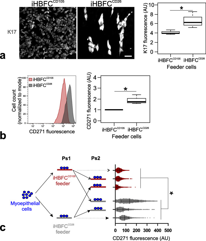
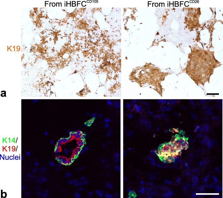
Methods: Primary lobular and interlobular fibroblasts were transduced to express human telomerase reverse transcriptase (hTERT). Differentiation of the established cell lines along lobular and interlobular pathways was determined by immunocytochemical staining and genome-wide RNA sequencing. Their functional properties were further characterized by analysis of mesenchymal stem cell (MSC) differentiation repertoire in culture and in vivo. The cells' physiological relevance for parenchymal differentiation was examined in heterotypic co-culture with fluorescence-activated cell sorting (FACS)-purified normal breast primary luminal or myoepithelial progenitors. The co-cultures were immunostained for quantitative assessment of epithelial branching morphogenesis, polarization, growth, and luminal epithelial maturation. In extension, myoepithelial progenitors were tested for luminal differentiation capacity in culture and in mouse xenografts. To unravel the significance of transforming growth factor-beta (TGF-β)-mediated crosstalk in TDLU-like morphogenesis and differentiation, fibroblasts were incubated with the TGF-β signaling inhibitor, SB431542, prior to heterotypic co-culture with luminal cells.

Results: hTERT immortalized fibroblast cell lines retained critical phenotypic traits in culture and linked to primary fibroblasts. Cell culture assays and transplantation to mice showed that the origin of fibroblasts determines TDLU-like and ductal-like differentiation of epithelial progenitors. Whereas lobular fibroblasts supported a high level of branching morphogenesis by luminal cells, interlobular fibroblasts supported ductal-like myoepithelial characteristics. TDLU-like morphogenesis, at least in part, relied on intact TGF-β signaling.

Conclusions: The significance of the most prominent cell type in normal breast stroma, the fibroblast, in directing epithelial differentiation is largely unknown. Through establishment of lobular and interlobular fibroblast cell lines, we here demonstrate that epithelial progenitors are submitted to stromal cues for site-specific differentiation. Our findings lend credence to considering stromal subtleties of crucial importance in the development of normal breast and, in turn, breast cancer.

Keywords: Breast; Differentiation; Epithelial progenitors; Fibroblast.


Conflict of interest statement

The authors declare that they have no competing interests.

Figures

Fig. 1
Fig. 1
Lineage specific markers are maintained in hTERT immortalized HBFCs. a Diagram depicting the cumulative population doublings of CD105+ and CD26+ HBFCs transduced with empty vector (evHBFCCD105; open squares and evHBFCCD26; open triangles) or hTERT (iHBFCCD105; closed squares and iHBFCCD26; closed triangles) and recorded between passages 10 (day zero) and 57 (day 412). Whereas iHBFCCD105 and iHBFCCD26 continued to proliferate, empty vector controls ceased to expand after around 18 and 24 population doublings, respectively. Also, note that iHBFCCD26 have an intrinsic growth advantage irrespective of immortalization. b iHBFCs were examined repeatedly for the expression of lineage markers CD105 and CD26 by immunoperoxidase staining (brown), here illustrated for cells in passage 50. Like their primary ancestors, iHBFCCD105 are CD105high/CD26low (left) and iHBFCCD26 are CD105low/CD26high (right). Nuclei are counterstained with hematoxylin (blue) (bar = 50 μm)
Fig. 2
Fig. 2
iHBFCs resemble HBFCs by gene expression profiles and CD140b and CD248 are lobular markers in situ. a Venn diagram showing the number of differentially expressed genes (DEGs; p < 0.05 and fold change ≥ 2) based on genome wide RNA-sequencing of CD105+ and CD26+ HBFCs and iHBFCs, respectively. Bar diagram shows the percent overlap of DEGs between the cells indicated. b Heatmap of expression values of DEGs annotated with a cluster of differentiation (CD) name represented in a for iHBFCCD105and iHBFCCD26. c Cryostat sections of normal breast biopsies stained with peroxidase (brown) for CD140b and CD248 selected based on the CD gene expression profile of iHBFCs. Note the relatively intense staining in TDLUs (left) versus ducts (right). Nuclei are counterstained with hematoxylin (blue) (bar = 100 μm)
Fig. 3
Fig. 3
iHBFCCD105 support luminal epithelial growth and TDLU-like branching morphogenesis. Comparison of the capacity of iHBFCCD105 and iHBFCCD26 to induce human breast epithelial morphogenesis. a Phase contrast micrographs of luminal breast epithelial cells co-cultured for 16 days on passage 40 iHBFCCD105 (left) or iHBFCCD26 (right) (bar = 100 μm). Only iHBFCCD105 facilitate elaborate TDLU-like branching morphogenesis. b Double immunofluorescence staining of luminal epithelial/iHBFC co-cultures with K19 (red) and MUC1 (green; bar = 100 μm). Note the staining of correctly polarized MUC1 in K19+ structures in both co-cultures. c Illustration of difference in induced branching morphogenesis by iHBFCCD105 and iHBFCCD26, respectively, by low magnification imaging and segmentation in ImageJ of branching morphogenesis in luminal epithelial/iHBFC co-cultures stained by peroxidase for K19 (brown). Segmented images show epithelial structures projected in black pixels (bar = 1000 μm). d Dot plot depicting the inductive capacity of seven pairs of iHBFCCD105 (left) and iHBFCCD26 (right) measured as the number of luminal epithelial structures per square unit area using luminal epithelial cells from five different biopsies. Consistently, iHBFCCD105 have higher inductive capacity (asterisk indicates significance at p < 0.05 by Wilcoxon signed-rank test)
Fig. 4
Fig. 4
iHBFCCD26 convey a ductal-like differentiation of myoepithelial cells. a Images showing FACS sorted CD271high/MUC1low breast primary myoepithelial cells in co-culture with iHBFCCD105 (left) and iHBFCCD26 (right), fluorescently labeled for K17 (white) and K14 (not shown) by immunocytochemistry. K14 staining was used as a guide in image analysis to identify K14+ myoepithelial cells prior to measuring myoepithelial K17 staining intensity. Box plot shows interquartile range and median of K17 mean fluorescence in arbitrary units (AU) of three biopsies (whiskers indicate upper and lower quartiles; asterisk indicates significance at p < 0.05 by Kruskal-Wallis rank-sum test). b Primary myoepithelial/fibroblast co-cultures (iHBFCCD105 (red), iHBFCCD26 (gray)), were single cell suspended and stained for CD271 before analysis by FACS. Histogram shows cell count normalized to mode versus myoepithelial CD271 staining intensity in arbitrary units (AU) of a single biopsy (left) and box plot shows the interquartile range and median of the mean of CD271 fluorescence intensity relative to iHBFCCD105 in arbitrary units of three biopsies (right; whiskers indicate upper and lower quartile, asterisk indicates significance at p < 0.05 by Kruskal-Wallis rank-sum test). c Schematic showing the experimental outline (left): primary CD271+ myoepithelial cells are plated onto confluent fibroblast feeders (passage 1 co-culture, Ps1), from which myoepithelial cells are isolated and then re-plated onto new fibroblast feeders (passage 2 co-culture, Ps2). Dot plot (right) shows normalized myoepithelial CD271 fluorescence in arbitrary units (AU) with mean values and standard deviations indicated by vertical bars as measured by FACS of 2250 cells in passage 1 and 2 co-cultures grouped according to feeder (iHBFCCD105 (red) or iHBFCCD26 (gray)). Note that the myoepithelial phenotype shifts as a consequence of a switch between fibroblasts (asterisk indicates significance at p < 0.05 by nested t test)
Fig. 5
Fig. 5
The luminal differentiation repertoire of myoepithelial progenitors is directed by interaction with specialized fibroblasts. a Comparison of capacity of fibroblasts to direct epithelial progenitor capacity. Myoepithelial cells co-cultured with iHBFCCD105 or iHBFCCD26 were passaged and subjected to luminal differentiation conditions at clonal density and peroxidase stained for K19. While the induced K19 appeared mainly scattered when derived from iHBFCCD105 co-culture (left), additional rather homogenous islets presented from iHBFCCD26 co-cultures (right). The distinct phenotypes were observed in five out of seven tests with absence of homogeneous islets from iHBFCCD26 in two tests (bar = 500 μm). b Representative multicolor confocal images (K19, red; K14, green; nuclei, blue) of cryostat sections of xenografted NOG mice 8 weeks after orthotopic injection of myoepithelial cells from primary co-culture with iHBFCCD105 or iHBFCCD26. Bilayered epithelial structures were obtained in 6/10 and 5/8 injections from iHBFCCD105 and iHBFCCD26, respectively, although at limited numbers, down to a few per transplant. Whereas iHBFCCD105 co-culture derived myoepithelial cells readily differentiated into luminal K14−/low/K19+ cells, co-culture with iHBFCCD26 resulted mainly in K14+/K19+ luminal cells (bar = 50 μm)
Fig. 6
Fig. 6
HBFCCD105 TGF-β signaling supports parenchymal morphogenesis. a Overview of experimental design in which HBFCs are plated and exposed to 10 μM SB431542 or vehicle (DMSO) from day 7 to day 10 at which SB431542 or vehicle are removed and primary CD271low/MUC1high luminal breast epithelial cells are added and co-cultured for 10 days prior to assessment of epithelial structure formation. b 15 tests representing recombinations of four fibroblast biopsies and five epithelial biopsies are presented in a paired dot plot and a representative set of micrographs, showing a significant reduction in epithelial structure formation per square unit area in response to SB431542 versus vehicle in HBFCCD105 co-cultures only (asterisk indicates significance at p < 0.05 by Wilcoxon signed-rank test, ns = not significant) (bar = 1000 μm)

References

    1. Driskell RR, Lichtenberger BM, Hoste E, Kretzschmar K, Simons BD, Charalambous M, Ferrori SR, Herault Y, Pavlovic G, Ferguson-Smith AC, et al. Distinct fibroblast lineages determine dermal architecture in skin development and repair. Nature. 2013;504:277–281. doi: 10.1038/nature12783. - DOI - PMC - PubMed
    1. Chen X, Song E. Turning foes to friends: targeting cancer-associated fibroblasts. Nature Rev Drug Discov. 2019;18:99–115. doi: 10.1038/s41573-018-0004-1. - DOI - PubMed
    1. Tlsty TD, Gascard P. Stromal directives can control cancer. Science. 2019;365:122–123. - PMC - PubMed
    1. Wellings SR, Jensen HM, Marcum RG. An atlas of subgross pathology of the human breast with special reference to possible precancerous lesions. J Natl Cancer Inst. 1975;55:231–273. - PubMed
    1. Cardiff RD, Wellings SR. The comparative pathology of human and mouse mammary glands. J Mammary Gland Biol Neoplasia. 1999;4(1):105–122. doi: 10.1023/A:1018712905244. - DOI - PubMed

Publication types

MeSH terms