

Epidemiology and genotyping of Anaplasma marginale and co-infection with piroplasms and other Anaplasmataceae in cattle and buffaloes from Egypt
- PMID: 32993778
- PMCID: PMC7526245
- DOI: 10.1186/s13071-020-04372-z
Epidemiology and genotyping of Anaplasma marginale and co-infection with piroplasms and other Anaplasmataceae in cattle and buffaloes from Egypt
Abstract
Background: Anaplasma marginale is an obligate intracellular bacterium and the main cause of bovine anaplasmosis in tropical and subtropical regions. In Egypt, data regarding the prevalence of A. marginale in ruminant hosts and of the circulating genotypes is lacking. This study therefore aimed to (i) investigate the presence, epidemiology and genotypes of A. marginale in cattle and buffaloes in Egypt, (ii) to evaluate suitable diagnostic tools and (iii) to identify co-infections of A. marginale with other selected tick-borne pathogens.
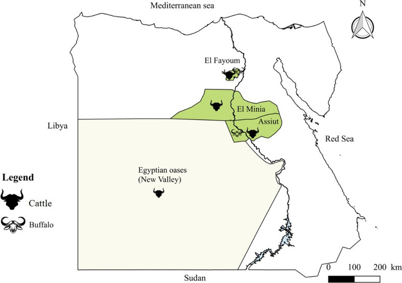
Methods: Blood samples were collected from 394 animals (309 cattle and 85 buffaloes) from three different areas in Egypt. For the detection of A. marginale infection, several tests were compared for their sensitivity and specificity: blood smear analysis, enzyme-linked immunosorbent assay (ELISA), PCR, real-time PCR and reverse line blot (RLB) assay. Co-infections with A. marginale, piroplasms and other Anaplasmataceae were surveyed by RLB while A. marginale genotypes were identified by amplifying and sequencing the partial msp1α gene.
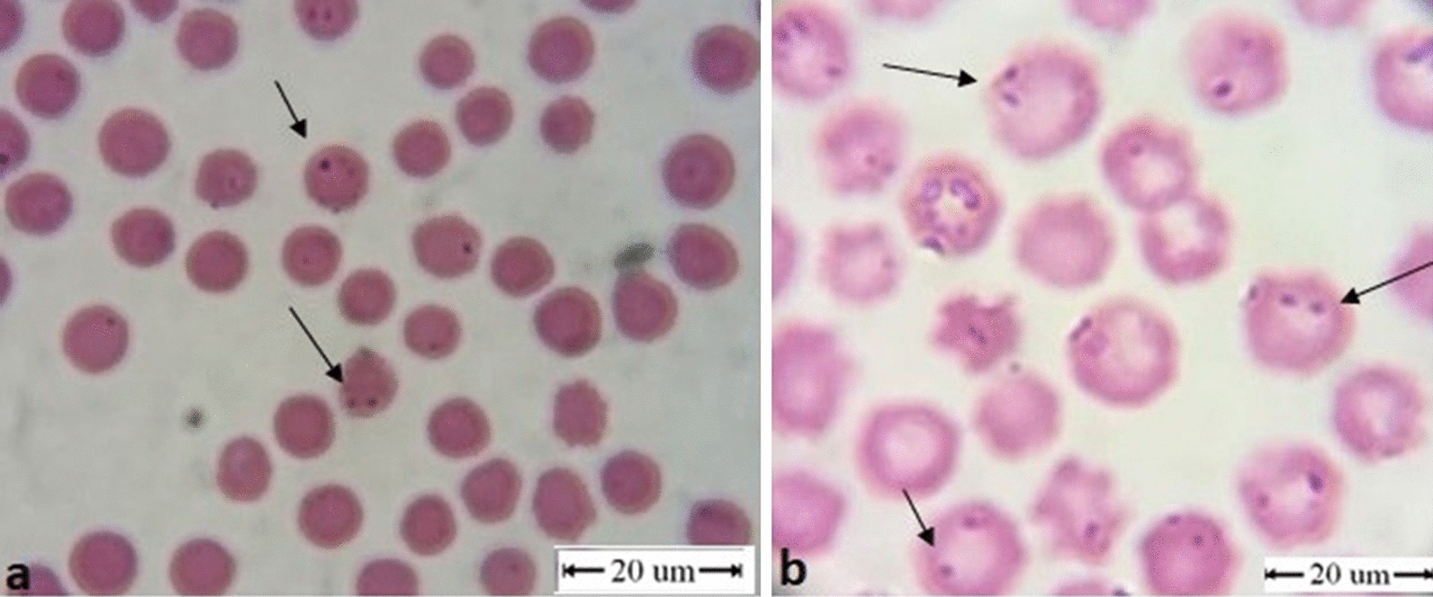
Results: Anaplasma marginale DNA was amplified by qPCR in 68.3% of cattle and 29.4% of buffaloes. RLB showed infection with A. marginale in 50.2% of cattle and 42.5% of buffaloes. Blood smear analysis detected this agent in 16.2% of cattle and 2.4% of buffaloes. ELISA showed specific antibodies against A. marginale in 54.9% of cattle. Anaplasma marginale was associated, in cattle and buffaloes, with several tick-borne pathogens (Theileria annulata, Babesia bovis, Babesia bigemina, Babesia occultans and Anaplasma platys). A significant difference of A. marginale infection level was noticed in cattle, where animals between 3-5-years-old had a higher prevalence (79.2%) compared to those older than 5 years (36.4%) and younger than 3 years (59.7%) and one year (64.5%), respectively (P = 0.002281). Microsatellite analysis identified 15 different genotypes.
Conclusions: The epidemiological findings revealed high prevalence of A. marginale in cattle and buffaloes in all the investigated areas. The circulation of diverse genotypes was observed, most of these A. marginale genotypes being specific for Egypt. The qPCR assay was confirmed to be the most sensitive tool for detection of A. marginale in cattle and buffaloes even in the carrier state, highlighting the importance of using suitable diagnostic tests.
Keywords: Anaplasma marginale; Buffaloes; Cattle; Co-infections; Diagnostic tools; Egypt; Genotypes.
Conflict of interest statement
The authors declare that they have no competing interests.
Figures
References
-
- AL-Hosary A, Ahmed L, Ahmed J, Nijhof A, Clausen P-H. Epidemiological study on tropical theileriosis (Theileria annulata infection) in the Egyptian Oases with special reference to the molecular characterization of Theileria spp. Ticks Tick-Borne Dis. 2018;9:1489–1493. doi: 10.1016/j.ttbdis.2018.07.008. - DOI - PubMed
-
- Al-Hosary A, Ahmed L, Seitzer U. First report of molecular identification and characterization of Theileria spp. from water buffaloes (Bubalus bubalis) in Egypt. Adv Anim Vet Sci. 2015;3:629–633. doi: 10.14737/journal.aavs/2015/3.12.629.633. - DOI
-
- Abdel-Rady A, Ahmed LS, Mohamed A, Al-Hosary A. Using polymerase chain reaction (PCR) for diagnosis of bovine theileriosis in Upper Egypt. IJAVMS. 2010;4:67–74.
MeSH terms
Grants and funding
LinkOut - more resources
Full Text Sources
