. 2020 Sep 29;13(651):eabe1202.
doi: 10.1126/scisignal.abe1202.

High-resolution structures of the SARS-CoV-2 2'- O-methyltransferase reveal strategies for structure-based inhibitor design

Affiliations

High-resolution structures of the SARS-CoV-2 2'- O-methyltransferase reveal strategies for structure-based inhibitor design

Monica Rosas-Lemus et al. Sci Signal. .

Abstract

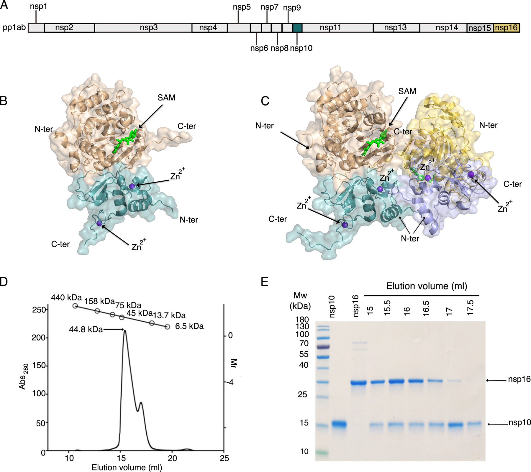
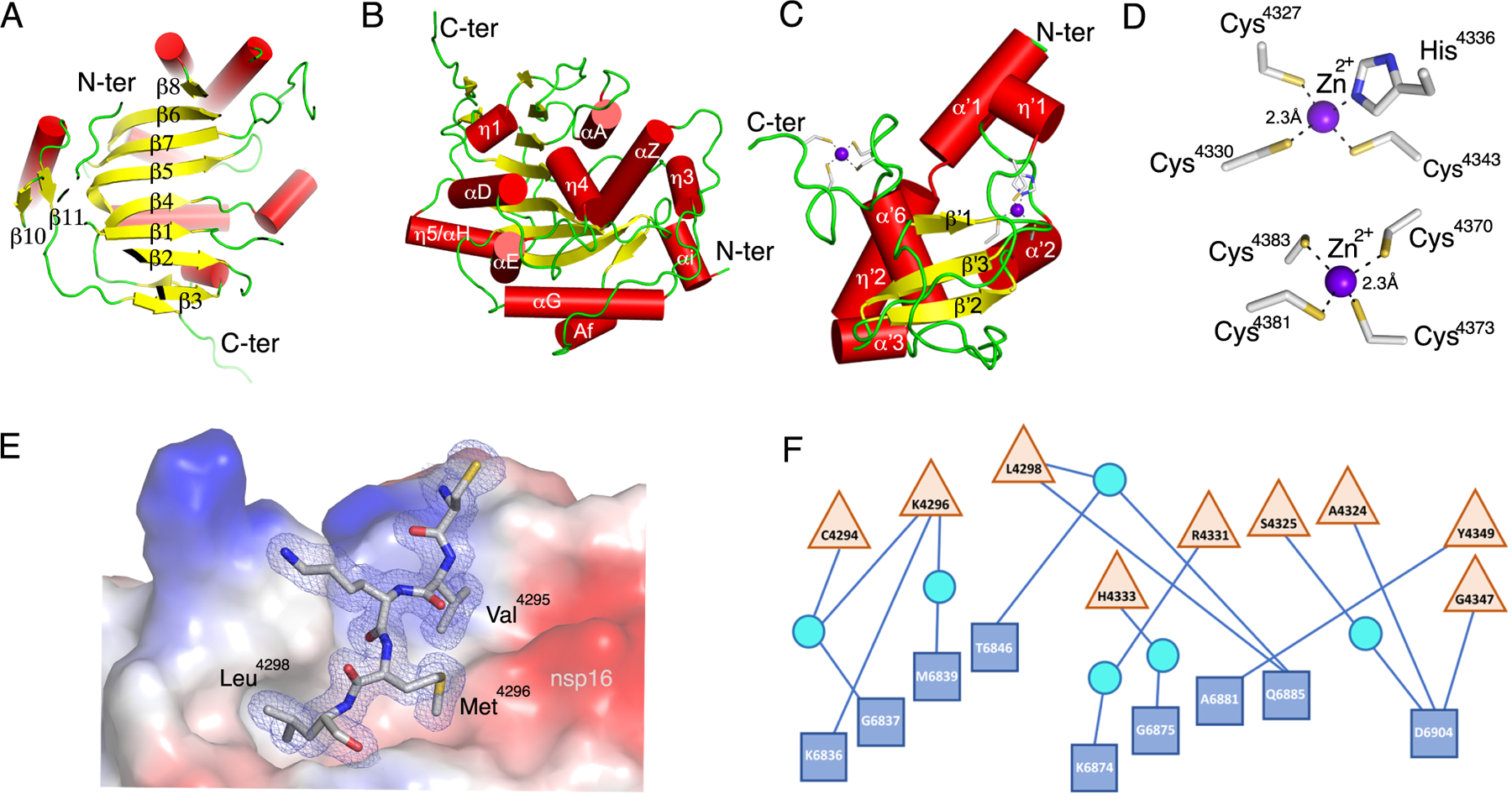
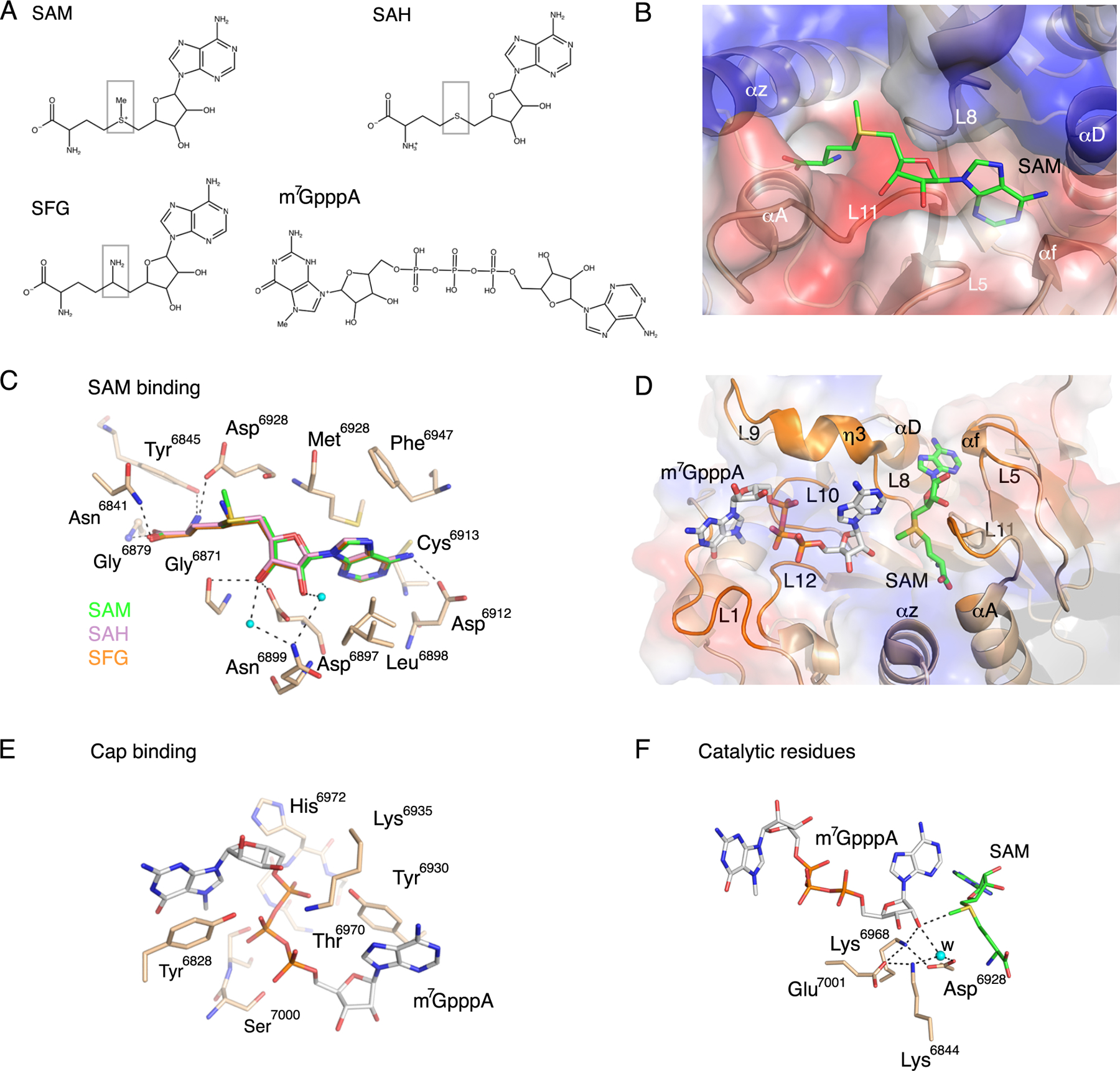
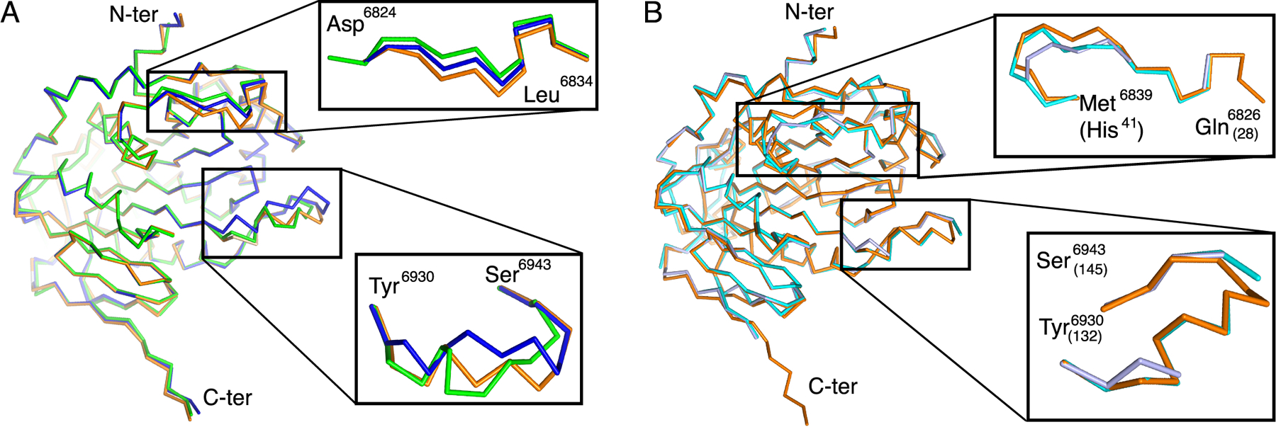
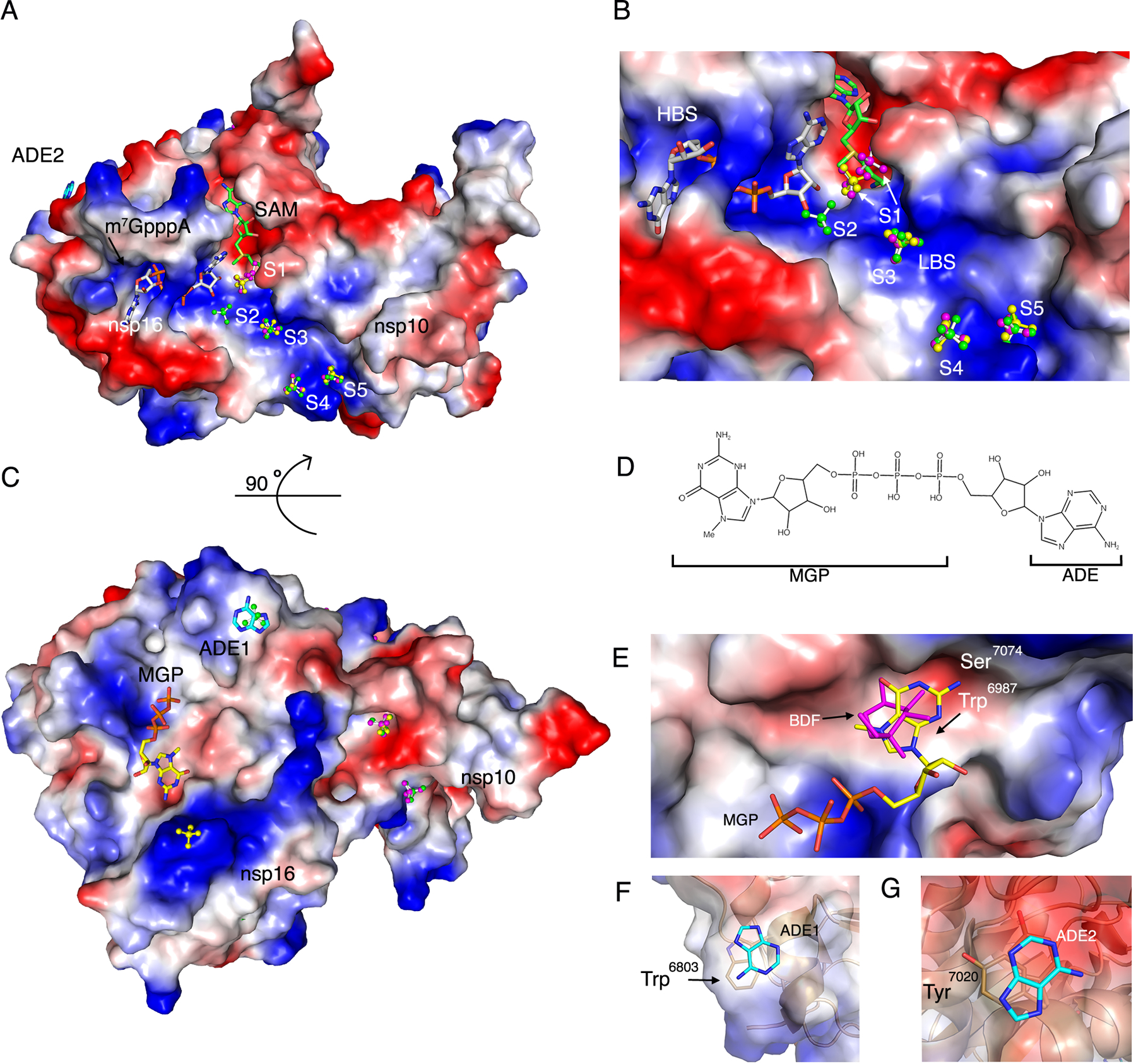
There are currently no antiviral therapies specific for SARS-CoV-2, the virus responsible for the global pandemic disease COVID-19. To facilitate structure-based drug design, we conducted an x-ray crystallographic study of the SARS-CoV-2 nsp16-nsp10 2'-O-methyltransferase complex, which methylates Cap-0 viral mRNAs to improve viral protein translation and to avoid host immune detection. We determined the structures for nsp16-nsp10 heterodimers bound to the methyl donor S-adenosylmethionine (SAM), the reaction product S-adenosylhomocysteine (SAH), or the SAH analog sinefungin (SFG). We also solved structures for nsp16-nsp10 in complex with the methylated Cap-0 analog m7GpppA and either SAM or SAH. Comparative analyses between these structures and published structures for nsp16 from other betacoronaviruses revealed flexible loops in open and closed conformations at the m7GpppA-binding pocket. Bound sulfates in several of the structures suggested the location of the ribonucleic acid backbone phosphates in the ribonucleotide-binding groove. Additional nucleotide-binding sites were found on the face of the protein opposite the active site. These various sites and the conserved dimer interface could be exploited for the development of antiviral inhibitors.


Conflict of interest statement

Competing interests: K.J.F.S. has a significant financial interest in Situ Biosciences, LLC, a contract research organization that conducts antimicrobial testing for industrial products including antiviral testing. This work has no overlap with the interests of the company. K.J.F.S. is a consultant for a healthcare firm on public health topics related to SARS-CoV-2 and COVID-19 that are unrelated to this article. The other authors declare that they have no competing interests.

Figures

Fig. 1.
Fig. 1.. Overall structure of the nsp16-nsp10 oligomers.
(A) Linear schematic of the orf1a/orf1b protein product pp1ab prior to proteolytic processing. (B) Cartoon representation of the nsp16-nsp10 heterodimer of the small unit cell crystal form (PDB code 6W4H). (C) Cartoon representation of the two nsp16-nsp10 heterodimers in the asymmetric unit of the large unit cell crystal form (PDB code 6W75). In (B) and (C), nsp16 is in shades of tan and yellow and nsp10 is in shades of teal and purple. Ligands are represented as sticks, with SAM in bright green and Zn2+ in purple. N-ter, N terminus; C-ter, C terminus. (D) Elution profile for analytical size-exclusion chromatography (SEC) with corresponding plot for molecular weight standards shown at the top. (E) Separation of elution fractions on a 4–15% gradient SDS-PAGE gel stained with Coomassie blue.
Fig. 2.
Fig. 2.. Detailed representation of nsp16, nsp10, and the heterodimer interface.
(A to C) Cartoon representations of two views of nsp16 featuring the canonical β-sheet (A) and overall secondary structure of nsp16 (B) and nsp10 (C). α-helices are shown as red cylinders, β-strands as yellow arrows, loops as green strands, and zinc ions as purple spheres. (D) Close-up view of the two Zn2+ binding sites in nsp10. (E) Interaction of Cys4294-Leu4298 (sequence CVKML, grey sticks) from nsp10 with the hydrophobic surface of nsp16 (colored by electrostatic potential). Oxygen, red sticks; nitrogen, blue sticks; sulfur, yellow sticks. (F) Schematic representation of residues from nsp16 (blue squares) and nsp10 (tan triangles) that interact through hydrogen bonds, represented as lines. Some interactions are mediated by water molecule (cyan circles). For panels A to D, structural representations are based on the structure of the nsp16-nsp10 complex with m7GpppA and SAM (PDB code 6WVN). E and F are based on the structure of the nsp16-nsp10 complex with SAM in the small unit crystal form (PDB code 6W4H).
Fig. 3.
Fig. 3.. Substrate interactions and catalytic site.
(A) Chemical structures of the methyl donor SAM; the product after methyl transfer, SAH; the SAH analog and inhibitor, SFG; and the Cap-0 analog, m7GpppA. Boxes highlight differences in the chemical structures of SAH and SFG compared to SAM. (B) Cartoon and surface charge representations of the nsp16 SAM binding cleft occupied by SAM (green sticks). (C) Close-up view of the overlay of nsp16 structures with the ligands SAM (green, PDB code 6W75), SAH (pink, PDB code 6WJT) and SFG (orange, PDB code 6WKQ). (D) Cartoon and surface charge representations of nsp16 (PDB code 6WVN) with the SAM binding cleft occupied by SAM (green sticks) and the Cap binding site occupied by m7GpppA (gray sticks). (E). Detailed view of the residues that coordinate m7GpppA in the Cap binding site as tan sticks. (F) Close-up view of the side chains of the catalytic residues showing the orientation of the methyl group in SAM in proximity to the acceptor 2’-OH group in m7GpppA. Dashed lines indicate the interactions between the residues in the active site, and small cyan dots indicate water molecules (w). Red sticks, oxygen; blue sticks, nitrogen; orange sticks, phosphate; yellow sticks, sulfur.
Fig. 4.
Fig. 4.. Structural alignment of nsp16 in the presence and absence of m7GpppA.
(A) Alignment of the C-α chain of nsp16 from SARS-CoV-2 in complex with SAM from the small unit cell (blue, PDB ID 6W4H), in complex with SAM from the large unit cell crystal form (green, PDB code 6W75), and in complex with SAM and m7GpppA (orange, PDB code 6WVN). Two flexible loops are enlarged in insets. (B) Alignment of the C-α chain of nsp16 from SARS-CoV-2 in complex with SAM and m7GpppA (orange) with the corresponding region of MERS-CoV nsp16 in complex with SAM alone (light blue, PDB code 5YN6) or in complex with SAM and m7GpppA (cyan, PDB code 5YNM). Numbering of MERS residues is indicated in parentheses.
Fig. 5.
Fig. 5.. Sulfate and nucleotide binding sites on nsp16.
(A) Surface charge representation of nsp16-nsp10 bound to SAM and m7GpppA (PDB code 6 WVN) with sulfates in balls and sticks along the nucleotide binding groove numbered from the catalytic core to the nsp10 extension (S1-S5). The sulfates in the overlayed structures are designated by color according with their corresponding PDB code: 6WRZ (green), 6WVN (yellow), and 6WQ3 (pink). m7GpppA is shown as gray sticks and SAM in green sticks. Positive charges are shown in blue and negative charges in red. ADE2, adenine moiety 2. (B) Close-up view of (A) showing m7GpppA, SAM and S1–S5 in the high-affinity binding site (HBS) and low-affinity binding site (LBS). (C) 90° rotation of the complex showing the secondary binding sites MGP and ADE1 along with additional sulfates. (D) Schematic representation of m7GpppA noting the MGP (m7GpppA guanine and phosphate) and ADE (adenine) moieties. (E) Surface charge representation of the nsp16 MGP binding site with MGP (yellow sticks) and BDF (pink sticks) from structure 6W4H. (F and G) Cartoon and surface charge representation of the adenine moieties (ADE1 and ADE2) bound to nsp16 from structure (PDB code 6WVN). Sticks, carbon; blue, nitrogen; red, oxygen; orange, phosphate; yellow, sulfate.

References

    1. Keni R, Alexander A, Nayak PG, Mudgal J, Nandakumar K, COVID-19: Emergence, Spread, Possible Treatments, and Global Burden. Front Public Health 8, 216 (2020). - PMC - PubMed
    1. Dong E, Du H, Gardner L, An interactive web-based dashboard to track COVID-19 in real time. Lancet Infect Dis, (2020). - PMC - PubMed
    1. Ye ZW, Yuan S, Yuen KS, Fung SY, Chan CP, Jin DY, Zoonotic origins of human coronaviruses. Int J Biol Sci 16, 1686–1697 (2020). - PMC - PubMed
    1. Chan JF, Kok KH, Zhu Z, Chu H, To KK, Yuan S, Yuen KY, Genomic characterization of the 2019 novel human-pathogenic coronavirus isolated from a patient with atypical pneumonia after visiting Wuhan. Emerg Microbes Infect 9, 221–236 (2020). - PMC - PubMed
    1. Petersen E, Koopmans M, Go U, Hamer DH, Petrosillo N, Castelli F, Storgaard M, Al Khalili S, Simonsen L, Comparing SARS-CoV-2 with SARS-CoV and influenza pandemics. Lancet Infect Dis, (2020). - PMC - PubMed

Publication types

MeSH terms

LinkOut - more resources