. 2020 Dec 25;295(52):17950-17972.
doi: 10.1074/jbc.RA120.014710. Epub 2020 Sep 28.

HIV-1 Gag release from yeast reveals ESCRT interaction with the Gag N-terminal protein region

Affiliations

HIV-1 Gag release from yeast reveals ESCRT interaction with the Gag N-terminal protein region

Birgit Meusser et al. J Biol Chem. .

Abstract

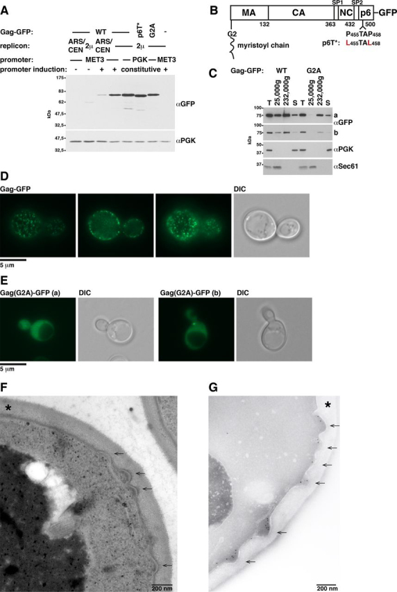
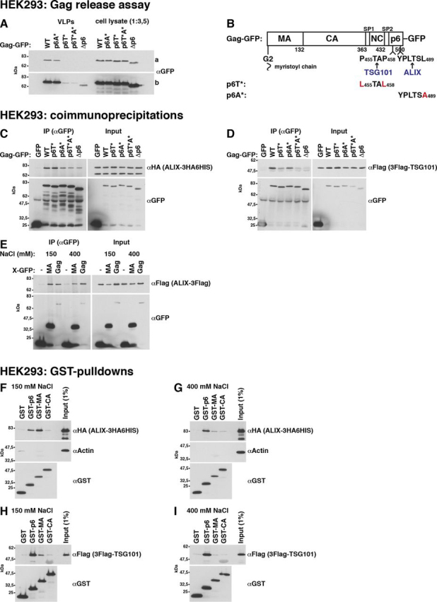
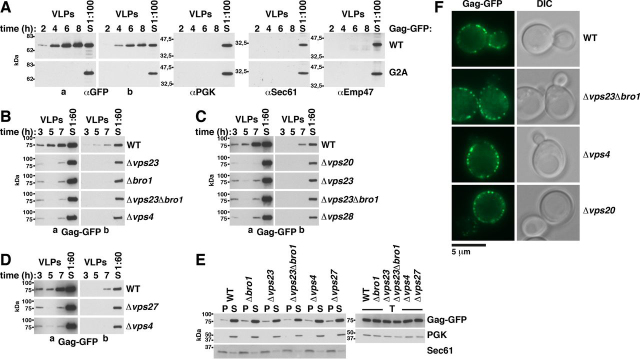
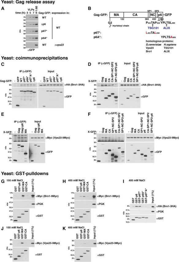
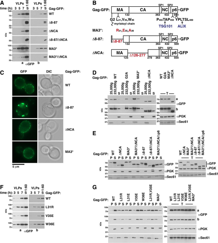
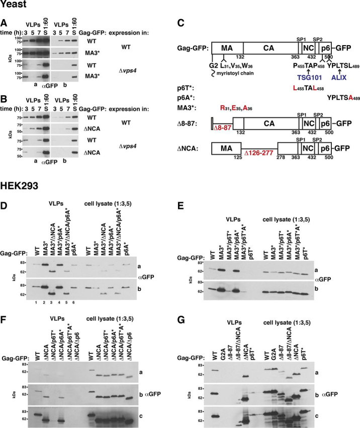
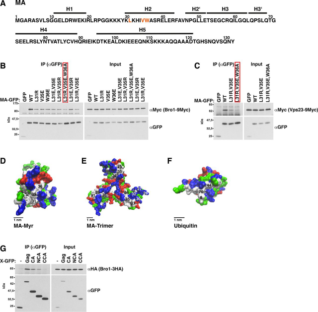
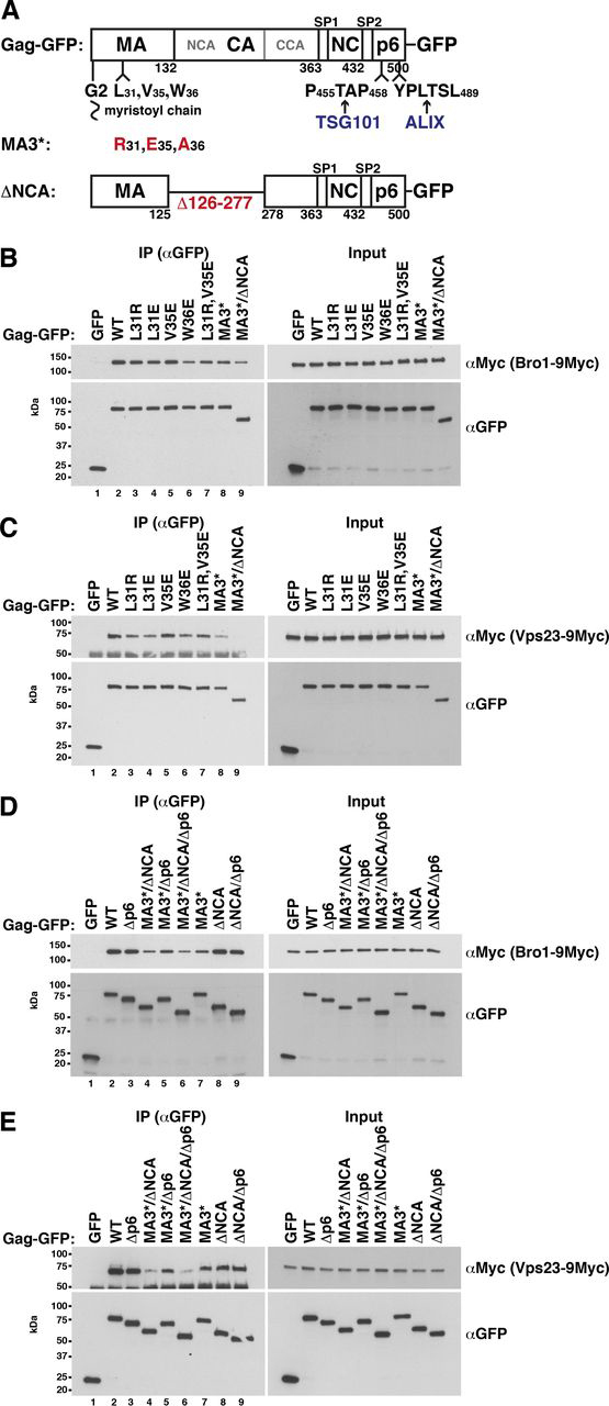
The HIV-1 protein Gag assembles at the plasma membrane and drives virion budding, assisted by the cellular endosomal complex required for transport (ESCRT) proteins. Two ESCRT proteins, TSG101 and ALIX, bind to the Gag C-terminal p6 peptide. TSG101 binding is important for efficient HIV-1 release, but how ESCRTs contribute to the budding process and how their activity is coordinated with Gag assembly is poorly understood. Yeast, allowing genetic manipulation that is not easily available in human cells, has been used to characterize the cellular ESCRT function. Previous work reported Gag budding from yeast spheroplasts, but Gag release was ESCRT-independent. We developed a yeast model for ESCRT-dependent Gag release. We combined yeast genetics and Gag mutational analysis with Gag-ESCRT binding studies and the characterization of Gag-plasma membrane binding and Gag release. With our system, we identified a previously unknown interaction between ESCRT proteins and the Gag N-terminal protein region. Mutations in the Gag-plasma membrane-binding matrix domain that reduced Gag-ESCRT binding increased Gag-plasma membrane binding and Gag release. ESCRT knockout mutants showed that the release enhancement was an ESCRT-dependent effect. Similarly, matrix mutation enhanced Gag release from human HEK293 cells. Release enhancement partly depended on ALIX binding to p6, although binding site mutation did not impair WT Gag release. Accordingly, the relative affinity for matrix compared with p6 in GST-pulldown experiments was higher for ALIX than for TSG101. We suggest that a transient matrix-ESCRT interaction is replaced when Gag binds to the plasma membrane. This step may activate ESCRT proteins and thereby coordinate ESCRT function with virion assembly.

Keywords: ALIX; Bro1; Gag; Saccharomyces cerevisiae; endosomal sorting complexes required for transport (ESCRT); human immunodeficiency virus (HIV); matrix; plasma membrane; virus release; yeast.


Conflict of interest statement

Conflict of interest—The authors declare that they have no conflicts of interest with the contents of this article.

Figures

Figure 1
Figure 1
HIV-1 Gag-GFP forms buds at the yeast PM.A, the Gag-GFP expression level was determined by cell extract immunoblotting. Gag-GFP, a version with mutated TSG101-binding site in p6 (p6T*), or a version with a mutated myristoylation site (G2A) was expressed in WT cells from a vector with constitutive PGK promoter and 2µ replicon or from a vector with MET3 promoter and 2µ or ARS/CEN replicon. Cells expressing MET3 promoter Gag-GFP were grown in medium containing 20 mg/liter methionine to repress the promoter. To induce the promoter, cells were shifted to medium lacking methionine for 4 h. PGK served as loading control. B, Gag-GFP schematic. C, Gag-GFPmembrane binding was analyzed by differential centrifugation, showing that the 25,000 × g pellet differentiates between PM-bound and cytosolic Gag. A 25,000 × g pellet, a 232,000 × g pellet, and a supernatant (S) were prepared from extracts (T) of WT cells expressing GFP-tagged Gag or Gag(G2A) from a 2µ vector with PGK promoter and analyzed by immunoblotting with the indicated antibodies. The pellets were concentrated (25,000 × g, 4×; 232,000 × g, 3.3×) compared with supernatant and extract samples. The cytosolic protein PGK and the integral ER membrane protein Sec61 served as references. A short (b) and a long (a) exposure are shown. D and E, the intracellular localization of GFP-tagged Gag or Gag(G2A) expressed from a 2µ vector with PGK promoter in WT cells was analyzed by fluorescence microscopy, showing that Gag accumulates in punctate structures at the PM dependent on its N-terminal myristoylation. Three layers of a yeast cell are shown for Gag. DIC, differential interference contrast. F and G, PM deformation (arrows) induced by Gag-GFP expression was analyzed by EM. Asterisks, cell wall. F, WT cells expressing Gag-GFP from a 2µ vector with MET3 promoter induced for 6 h were embedded in Epon. G, cryosections of WT cells (SUB62) expressing Gag-GFP from a 2µ vector with PGK promoter were prepared, and GFP was labeled with immunogold.
Figure 4
Figure 4
ALIX and TSG101 bind to MA. The relative affinity for MA compared with p6 is higher for ALIX.A, Gag-GFP-VLP release from HEK293 cells recapitulating published data for p6 dependence of HIV-1 release. 2 days after transfection with CMV promoter expression vectors for the indicated Gag versions, VLPs were harvested from the culture medium, and cell lysates were prepared. Gag was detected by immunoblotting with anti-GFP antibodies. A short (a) and a long (b) exposure are shown. B, Gag-GFP schematic. C–E, coimmunoprecipitations (IP) of epitope-tagged ALIX or TSG101 with GFP-tagged Gag versions or MA expressed from CMV promoter vectors in HEK293 cells, indicating that ALIX binding to Gag is reduced but not prevented by p6 deletion and that ALIX binds to MA. TSG101 binding to Gag more strongly depends on p6. GFP-tagged proteins were immunoprecipitated with anti-GFP antibodies in the presence of 150 mm NaCl (C and D) or as indicated (E), and coimmunopreciptated ALIX or TSG101 was detected with antibodies recognizing the epitope tag. F–I, pulldown experiments showing that ALIX and TSG101 bind to GST-MA expressed in E. coli. The relative affinity for MA compared with p6 is higher for ALIX. GST-tagged Gag fragments (MA (aa 1–132), CA (aa 133–363), and p6 (aa 448–500)) were bound to GSH-Sepharose and incubated with extract of HEK293 cells expressing epitope-tagged ALIX or TSG101 from a CMV promoter vector. Bead-bound proteins were analyzed by immunoblotting with the indicated antibodies. α-Actin served as control, showing specific ESCRT protein binding.
Figure 2
Figure 2
Gag-GFP release from yeast spheroplasts depends on ESCRT proteins.A, Gag-GFP or Gag(G2A)-GFP release from WT yeast spheroplasts was analyzed by immunoblotting of high-speed centrifugation sediments derived from the incubation medium with anti-GFP antibodies (VLPs). VLPs were harvested every second hour after spheroplast preparation over a period of 8 h. Spheroplasts were incubated in fresh medium after each harvest. Release increased from the first to the third time point and required Gag myristoylation. Immunoblots with antibodies detecting PGK (cytosolic protein), Sec61 (ER membrane protein), or Emp47 (Golgi and COPII membrane protein) served as control for specific Gag release. Long (a) and short (b) exposures are shown. S, lysate of spheroplasts prepared at the final VLP harvest. B–D, same as A except that Gag-GFP was expressed in the WT or the indicated mutants and VLPs were harvested at the indicated times, showing ESCRT-dependent release. E and F, ESCRT deletion does not affect Gag-GFP-PM accumulation. E, Gag-GFP-membrane binding was analyzed by immunoblotting of membrane-containing 25,000 × g pellets (P) and cytosol-containing supernatants (S) derived from cell extracts (T). Sec61 and PGK served as references. F, Gag-GFP accumulation in punctate structures at the PM was analyzed by fluorescence microscopy. DIC, differential interference contrast. A–F, WT cells or the indicated mutants expressed Gag-GFP or Gag(G2A)-GFP from a 2µ vector with PGK promoter.
Figure 3
Figure 3
Yeast ESCRT proteins bind to the Gag N-terminal protein region.A, release of Gag-GFP or the indicated mutants from WT yeast spheroplasts or a Δvps23 mutant was analyzed by immunoblotting of high-speed centrifugation sediments derived from the incubation medium with anti-GFP antibodies (VLPs), showing that p6 is not required for Gag-GFP release from yeast. Gag-GFP versions were expressed from a 2µ vector with PGK promoter. S, lysate of spheroplasts prepared at the final VLP harvest. B, Gag-GFP schematic. C–F, coimmunoprecipitation (IP) experiments with GFP-tagged Gag versions or Gag fragments expressed from a 2µ vector with induced MET3 promoter and genomically epitope-tagged Bro1 or Vps23, indicating that Bro1 and Vps23 bind to Gag via MA and CA. Gag versions or Gag fragments (MA (aa 1–132), CA (aa 133–363), p6 (aa 448–500), CA-SP1-NC-SP2 (aa 133–447), SP1-NC-SP2 (aa 364–447), SP1-NC-SP2-p6 (aa 364–500), and GagΔp6 (aa 1–447)) were immunoprecipitated with anti-GFP antibodies in the presence of 400 mm NaCl, and coimmunoprecipitated Bro1 or Vps23 was detected by immunoblotting with anti-HA or anti-Myc antibodies. C, Bro1 binds to Gag independent of p6. D, Bro1 binds to Gag fragments containing MA or CA. E, Vps23 binds to Gag independent of p6 and to MA. F, Vps23 binds to Gag fragments containing MA or CA. G–K, pulldown experiments showing that genomically epitope-tagged Bro1 and Vps23 bind to GST-MA expressed in E. coli and that Bro1 additionally binds to GST-p6 dependent on the ALIX-binding motif. GST-tagged Gag fragments (MA (aa 1–132), CA (aa 133–363), and p6 (aa 448–500)) were bound to GSH-Sepharose and incubated with yeast extract. Bead-bound proteins were analyzed by immunoblotting with the indicated antibodies. PGK served as control, showing specific ESCRT protein binding. G and J, binding buffer contained 150 mm NaCl. H, I, and K, binding buffer contained 400 mm NaCl.
Figure 7
Figure 7
An MA hydrophobic patch mutation (MA3*) increases Gag-GFP-membrane binding and enhances Gag-GFP release from yeast.A, release of Gag-GFP versions from yeast spheroplasts was analyzed by immunoblotting of high-speed centrifugation sediments derived from the incubation medium with anti-GFP antibodies (VLPs), showing that MA3* increases Gag release. ΔNCA reduces Gag release and is dominant over MA3*. MA globular head deletion (Δ8–87) abrogates Gag release. S, lysate of spheroplasts prepared at the final VLP harvest. Long (a) and short (b) exposures are shown. B, Gag-GFP schematic. C, PM accumulation of Gag-GFP versions was analyzed by fluorescence microscopy, showing that Gag(MA3*) and Gag(ΔNCA) form punctate structures similar to Gag, whereas Gag(Δ8-87) forms PM-associated aggregates with larger diameter. DIC, differential interference contrast. D, membrane binding of Gag-GFP versions was analyzed by differential centrifugation, showing that MA3* increases specifically the Gag amount that sediments with the 25,000 × g membrane pellet. Gag(ΔNCA) sediments similarly to Gag. A 25,000 × g pellet, a 232,000 × g pellet, and a supernatant (S) were prepared from cell extracts (T) and analyzed by immunoblotting with the indicated antibodies. The pellets were concentrated (25,000 × g, 4×; 232,000 × g, 3.3×) compared with supernatant and extract samples. The cytosolic protein PGK and the integral ER membrane protein Sec61 served as references. A short (a) and long (b) exposure are shown. E, membrane-containing 25,000 × g pellets (P) and cytosol-containing supernatants (S) derived from cell extracts (T) were analyzed as in D, showing that increased amounts of Gag versions carrying the MA3* mutation sediment with membranes compared with WT Gag, whereas the ΔNCA mutation does not affect the membrane association. G, same as E, except that cells expressed the indicated Gag-GFP versions, showing that Leu-31 and Trp-36 mutations increase Gag-membrane binding. A long (a) and a short (b) exposure are shown. F, same as A, except that the indicated Gag-GFP versions were expressed, showing that Leu-31 and Trp-36 mutations increase Gag release. A and C–G, Gag-GFP versions were expressed from a 2µ vector with PGK promoter in WT yeast.
Figure 9
Figure 9
Increased Gag(MA3*)-GFP release from yeast depends on ESCRT proteins; MA3* increases Gag-GFP release from HEK293 cells dependent on NCA and the ALIX-binding site in p6.A and B, release of Gag-GFP versions from yeast spheroplasts was analyzed by immunoblotting of high-speed centrifugation sediments derived from the incubation medium with anti-GFP antibodies (VLPs). Gag-GFP versions were expressed from a 2µ vector with PGK promoter. S, lysate of spheroplasts prepared at the final VLP harvest. Long (a) and short (b) exposures are shown. A, VPS4 deletion abrogates the release-increasing MA3* effect after 5 and 7 h of incubation. B, Gag(ΔNCA)-GFP release from Δvps4 spheroplasts is slightly decreased compared with Gag-GFP. D–G, Gag-GFP release from HEK293 cells 2 days after transfection with CMV promoter expression vectors for the indicated Gag versions. VLPs were harvested from the culture medium, and cell lysates were prepared. Gag-GFP was detected by immunoblotting with anti-GFP antibodies. Several exposures (a–c) are shown. D, the MA3* mutation increases Gag-GFP release. A combination of ΔNCA and mutation of the ALIX-binding site in p6 (p6A*) abrogates this increase, whereas an isolated p6A* mutation does not impair Gag-GFP release. E, similar to Gag-GFP, mutating the TSG101-binding site in p6 (p6T*) strongly reduces Gag(MA3*)-GFP release. F and G, Gag(ΔNCA) is efficiently released. F, similar to Gag-GFP, p6T* strongly reduces Gag(ΔNCA)-GFP release. G, MA globular head (Δ8–87) deletion strongly impairs the release. C, Gag-GFP schematic.
Figure 5
Figure 5
Mutation of an MA hydrophobic patch consisting of Leu-31, Val-35, and Trp-36 reduces Bro1 and Vps23 binding to MA; Bro1 binds via NCA to CA.A, MA protein sequence derived from pGag-EGFP (130) used as template in this study. Leu-31, Val-35, and Trp-36, the mutation of which reduced binding to Bro1, are marked in orange. Helix (H) assignments are derived from the MA X-ray structure (11). B and C, GFP-tagged MA versions were expressed from a 2µ vector with induced MET3 promoter in yeast cells carrying genomically 9Myc-tagged Bro1 or Vps23. MA was immunoprecipitated (IP) in the presence of 400 mm NaCl with anti-GFP antibodies, and coimmunoprecipitated Bro1 or Vps23 was detected by immunoblotting with anti-Myc antibodies. The red box indicates mutant MA3* that was chosen in subsequent experiments to diminish the ESCRT-MA interaction. B, Leu-31, Val-35, or Trp-36 mutation reduces the MA-Bro1 interaction. C, MA mutants that reduce the binding to Bro1 also diminish the MA-Vps23 interaction. D–F, molecular surface structures visualized with the VMD software. White, nonpolar aa; blue, basic aa; red, acidic aa; green, polar aa; black, myristoyl residue. D, MA NMR structure (PDB entry 2H3I) (19), showing that Leu-31, Val-35, and Trp-36 form a hydrophobic patch on the MA surface. E, MA trimer X-ray structure (PDB entry 1HIW) (11), showing that Leu-31, Val-35, and Trp-36 are located on the MA side that is exposed to the PM and are not part of the trimerization interface. Basic aa 26, 27, 30, and 32 were proposed to be involved in MA binding to phospholipids (13, 99, 101). F, human ubiquitin X-ray structure (PDB entry 1UBQ) (135), showing that the hydrophobic patch in MA consisting of Leu-31, Val-35, and Trp-36 resembles the hydrophobic ubiquitin patch consisting of Leu-8, Ile-44, and Val-70 which is involved in ubiquitin binding to Vps23, Bro1, TSG101, and ALIX (58, 67, 102). G, Bro1 coimmunoprecipitation with GFP-tagged CA versions, showing that Bro1 binds to the N-terminal CA domain; same as B except that GFP-tagged CA, NCA (aa 133–278), or CCA (aa 279–363) was expressed in a yeast strain carrying genomically 3HA-tagged Bro1 and that coimmunoprecipitated Bro1 was detected by immunoblotting with anti-HA antibodies.
Figure 6
Figure 6
Mutations in the Gag N-terminal protein region reduce Gag-Bro1 and Gag-Vps23 interaction.B–E, coimmunoprecipitation (IP) experiments with Gag-GFP versions expressed from a 2µ vector with induced MET3 promoter in yeast cells carrying genomically 9Myc-tagged Bro1 or Vps23, showing that MA mutations in Leu-31 and Trp-36 slightly reduce the Gag-ESCRT binding. aa126-277 deletion (ΔNCA) enhances this effect (Gag(MA3*)-GFP versus Gag(MA3*/ΔNCA-GFP). V35E does not reduce the Gag-ESCRT coimmunoprecipitation. Gag was immunoprecipitated with anti-GFP antibodies in the presence of 400 mm NaCl, and coimmunoprecipitated Bro1 or Vps23 was detected by immunoblotting with anti-Myc antibodies. A, Gag-GFP schematic.
Figure 8
Figure 8
MA3* increases MA-GFP-PM binding, whereas ESCRT deletion does not.A and B, WT yeast cells or the indicated ESCRT mutants expressed MA-GFP or MA3*-GFP from a 2µ vector with induced MET3 promoter. A, MA-GFP binding to the PM was analyzed by fluorescence microscopy, showing PM rim staining for MA3*. DIC, differential interference contrast. B, membrane-containing 232,000 × g pellets (P) and cytosol-containing supernatants (S) derived from cell extracts (T) were analyzed by immunoblotting with anti-GFP antibodies, showing that increased MA3* amounts sediment compared with MA, whereas similar MA amounts sediment from WT and ESCRT mutant cell extracts. The pellet samples were 5× concentrated compared with the supernatant and extract samples. The cytosolic protein PGK and the integral ER membrane protein Sec61 served as references.

References

    1. Freed E.O. HIV-1 assembly, release and maturation. Nat. Rev. Microbiol. 2015;13:484–496. doi: 10.1038/nrmicro3490. 26119571. - DOI - PMC - PubMed
    1. Gheysen D., Jacobs E., de Foresta F., Thiriart C., Francotte M., Thines D., De Wilde M. Assembly and release of HIV-1 precursor Pr55gag virus-like particles from recombinant baculovirus-infected insect cells. Cell. 1989;59:103–112. doi: 10.1016/0092-8674(89)90873-8. 2676191. - DOI - PubMed
    1. Kutluay S.B., Bieniasz P.D. Analysis of the initiating events in HIV-1 particle assembly and genome packaging. PLoS Pathog. 2010;6 doi: 10.1371/journal.ppat.1001200. 21124996. - DOI - PMC - PubMed
    1. Datta S.A., Curtis J.E., Ratcliff W., Clark P.K., Crist R.M., Lebowitz J., Krueger S., Rein A. Conformation of the HIV-1 Gag protein in solution. J. Mol. Biol. 2007;365:812–824. doi: 10.1016/j.jmb.2006.10.073. 17097677. - DOI - PMC - PubMed
    1. Datta S.A., Heinrich F., Raghunandan S., Krueger S., Curtis J.E., Rein A., Nanda H. HIV-1 Gag extension: conformational changes require simultaneous interaction with membrane and nucleic acid. J. Mol. Biol. 2011;406:205–214. doi: 10.1016/j.jmb.2010.11.051. 21134384. - DOI - PMC - PubMed

Uncited reference

    1. Seufert W., McGrath J.P., Jentsch S. UBC1 encodes a novel member of an essential subfamily of yeast ubiquitin-conjugating enzymes involved in protein degradation. EMBO J. 1990;9:4535–4541. doi: 10.1002/j.1460-2075.1990.tb07905.x. 2265617. - DOI - PMC - PubMed
    1. Finley D., Özkaynak E., Varshavsky A. The yeast polyubiquitin gene is essential for resistance to high temperatures, starvation, and other stresses. Cell. 1987;48:1035–1046. doi: 10.1016/0092-8674(87)90711-2. 3030556. - DOI - PubMed

Publication types

MeSH terms

Substances