. 2020 Dec;69(12):2630-2641.
doi: 10.2337/db20-0384. Epub 2020 Sep 29.

STAT1 Dissociates Adipose Tissue Inflammation From Insulin Sensitivity in Obesity

Affiliations

STAT1 Dissociates Adipose Tissue Inflammation From Insulin Sensitivity in Obesity

Aaron R Cox et al. Diabetes. 2020 Dec.

Abstract

Obesity fosters low-grade inflammation in white adipose tissue (WAT) that may contribute to the insulin resistance that characterizes type 2 diabetes. However, the causal relationship of these events remains unclear. The established dominance of STAT1 function in the immune response suggests an obligate link between inflammation and the comorbidities of obesity. To this end, we sought to determine how STAT1 activity in white adipocytes affects insulin sensitivity. STAT1 expression in WAT inversely correlated with fasting plasma glucose in both obese mice and humans. Metabolomic and gene expression profiling established STAT1 deletion in adipocytes (STAT1 a-KO ) enhanced mitochondrial function and accelerated tricarboxylic acid cycle flux coupled with reduced fat cell size in subcutaneous WAT depots. STAT1 a-KO reduced WAT inflammation, but insulin resistance persisted in obese mice. Rather, elimination of type I cytokine interferon-γ activity enhanced insulin sensitivity in diet-induced obesity. Our findings reveal a permissive mechanism that bridges WAT inflammation to whole-body insulin sensitivity.

Trial registration: ClinicalTrials.gov NCT02792400.


Figures

Figure 1
Figure 1
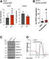
Higher STAT1 levels correspond with impaired adipocyte lipid metabolism. A: Relative mRNA expression of Stat1 in lean (gray) or HFD-induced obese (DIO; red) wild-type mice in iWAT (n = 11/group) and eWAT (n = 3/group). *P < 0.05. Data are represented as mean ± SD. B: Relative STAT1 expression was measured in human subcutaneous (sq) adipose tissue biopsied from subjects with prediabetes (red; n = 11) compared with those with normal glucose tolerance (NGT; gray; n = 18). #P < 0.09. Human subcutaneous preadipocytes were differentiated for 8 days and then transfected with STAT1 or control vector (pcDH). C: To confirm STAT1 expression, immunoblotting of total STAT1 and FLAG was performed, along with markers of mature adipocytes and mitochondrial proteins. D: Respiration (as OCR) was measured in human adipocytes expressing control vector (pcDH; black dashed line) or STAT1 (red dashed line) over time with the addition of oligomycin (α), carbonyl cyanide-4-(trifluoromethoxy) phenylhydrazone (β), and antimycin-A/rotenone (γ). *P < 0.05 indicates changes in maximal respiration. Data are represented as the mean from 15 wells collected over 3 independent experiments. rel, relative.
Figure 2
Figure 2
STAT1 deletion restores adipocyte metabolism in human and mouse adipocytes. Human subcutaneous preadipocytes were differentiated for 10 days and then transfected with STAT1 siRNA or scRNA (control) for 48 h. After transfection, cells were treated with (+) or without (−) 100 ng/mL IFN-γ for 24 h. A: Relative mRNA expression of STAT1, IRF1, UCP1, and ADIPOQ (n = 9) (scRNA, gray; STAT1 siRNA, red). *P < 0.05 vs. scRNA; #P < 0.05 vs. IFN-γ vehicle treated. Data are represented as mean ± SD. B: OCR in differentiated human adipocytes after exposure to IFN-γ with addition of oligomycin (α), carbonyl cyanide-4-(trifluoromethoxy) phenylhydrazone (β), and antimycin-A/rotenone (γ) (scRNA vehicle [veh], black; scRNA + IFN-γ [IFNG], green; STAT1 siRNA vehicle, blue; and STAT1 siRNA + IFN-γ, red) (n = 12). *P < 0.06 vs. scRNA; #P < 0.06 vs. IFN-γ vehicle treated for maximal respiration. 3T3-L1 cells were transfected with Cas9 and Stat1 single gRNA or a nonmammalian targeting control (ntCR1) gRNA. C: Immunoblots of total lysates from ntCR1 or g3 Stat1 (gStat1) cells with or without differentiation (diff) for 10 days. D: Relative (rel) mRNA expression of Stat1, Pparγ2, AdipoQ, and Pgc1a from ntCR1 and g3 Stat1 cells with or without differentiation for 10 days (n = 3). *P < 0.05 vs. ntCR1; #P < 0.05 vs. no differentiation. Data are represented as mean ± SD. E: Differentiated ntCR1 and gSTAT1 cells were stained to identify mitochondria (red), lipids (green), and nuclei (blue). Scale bars, 25 μm. F: Differentiated ntCR1 and gSTAT1 cells were treated with or without 100 ng/mL IFN-γ for 24 h and then harvested for quantification of relative mRNA for inflammatory (Stat1 and Irf1), adipocyte marker (Pparγ2 and AdipoQ), and lipid metabolism genes (Pgc1a, Acly, and Aspa) (n = 3–6/group) (ntCR1, gray; gSTAT1, red). *P < 0.05 vs. ntCR1; #P < 0.05 vs. IFN-γ vehicle. Data are represented as mean ± SD.
Figure 3
Figure 3
STAT1 deletion in adipocytes reduces subcutaneous fat cell size. Body mass (A) and composition (percent body mass) (B) for STAT1fl/fl and STAT1a-KO mice on HFD for 18 weeks (n = 9–11/group). C: Tissue weights from STAT1fl/fl and STAT1a-KO mice on HFD (n = 7/group). Glucose tolerance tests (GTT; n = 8/group) (D) and insulin tolerance tests (ITT; n = 12–13/group) (E) with corresponding overnight fasting serum insulin (n = 8/group) (F) in STAT1fl/fl and STAT1a-KO mice on HFD. eWAT and iWAT staining for macrophages (Mac3; brown) (G) and relative (rel) mRNA expression of inflammatory genes (n = 4–8/group) (H). Scale bars, 100 μm. Adipocyte cell size distribution (percentage of total cells), average size (I), and number of adipocytes (per cm2) (J) tabulated across four magnification ×20 fields of view per mouse fat depot (n = 5–6/group) (STAT1fl/fl, gray; STAT1a-KO, red). *P < 0.05; #P < 0.1. Data are represented as mean ± SD. BAT, brown adipose tissue; BW, body weight.
Figure 4
Figure 4
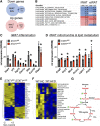
iWAT from STAT1a-KO mice exhibit enrichment of mitochondrial genes and TCA cycle improvements. RNA-Seq (A) coupled with GSEA (B) identified gene signatures altered by STAT1a-KO in the eWAT and iWAT of obese mice. Relative (rel) mRNA expression of key genes that validate the anti-inflammatory (C) and metabolic (D) gene signatures of Stat1 deletion in iWAT (n = 5/group) (STAT1fl/fl, gray; STAT1a-KO, red). Heat maps representing hierarchical clustering of altered metabolites in iWAT between obese STAT1fl/fl and STAT1a-KO mouse models (n = 5/group; false discovery rate <0.25) (E) and lean or diet-induced obese wild-type (WT) mice (n = 4–6/group) (F) assessed using mass spectrometry. G: Metabolomics analysis of iWAT establish HFD feeding in WT mice reduced (red) TCA cycle metabolites that become rescued (green stars) in STAT1a-KO mice. *P < 0.05. Data are represented as mean ± SD. 2PG, 2-phosphoglycerate; 3PG, 3-phosphoglycerate; Ac-CoA, acetyl-CoA; F6P, fructose-6-phosphate; FBP, fructose-1,6-bisphosphate; G3P, glyceraldehyde 3-phosphate; G6P, glucose-6-phosphate; GBP, glucose-1,6-bisphosphate; PEP, phosphoenolpyruvate; α-KG, α-ketoglutarate; RPKM, reads per kilobase per million.
Figure 5
Figure 5
Complete disruption of IFN-γ signaling restores metabolic homeostasis in adipocytes and insulin sensitivity in diet-induced obese mice. Body weight gain (percent initial; n = 4–5/group) (A), body composition (MRI; n = 9/group) (B), and tissue weights (n = 13–14/group) (C) measured after 12 weeks of HFD. *P < 0.05. D: Insulin sensitivity was determined by insulin tolerance tests (ITT) in obese IFNGR1+/+ and IFNGR1−/− mice (n = 9 mice/group). *P < 0.05. Serum insulin (E) and leptin (F) levels were assessed in obese mice fasted 4 h (n = 9 mice/group). *P < 0.05. iWAT hematoxylin and eosin (H/E) (G) was used to measure adipocyte cell size distribution (percent total cells), average adipocyte size (H), and number of adipocytes (per cm2) (I) tabulated across four magnification ×20 fields of view per mouse fat depot (n = 4–5/group) (IFNGR1+/+, gray; IFNGR1−/−, red). *P < 0.05. Scale bars, 100 μm. J: IFN-γ–STAT1 inflammation and metabolism genes from iWAT of IFNGR1+/+ and IFNGR1−/− mice on HFD (n = 10–14/group). *P < 0.05; #P < 0.1. K: Metabolite levels in iWAT of obese IFNGR1+/+ (gray) and IFNGR1−/− (red) mice (n = 4–5/group) were assessed using mass spectrometry. *P < 0.05. Diagram shows red metabolites decreased by HFD in wild-type mice; Ifngr1 deletion rescued (green stars) and reduced (blue) metabolites. L: Validation of Ifngr1 deletion and impaired Stat1 signaling by Western blot analysis of total cell lysates from IFNGR1+/+ and IFNGR1−/− SVF-derived adipocytes after 24-h exposure to IFN-γ. M: Relative (rel) mRNA expression of Stat1, Oas1, AdipoQ, and Ucp1 from IFNGR1+/+ (gray) and IFNGR1−/− (red) adipocytes after exposure to IFN-γ (n = 3). *P < 0.05 vs. IFNGR1+/+; #P < 0.05 vs. vehicle (veh). N: Respiration (as OCR) was measured in IFNGR1+/+ and IFNGR1−/− adipocytes after IFN-γ treatment and during oligomycin (α), carbonyl cyanide-4-(trifluoromethoxy)phenylhydrazone (β), and antimycin-A/rotenone (γ) (n = 5) additions. *P < 0.05 vs. IFNGR1+/+; #P < 0.05 vs. vehicle. IFNGR1+/+ vehicle, black; IFNGR1+/+ + IFN-γ, blue; IFNGR1−/− vehicle, green; IFNGR1−/− + IFN-γ, red. All data are represented as mean ± SD. BAT, brown adipose tissue; BW, body weight.

References

    1. Afshin A, Forouzanfar MH, Reitsma MB, et al. .; GBD 2015 Obesity Collaborators . Health effects of overweight and obesity in 195 countries over 25 years. N Engl J Med 2017;377:13–27 - PMC - PubMed
    1. Smith GI, Mittendorfer B, Klein S. Metabolically healthy obesity: facts and fantasies. J Clin Invest 2019;129:3978–3989 - PMC - PubMed
    1. Patsouris D, Li PP, Thapar D, Chapman J, Olefsky JM, Neels JG. Ablation of CD11c-positive cells normalizes insulin sensitivity in obese insulin resistant animals. Cell Metab 2008;8:301–309 - PMC - PubMed
    1. Lee YS, Li P, Huh JY, et al. . Inflammation is necessary for long-term but not short-term high-fat diet-induced insulin resistance. Diabetes 2011;60:2474–2483 - PMC - PubMed
    1. Kanda H, Tateya S, Tamori Y, et al. . MCP-1 contributes to macrophage infiltration into adipose tissue, insulin resistance, and hepatic steatosis in obesity. J Clin Invest 2006;116:1494–1505 - PMC - PubMed

Publication types

MeSH terms

Associated data