. 2020 Nov 24;13(11):dmm044354.
doi: 10.1242/dmm.044354.

Differential physiological roles for BIN1 isoforms in skeletal muscle development, function and regeneration

Affiliations

Differential physiological roles for BIN1 isoforms in skeletal muscle development, function and regeneration

Ivana Prokic et al. Dis Model Mech. .

Abstract

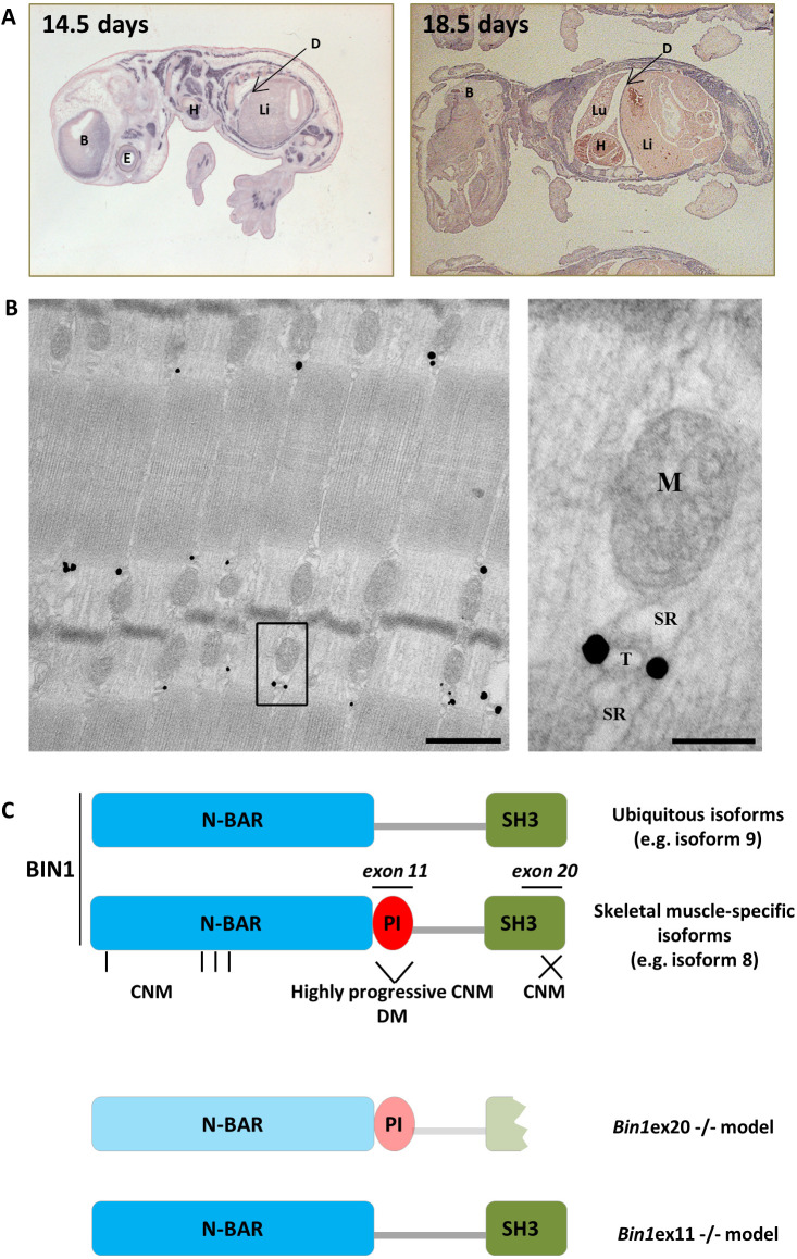
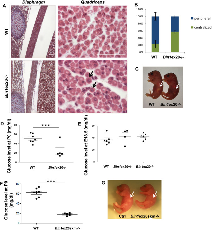
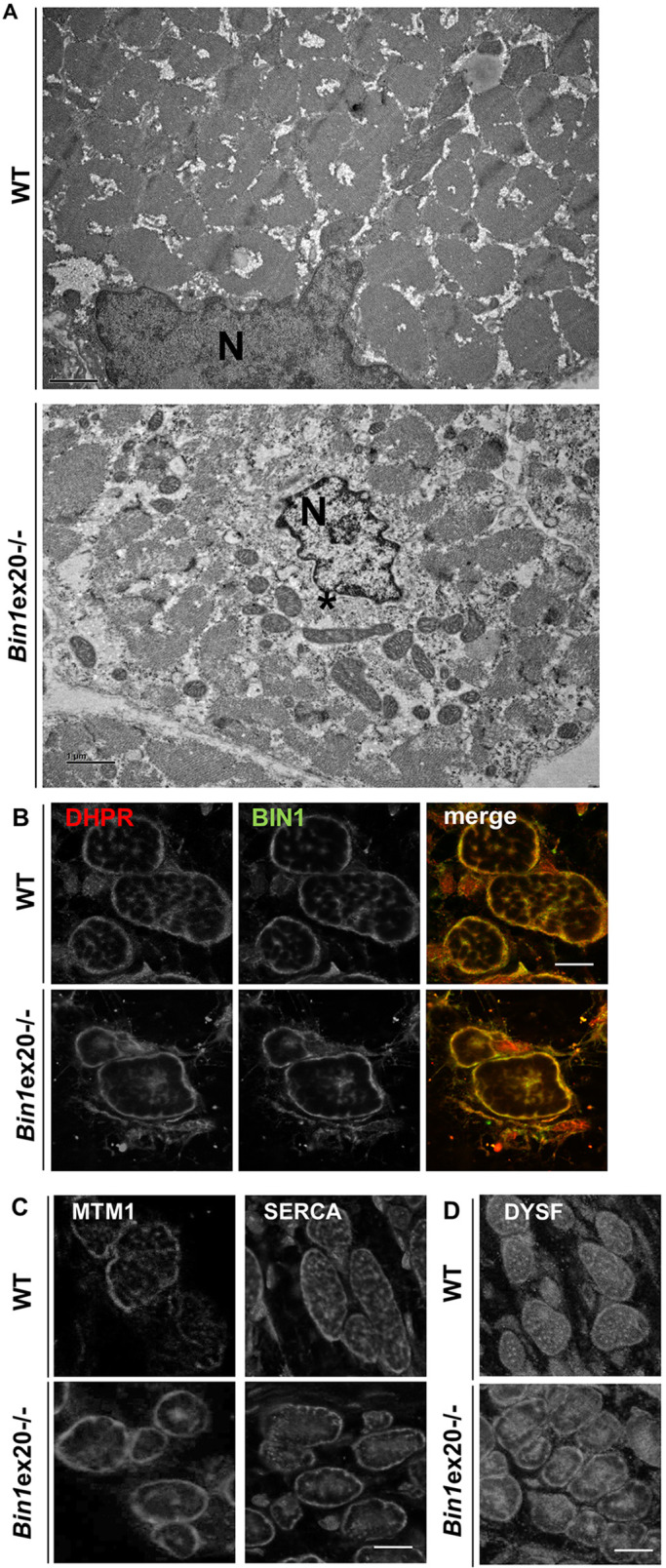
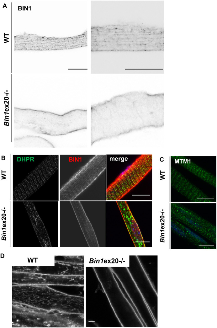
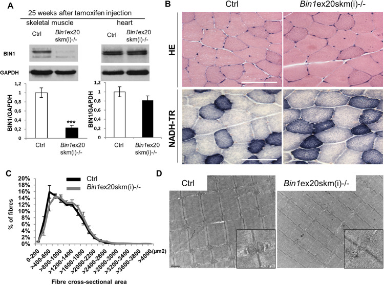
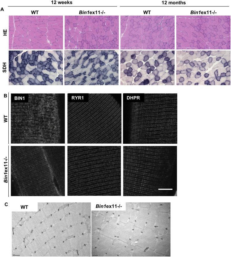
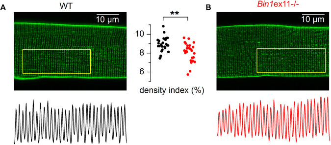
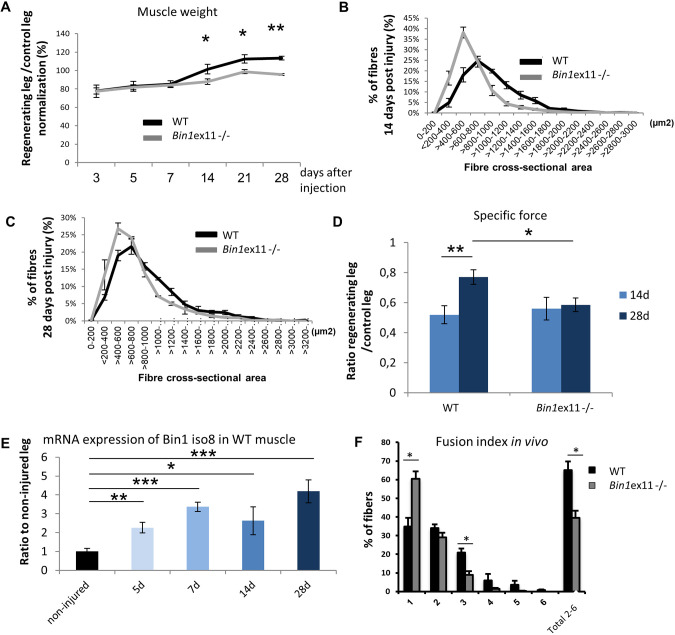
Skeletal muscle development and regeneration are tightly regulated processes. How the intracellular organization of muscle fibers is achieved during these steps is unclear. Here, we focus on the cellular and physiological roles of amphiphysin 2 (BIN1), a membrane remodeling protein mutated in both congenital and adult centronuclear myopathies (CNM), that is ubiquitously expressed and has skeletal muscle-specific isoforms. We created and characterized constitutive muscle-specific and inducible Bin1 homozygous and heterozygous knockout mice targeting either ubiquitous or muscle-specific isoforms. Constitutive Bin1-deficient mice died at birth from lack of feeding due to a skeletal muscle defect. T-tubules and other organelles were misplaced and altered, supporting a general early role for BIN1 in intracellular organization, in addition to membrane remodeling. Although restricted deletion of Bin1 in unchallenged adult muscles had no impact, the forced switch from the muscle-specific isoforms to the ubiquitous isoforms through deletion of the in-frame muscle-specific exon delayed muscle regeneration. Thus, ubiquitous BIN1 function is necessary for muscle development and function, whereas its muscle-specific isoforms fine tune muscle regeneration in adulthood, supporting that BIN1 CNM with congenital onset are due to developmental defects, whereas later onset may be due to regeneration defects.

Keywords: Animal model; BAR domain; Centronuclear myopathy; Dynamin; Myoblast fusion; Myotonic dystrophy; Myotubular myopathy; SH3 domain; Triad; XLMTM.


Conflict of interest statement

Competing interestsB.S.C. and J. Laporte are co-founders of Dynacure, and B.S.C. is a Dynacure employee.

Figures

Fig. 1.
Fig. 1.
BIN1 expression and localization. (A) In situ Bin1 labeling in wild-type mouse at E14.5 and E18.5. (B) Immunogold labeling using anti-PI domain-specific antibody on a TA muscle from a wild-type adult mouse showing specific triad localization. Scale bars: 500 nm (left panel); 100 nm (right panel). (C) BIN1 functional domains encompass the N-BAR (N-terminal amphipathic helix with Bin-Amph-Rvs sequences), the PI (phosphoinositides binding), and the SH3 (src homology) domains. Exon 11 encodes the muscle-specific PI domain, whereas exon 20 encodes the second half of the ubiquitous SH3 domain. Missense mutations in the N-BAR (vertical bars) or truncation of the SH3 domains (cross) lead to recessive CNM, whereas skipping of the PI domain is linked to highly progressive CNM and DM. Bottom: Bin1ex20−/− mice express a strongly reduced and truncated BIN1, whereas Bin1ex11−/− mice express the ubiquitous isoforms in muscle. B, brain; D, diaphragm; E, eye; H, heart; Li, liver; Lu, lung, M, mitochondria; SR, sarcoplasmic reticulum; T, T-tubule.
Fig. 2.
Fig. 2.
Muscle-related lethality in Bin1ex20−/− mice. (A) Histology of the diaphragm and quadriceps in newborn wild-type (WT) and Bin1ex20−/− mice showing centralized nuclei in the quadriceps (arrows). Scale bars: 200 µm. (B) Quantification of centralized nuclei versus peripheral nuclei in quadriceps. (C) Bin1ex20−/− mice had an empty stomach (arrows) compared to wild-type littermates. (D) Blood glucose level in P0 newborns. Most Bin1ex20−/− mice showed a threefold reduction in glucose level compared to wild-type littermates (n≥5 mice per group). (E) Blood glucose level in E18.5 embryos showing no difference in the glucose level between the wild-type, Bin1ex20+/− and constitutive Bin1ex20−/− mice (n≥4 mice per group). (F) Glucose measurement at P0 in muscle-specific Bin1ex20skm−/− mice showed a threefold reduction compared to wild-type littermates. (G) Bin1ex20skm−/− mice had an empty stomach (arrows) compared to wild-type littermates. (n≥5 mice per group). Unpaired Student's t-test. Data are mean±s.e.m. ***P<0.001.
Fig. 3.
Fig. 3.
Alterations in intracellular organization and triads upon BIN1 defect. (A) Electron microscope visualized ultrastructure showed a general disorganization of muscle from newborn Bin1ex20−/− mice with a central collapse of nuclei (N), surrounded by an area devoid of myofibrils and filled with mitochondria and amorphous materials (*). (B,C) BIN1 detected with a pan-isoform antibody colocalized with the T-tubule marker DHPR in newborn muscle fiber (transversal view). Markers of T-tubules (DHPR in B), junctional sarcoplasmic reticula (MTM1 in C) longitudinal sarcoplasmic reticula (SERCA in C) were collapsed to the center of myofibers in the Bin1ex20−/− mice. (D) The marker of premature T-tubules (dysferlin) was collapsed to the center in Bin1ex20−/− mice. Of note, muscle fibers in wild-type newborns are usually less polygonal than in adults. Immunolabeling of 8 µm transversal section. Scale bars: 1 µm (A); 10 µm (B-D).
Fig. 4.
Fig. 4.
Defects in T-tubule and triad formation. T-tubule and BIN1 localization finish to mature around birth, and BIN1 is still partially longitudinal in newborn mice. (A) BIN1 detected with a pan-isoform antibody localizes on intracellular longitudinal tubules in newborn isolated myofibers, a pattern lost in Bin1ex20−/− mice. Signal is shown in black over white. (B) DHPR aggregation in Bin1ex20−/− isolated myofibers from newborn mice. (C) MTM1 localization was disrupted in Bin1ex20−/− isolated myofibers. (D) Primary muscle cells differentiated into myotubes in culture and with FM4-64 staining of sarcolemma-connected membrane tubules; Bin1ex20−/− myotubes lack the dense tubules network. Scale bars: 5 µm.
Fig. 5.
Fig. 5.
BIN1 is not essential for adult muscle maintenance. Characterization of Bin1ex20skm(i)−/− mice 25 weeks after tamoxifen injection. (A) BIN1 protein level 25 weeks after intraperitoneal tamoxifen injection (n≥3 mice per group). Unpaired Student's t-test. ***P<0.001. (B) Histology of TA muscles stained with H&E (upper panel) and NADH-tetrazolium reductase (NADH-TR, lower panel). (C) Fiber cross-sectional areas of TA muscles were grouped into 200 µm² intervals, and represented as the percentage of total fibers in each group (n≥5 mice). (D) Electron microscopy images of TA muscles with zoomed images of triads (insets). Data are mean±s.e.m. Scale bars: 100 µm (B); 1 µm (D).
Fig. 6.
Fig. 6.
Lack of BIN1 muscle-specific isoforms correlates with normal histology and triad structure. (A) Histological features in Bin1ex11−/− mice at 12 weeks and 12 months of age with H&E or SDH. (B) Localization of BIN1 without the PI domain and triad markers in isolated fibers stained with BIN1 (pan-isoform antibody), RYR1 (sarcoplasmic reticula), or DHPR (T-tubules) antibodies. (C) Electron micrograph of 12-week-old wild-type (WT) and Bin1ex11−/− muscles labeled with potassium ferrocyanide for T-tubules. Scale bars: 200 µm (A); 10 µm (B); 0.5 µm (C).
Fig. 7.
Fig. 7.
T-tubule network in muscle fibers from WT and Bin1ex11−/− mice. (A,B) x,y fluorescence images of di-8-anepps staining in fibers from a wild-type (WT) mouse (A) and from a Bin1ex11−/− mouse (B). The longitudinal profile of fluorescence along the outlined region of interest is shown below each image. The graph in the middle shows values for the T-tubule density index in fibers from wild-type mice (black points) and Bin1ex11−/− mice (red points). Unpaired Student's t-test. **P<0.01.
Fig. 8.
Fig. 8.
Muscle regeneration is altered in Bin1ex11−/− mice. Right TA muscles of 12 weeks mice were injected with notexin and left TA muscles were used as control. (A) Regenerative capacity, estimated through normalization of the muscle mass of injected leg to uninjected contralateral leg at different time points; represented as the percentage of recovery. (B,C) TA muscle fiber cross-sectional area at 14 days (B) and 28 days (C) after notexin injection. Only regenerating fibers (with centralized nuclei) were taken into account and fiber cross-sectional area is grouped into 200 µm² intervals, and represented as the percentage of total fibers (n=5-7 mice). (D) Specific maximal force of TA muscles expressed as the ratio between regenerating and control legs. Unlike wild-type (WT), Bin1ex11−/− muscle force does not increase with time. (E) Bin1 iso8 (muscle-specific) mRNA expression in wild-type mice in non-injured muscle and during regeneration. (F) Fusion index 14 days after notexin injection in wild-type and Bin1ex11−/− TA muscles. Number of nuclei per fiber was counted on transversal sections. n=3 mice per group. Data are mean±s.e.m. Unpaired Student's t-test. *P<0.05,**P<0.01,***P<0.001.

Similar articles

Cited by

References

    1. Al-Qusairi L., Weiss N., Toussaint A., Berbey C., Messaddeq N., Kretz C., Sanoudou D., Beggs A. H., Allard B., Mandel J.-L. et al. (2009). T-tubule disorganization and defective excitation-contraction coupling in muscle fibers lacking myotubularin lipid phosphatase. Proc. Natl. Acad. Sci. USA 106, 18763-18768. 10.1073/pnas.0900705106 - DOI - PMC - PubMed
    1. Amoasii L., Bertazzi D. L., Tronchère H., Hnia K., Chicanne G., Rinaldi B., Cowling B. S., Ferry A., Klaholz B., Payrastre B. et al. (2012). Phosphatase-dead myotubularin ameliorates X-linked centronuclear myopathy phenotypes in mice. PLoS Genet. 8, e1002965 10.1371/journal.pgen.1002965 - DOI - PMC - PubMed
    1. Baghdadi M. B. and Tajbakhsh S. (2018). Regulation and phylogeny of skeletal muscle regeneration. Dev. Biol. 433, 200-209. 10.1016/j.ydbio.2017.07.026 - DOI - PubMed
    1. Blondelle J., Ohno Y., Gache V., Guyot S., Storck S., Blanchard-Gutton N., Barthélémy I., Walmsley G., Rahier A., Gadin S. et al. (2015). HACD1, a regulator of membrane composition and fluidity, promotes myoblast fusion and skeletal muscle growth. J. Mol. Cell Biol. 7, 429-440. 10.1093/jmcb/mjv049 - DOI - PMC - PubMed
    1. Böhm J., Yis U., Ortac R., Cakmakci H., Kurul S. H., Dirik E. and Laporte J. (2010). Case report of intrafamilial variability in autosomal recessive centronuclear myopathy associated to a novel BIN1 stop mutation. Orphanet J. Rare Dis. 5, 35 10.1186/1750-1172-5-35 - DOI - PMC - PubMed

Publication types

MeSH terms