. 2020 Sep 29;10(1):16030.
doi: 10.1038/s41598-020-72539-w.

Antibodies from Sierra Leonean and Nigerian Lassa fever survivors cross-react with recombinant proteins representing Lassa viruses of divergent lineages

Affiliations

Antibodies from Sierra Leonean and Nigerian Lassa fever survivors cross-react with recombinant proteins representing Lassa viruses of divergent lineages

Megan L Heinrich et al. Sci Rep. .

Abstract

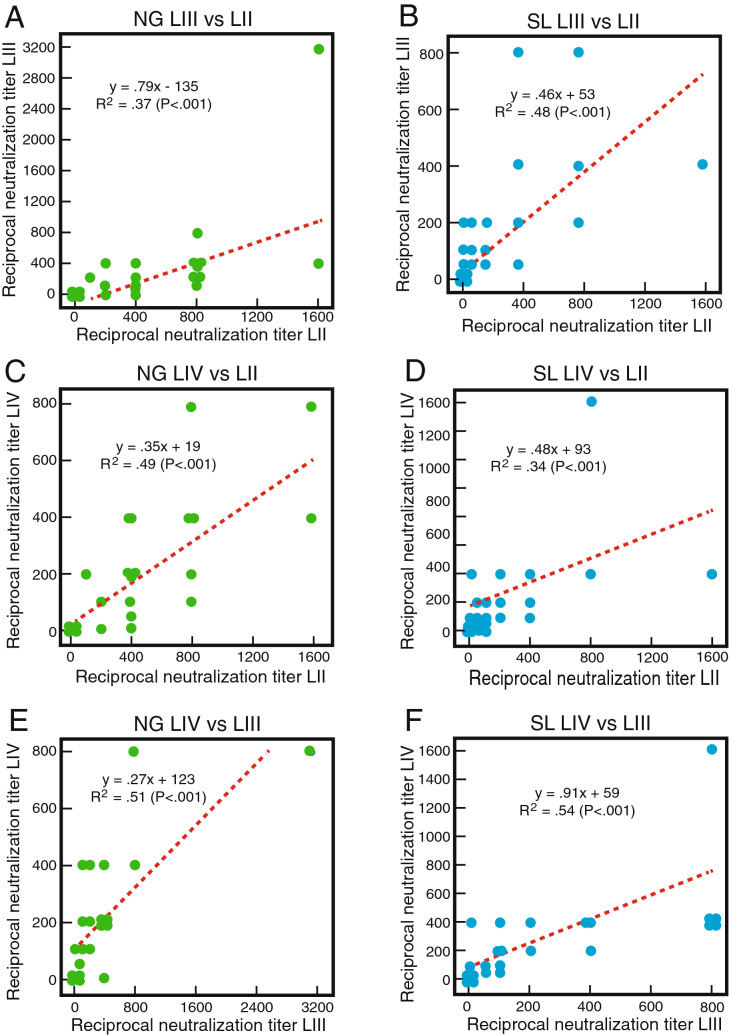
Lassa virus (LASV) is the causative agent of Lassa fever, an often-fatal hemorrhagic disease that is endemic in West Africa. Seven genetically distinct LASV lineages have been identified. As part of CEPI's (Coalition for Epidemic Preparedness Innovations) Lassa vaccine development program, we assessed the potential of the human immune system to mount cross-reactive and cross-protective humoral immune responses to antigens from the most prevalent LASV lineages, which are lineages II and III in Nigeria and lineage IV in Sierra Leone. IgG and IgM present in the blood of Lassa fever survivors from Nigeria or Sierra Leone exhibited substantial cross-reactivity for binding to LASV nucleoprotein and two engineered (linked and prefusion) versions of the glycoproteins (GP) of lineages II-IV. There was less cross-reactivity for the Zinc protein. Serum or plasma from Nigerian Lassa fever survivors neutralized LASV pseudoviruses expressing lineage II GP better than they neutralized lineage III or IV GP expressing pseudoviruses. Sierra Leonean survivors did not exhibit a lineage bias. Neutralization titres determined using LASV pseudovirus assays showed significant correlation with titres determined by plaque reduction with infectious LASV. These studies provide guidance for comparison of humoral immunity to LASV of distinct lineages following natural infection or immunization.


Conflict of interest statement

Tulane University and its various academic and industry partners have filed US and foreign patent applications for countermeasures to emerging viruses. Technical information may also be kept as trade secrets. If commercial products are developed, including Lassa diagnostics, MLH, MLB, DSKN, DJB, RWC, APK, KMH, MMR, IA, SK, RL, JGS, JSS, TWG, EOS, CTH, DSG, RFG and LMB may receive royalties or profits. This does not alter our adherence to all policies of the NIH and Scientific Reports on sharing data and materials. The authors declare no competing interests.

Figures

Figure 1
Figure 1
Recombinant LASV proteins representing lineages II–IV and their use in ELISA. Panel (A) Nucleoproteins (NP) from LASV representing lineages II–IV were expressed in E. coli, purified and resolved by sodium dodecyl-sulfate polyacrylamide gel electrophoresis (SDS-PAGE). Vertical lines indicate removal of lanes from a single gel. Circles: minor contaminants. Asterisk: monomeric NP. Square: NP breakdown product. Panel (B): LASV Zinc protein was expressed in E. coli, purified and resolved by SDS-PAGE and analyzed by western blotting. Monomeric and multimeric forms of the protein (asterisks) are detected. Panel (C) LASV linked glycoprotein was expressed in Drosophila S2 cells, and purified and resolved by SDS-PAGE in the presence and absence of dithiothreitol (DTT). Because most of the GP1 and 2 subunits are covalently linked, reduction by DTT does not affect the mobility of the GP protomer. Panel (D): LASV prefusion glycoprotein (Pf-GP) was expressed in Drosophila S2 cells, purified and resolved by SDS-PAGE in the presence and absence of dithiothreitol (DTT). Reduction by DTT converts GP protomer to GP1 and 2 subunits that have similar molecular weights. Panels (C,D) were prepared from three separate gels. Uncropped polyacrylamide gels and the western blot used to prepare panels (AD) are shown in Fig. S1. Panel (E): ReLASV Pan-Lassa NP ELISA configured with a mixture of recombinant NP from LASV representing lineages II–IV (LII–IV) and Pan-Lassa Pf-GP ELISA configured with a mixture of recombinant Pf-GP from LASV representing lineages II–IV were used to compare reactivity of IgG and IgM from control samples from heathy United States blood donors (n = 28) versus and Sierra Leonean Lassa fever survivors (n = 58). Data was analyzed using Prism (version 6.07, GraphPad Software, Inc., San Diego, CA) and the figure was compiled using Adobe Illustrator (version 15.1.0, San Jose, CA). Box and whisker plots representing means and standard deviations of samples are indicated. P values for pairwise comparisons are indicated.
Figure 2
Figure 2
Screening of plasma and serum from Lassa fever survivors for IgG and IgM reactivity to nucleoproteins and glycoprotein from LASV representing lineages II, III and IV. Binding of IgG or IgM in serum or plasma samples from Nigerian (Panel A, n = 140) or Sierra Leonean (Panel B, n = 80) Lassa fever survivors (1:100 dilution) to nucleoprotein (NP) or Prefusion glycoprotein (Pf-GP) was quantified using the Pan-Lassa NP or Pan-Lassa Prefusion-GP ELISA. Data was analyzed using Microsoft Excel (version 16.39, Microsoft, Redmond, WA) and JMP software (version 13.0.0, SAS Institute, Inc., Cary, NC). The figure was compiled using Adobe Illustrator (version 15.1.0, San Jose, CA). Blue dotted lines are linear regression plots of IgG seroreactivity of a mixture of NP representing lineages II–IV versus a mixture of Pf-GP representing lineages II–IV. Orange dotted lines are linear regression plots comparing IgM seroreactivity for the same mixtures.
Figure 3
Figure 3
Cross-reactivity of IgG in plasma and serum from Lassa fever survivors for recombinant proteins from LASV of lineages II–IV. Binding of IgG in serum or plasma samples (1:100 dilution) from Nigerian (NG, Panels A,C,E,G; n = 40) or Sierra Leonean (SL, Panels B,D,F,H; n = 61) Lassa fever survivors quantified using ELISA coated individually with NP, linked GP, Pf-GP or Z from LASV representing lineages II–IV (LII, LIII, LIV) as indicated. Data was analyzed using Microsoft Excel (version 16.39, Microsoft, Redmond, WA) and JMP software (version 13.0.0, SAS Institute, Inc., Cary, NC). The figure was compiled using Adobe Illustrator (version 15.1.0, San Jose, CA). Green dotted lines are linear regression plots of seroreactivity of LASV lineage II antigens versus lineage III antigens. Blue dotted lines are linear regression plots of seroreactivity of lineage II LASV antigens versus lineage IV antigens.
Figure 4
Figure 4
Plasma or serum endpoint dilutions demonstrating cross-reactivity of recombinant proteins from LASV of lineages II–IV. Examples of endpoint dilutions of serum or plasma samples from Nigerian or Sierra Leonean Lassa fever survivors demonstrating reactivity antigens representing LASV lineages II–IV (LII, LII, LIV). Panels A,D,G,J: Nigerian survivor 5 (NG5). Panels B,E,H,K: Sierra Leonean survivor 9 (SL9). Panels C,F,I,L: Sierra Leonean survivor 9 (SL9). ELISA were coated individually with NP, linked GP, Pf-GP or Z from LASV lineages II–IV as indicated. Data was analyzed using Microsoft Excel (version 16.39, Microsoft, Redmond, WA) and JMP software (version 13.0.0, SAS Institute, Inc., Cary, NC). The figure was compiled using Adobe Illustrator (version 15.1.0, San Jose, CA). Blue symbols: lineage II. Red symbols: lineage III. Green symbols: lineage IV. Error bars represent standard deviations, which in most cases were smaller than the symbols.
Figure 5
Figure 5
Cross-reactivity of IgM in plasma and serum of Lassa fever survivors for recombinant proteins from LASV of lineages II–IV. Cross-reactivity study for IgM binding conducted as in Fig. 3.
Figure 6
Figure 6
Neutralization of pseudoviruses expressing Lassa virus glycoprotein complex representing lineages II, III and IV by plasma or serum samples of Lassa fever survivors. 50% reciprocal neutralization titres of serum or plasma from Nigerian and Sierra Leonean Lassa fever survivors were determined using LASV pseudoviruses (LASVpv) expressing LASV glycoprotein complexes representing lineages II–IV (LII, LII, LIV). Panel (A): Lassa fever survivors from Nigeria (n = 37). Panel (B): Lassa fever survivors from Sierra Leone (n = 39). Note that not all samples from the cohorts had sufficient volume to perform the three pv assays. Panel (C): Mean of the 50% reciprocal neutralization titres represented in panels (A,B). Panel (D): neutralization curve for Nigerian subject 2 (NG2). Panel (E): neutralization curve for Sierra Leonean subject 20 (SL20). Blue bars or symbols are pp representing lineage II. Red bars or symbols are pv representing lineage III. Green bars or symbols are pv representing lineage IV. Data was analyzed using Microsoft Excel (version 16.39, Microsoft, Redmond, WA), JMP software (version 13.0.0, SAS Institute, Inc., Cary, NC) and Prism (version 6.07, GraphPad Software, Inc., San Diego, CA). The figure was compiled using Adobe Illustrator (version 15.1.0, San Jose, CA).Error bars represent standard error of the mean, which is smaller than the symbols in some cases. Asterisks in panels (D,E) represent enhancement at the indicated dilutions.
Figure 7
Figure 7
Cross-neutralization by plasma or serum samples of Lassa fever survivors. Linear correlations (dotted red lines) for the neutralization of LASV pseudoviruses expressing LASV glycoprotein complexes representing lineages II, III and IV (LII, LIII and LIV) are indicated. Panels (A,C,E): Nigerian (NG) Lassa fever survivors (n = 37). Panels (B,D,F): Sierra Leonian (SL) Lassa fever survivors (N = 39). Data was analyzed using Microsoft Excel (version 16.39, Microsoft, Redmond, WA) and JMP software (version 13.0.0, SAS Institute, Inc., Cary, NC). The figure was compiled using Adobe Illustrator (version 15.1.0, San Jose, CA). Note that multiple samples had the same 50% neutralization titres for a pair of lineages producing overlapping data points.
Figure 8
Figure 8
Comparison of Plaque Reduction neutralization to pseudovirus neutralization or ELISA binding. Panel (A): Comparison of 50% plaque reduction neutralization titres (PRNT) using LASV lineage II (0043/LV/14) versus PRNT using LASV lineage IV (Josiah) at Biosafety level-4. Panel (B): Comparison of 50% reciprocal neutralization titres using LASV pseudoviruses (pv) expressing the glycoprotein complex (GPC) of LASV lineage II versus LASVpv expressing GPC of LASV lineage IV (Josiah). Panel (C): Comparison of PRNT reciprocal titres using LASV lineage II to LASV pseudoviruses expressing LASV lineage II GPC reciprocal neutralization titres. Panel (D): Comparison of PRNT reciprocal titres using LASV lineage IV to LASV pseudoviruses expressing LASV lineage IV GPC reciprocal neutralization titres. Panel (E): Comparison of PRNT reciprocal titres using LASV lineage II to 50% reciprocal binding titres to prefusion glycoprotein (Pf-GP) of LASV lineage II. Panel (F): Comparison of PRNT reciprocal titres using LASV lineage IV to 50% reciprocal binding titres to Pf-GP of LASV lineage IV. Panel (G): Comparison of LASV pseudoviruses expressing lineage II GPC reciprocal titres to 50% reciprocal binding titres to Pf-GP of LASV lineage II. Panel (H): Comparison of LASV pseudoviruses expressing lineage IV GPC reciprocal titres to 50% reciprocal binding titres to Pf-GP of LASV lineage IV. Data was analyzed using Microsoft Excel (version 16.39, Microsoft, Redmond, WA) and JMP software (version 13.0.0, SAS Institute, Inc., Cary, NC). The figure was compiled using Adobe Illustrator (version 15.1.0, San Jose, CA). Note that multiple samples had the same 50% reciprocal titres producing overlapping data points.

References

    1. Akpede GO, et al. Caseload and case fatality of Lassa fever in Nigeria, 2001–2018: A specialist center's experience and its implications. Front. Public Health. 2019;7:170. doi: 10.3389/fpubh.2019.00170. - DOI - PMC - PubMed
    1. Ilori EA, et al. Epidemiologic and clinical features of Lassa fever outbreak in Nigeria, January 1–May 6, 2018. Emerg. Infect. Dis. 2019;25:1066–1074. doi: 10.3201/eid2506.181035. - DOI - PMC - PubMed
    1. Shaffer JG, et al. Lassa fever in post-conflict Sierra Leone. PLoS Negl. Trop. Dis. 2014;8:e2748. doi: 10.1371/journal.pntd.0002748. - DOI - PMC - PubMed
    1. McCormick JB. Epidemiology and control of Lassa fever. Curr. Top Microbiol. Immunol. 1987;134:69–78. - PubMed
    1. Arnold RB, Gary GW. A neutralization test survey for Lassa fever activity in Lassa, Nigeria. Trans. R. Soc. Trop. Med. Hyg. 1977;71:152–154. doi: 10.1016/0035-9203(77)90085-2. - DOI - PubMed

Publication types