Review
. 2020;22(11):37.
doi: 10.1007/s11940-020-00647-z. Epub 2020 Sep 25.

Unraveling the Possible Routes of SARS-COV-2 Invasion into the Central Nervous System

Affiliations
Review

Unraveling the Possible Routes of SARS-COV-2 Invasion into the Central Nervous System

Maria Lima et al. Curr Treat Options Neurol. 2020.

Abstract

Purpose of review: To describe the possible neuroinvasion pathways of Severe Acute Respiratory Syndrome-related Coronavirus-2 (SARS-CoV-2), the virus responsible for the Coronavirus disease-19 (Covid-19) pandemic.

Recent findings: We present data regarding the family of Coronaviruses (CoVs) and the central nervous system (CNS), and describe parallels between SARS-CoV-2 and other members of the family, which have been investigated in more depth and combine these findings with the recent advancements regarding SARS-CoV-2.

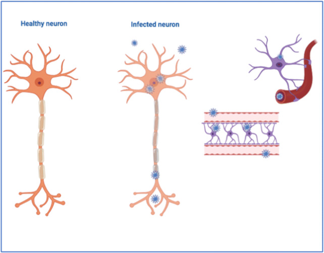
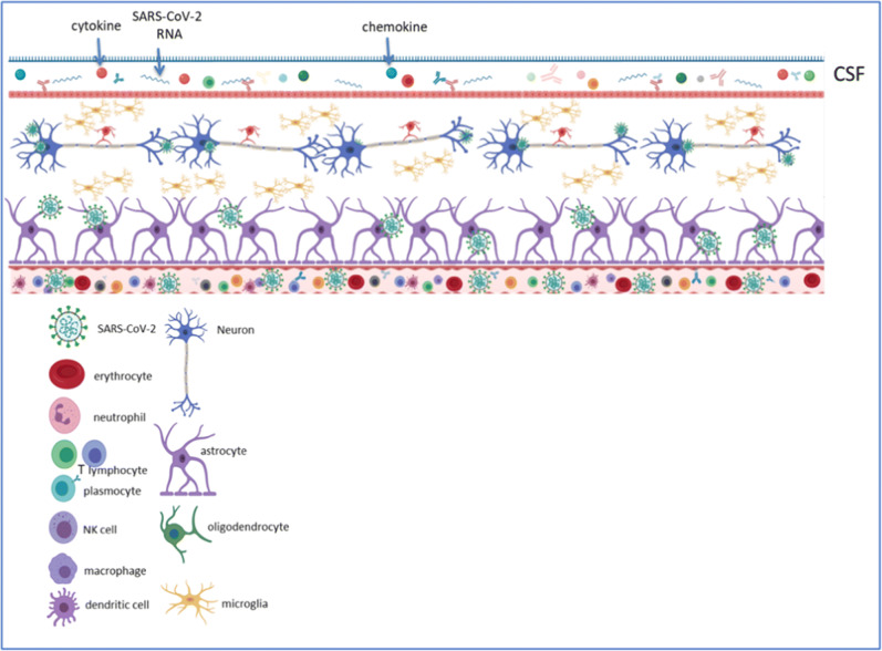
Summary: SARS-CoV-2 like other CoVs is neuroinvasive, neurotropic and neurovirulent. Two main pathways of CNS penetration seem to be the strongest candidates, the hematogenous and the neuronal. Τhe olfactory route in particular appears to play a significant role in neuroinvasion of coronaviruses and SARS-CoV-2, as well. However, existing data suggest that other routes, involving the nasal epithelium in general, lymphatic tissue and the CSF may also play roles in SARS-CoV-2 invasion into the CNS.

Keywords: CNS penetration; COVID-19; Coronaviruses (CoVs); Neuroinvasion; Neurological manifestations; Neurotropism; Neurovirulence; Pandemic; SARS-2; SARS-CoV-2.


Conflict of interest statement

Conflict of InterestThe authors declare that they have no conflict of interest.

Figures

Fig. 1
Fig. 1
A normal neuron and a neuron and astrocytes as infected with coronavirus.
Figure 2
Figure 2
Entry of the virus into the host CNS-related cell and further spread. The single-stranded RNA (ssRNA) genome of Severe Acute Respiratory Syndrome-related Coronavirus-2 (SARS-CoV-2) encodes large polyproteins, which are proteolytically cleaved into 16 non-structural proteins (nsps). In addition, 9–12 ORFs are encoded through the transcription of a nested set of subgenomic RNAs. The virus forms spherical particles consisting of four structural proteins: the envelope glycoprotein spike (S), the envelope (E) and membrane (M) transmembrane glycoproteins incorporated in the virion, and the protein nucleocapsid (N). The surface protein of the virus binds to its receptors, such as ACE 2. Following the entry of the virus into the host cell (such as the neuron), and the release of the viral genome, the viral RNA is uncoated in the cytoplasm. Translation of the viral polymerase protein is followed by RNA replication and subgenomic transcription; ORF1a and ORF1ab are translated to create pp1a and pp1ab, which are cleaved by the proteases that are encoded by ORF1a to produce 16 nsps that form the RNA replicase–transcriptase complex. During the phase of replication, full-length (−)RNA copies of the genome are shaped and form templates for full-length (+)RNA genomes. Subgenomic (−)RNAs are transcribed into subgenomic (+)mRNAs. The step of translation is followed by the assembling of the resulting structural proteins into the nucleocapsid and viral envelope at the ER–Golgi intermediate compartment (ERGIC), followed by release of the nascent virion from the infected neuron cell. Created with a modified Biorender template (under license) as per de Wit et al. Nature Reviews Microbiology volume 14, pages523–534(2016).
Fig. 3
Fig. 3
The CNS microenvironment following SARS-CoV-2 infection. Spread of SARS-CoV-2 from lungs to the CNS can most likely be achieved through the haematogenous route. In addition, it can enter the CNS through the olfactory bulb, and once the infectious agent persists due to the inability of the immune system to control/suppress viral replication, the virus may reach the whole brain and the CSF, and participate in demyelination. In the hematogenous route, SARS-CoV-2 may gain access by infecting endothelial cells of the blood-brain-barrier, epithelial cells of the blood-cerebrospinal fluid barrier in the choroid plexus, or it may indeed use inflammatory cells as “Trojan horse” to obtain access into the CNS. Experimental data suggest that primary glial cultures can secrete a series of inflammatory cytokines participating in the perpetuation of viral infection and further inflicting CNS tissue damage. The role of astrocytes in the machinery of SARS-CoV-2 mediated CNS pathology is yet undetermined and remains to be defined.

Similar articles

Cited by

References

References and Recommended Reading

Papers of particular interest, published recently, have been highlighted as: • Of importance
    1. Vabret A, Dina J, Brison E, Brouard J, Freymuth F. Human Coronaviruses. Pathol Biol (Paris) 2009;57(2):149–160. doi: 10.1016/j.patbio.2008.02.018. - DOI - PMC - PubMed
    1. Nitulescu GM, Paunescu H, Moschos SA, Petrakis D, Nitulescu G, Ion GND, Spandidos D, Nikolouzakis T, Drakoulis N, Tsatsakis A. Comprehensive analysis of drugs to treat Sars-Cov-2 infection: mechanistic insights into current Covid-19 therapies (review) Int J Mol Med. 2020;46(2):467–488. doi: 10.3892/ijmm.2020.4608. - DOI - PMC - PubMed
    1. Tsatsakis A, Petrakis D, Nikolouzakis TK, Docea AO, Calina D, Vinceti M, Goumenou M, Kostoff RN, Mamoulakis C, Aschner M, Hernández AF. Covid-19, an opportunity to reevaluate the correlation between long-term effects of anthropogenic pollutants on viral epidemic/pandemic events and prevalence. Food Chem Toxicol. 2020;141:111418. doi: 10.1016/j.fct.2020.111418. - DOI - PMC - PubMed
    1. Chan JF, Lau SK, To KK. Cheng VC, Woo PC, Yuen KY. Middle East respiratory syndrome coronavirus: another zoonotic Betacoronavirus causing Sars-like disease. Clin Microbiol Rev. 2015;28(2):465–522. doi: 10.1128/cmr.00102-14. - DOI - PMC - PubMed
    1. Calina D, Docea AO, Petrakis D, Egorov AM, Ishmukhametov AA, Gabibov AG, Shtilman M, Kostoff R, Carvalho F, Vinceti M, Spandidos D, Tsatsakis A. Towards effective Covid-19 vaccines: updates, perspectives and challenges (review) Int J Mol Med. 2020;46(1):3–16. doi: 10.3892/ijmm.2020.4596. - DOI - PMC - PubMed