

Dermoscopic diagnosis of subungual hematoma: new observations
- PMID: 32994768
- PMCID: PMC7507148
- DOI: 10.5114/ada.2020.98235
Dermoscopic diagnosis of subungual hematoma: new observations
Abstract
Introduction: There are very few studies focusing on the dermoscopic features of subungual hematoma which is one of the major imitators of subungual melanoma.
Aim: To identify the dermoscopic findings of subungual hematoma, which will facilitate the diagnostic process by reducing the use of more invasive diagnostic methods like nail avulsion or biopsy.
Material and methods: In this study, clinical and dermoscopic findings of the cases were reviewed. The diagnosis of subungual hematoma was confirmed by observing progression of the colour change to the distal edge of the nail plate in all the cases.
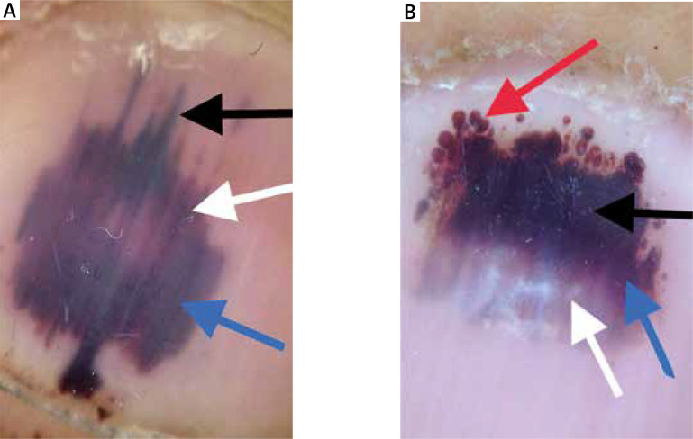
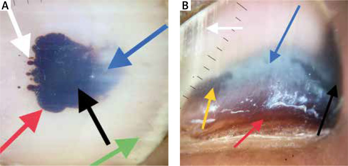
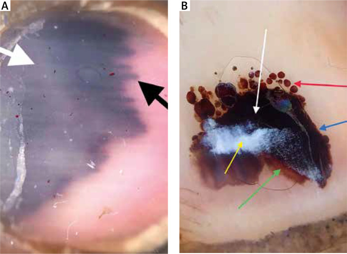
Results: A total of 47 subungual hematomas were enrolled in the study. The most common colour was purple-black (53%). Blue-white colour was observed in 12 (26%) lesions. 9 (19%) lesions showed granular leukonychia. All of the lesions had a homogenous pattern. In 25 (53%) lesions, a globular pattern was observed. 14 (30%) lesions showed a streaks pattern. Peripheral fading and periungual haemorrhage were present in 14 (30%) and 9 (9%) lesions, respectively.
Conclusions: We detected two new findings which have not been described previously for subungual hematoma: the first one is "blue-white colour" which is known as an important clue to melanoma. The second one is granular leukonychia localized on the hematoma. We suggest that in any case of the nail discoloration, a thorough dermoscopic examination should be performed. Moreover, progression of the colour change to the distal edge should be observed to ensure that a possible melanoma is not overlooked.
Keywords: dermoscopy; hematoma; subungual.
Copyright: © 2020 Termedia Sp. z o. o.
Conflict of interest statement
The authors declare no conflict of interest.
Figures
References
-
- Mun JH, Kim GW, Jwa SW, et al. Dermoscopy of subungual haemorrhage: its usefulness in differential diagnosis from nail-unit melanoma. Br J Dermatol. 2013;68:1224–9. - PubMed
-
- Pierre M. The Nail. 1st edn. Edinburgh, New York: Churchill Livingstone; 1981.
-
- Braun RP, Baran R, Le Gal FA, et al. Diagnosis and management of nail pigmentations. J Am Acad Dermatol. 2007;56:835–47. - PubMed
-
- Zaias N. The Nail in Health and Disease. 2nd edn. Norwalk, Connecticut: Appleton & Lange; 1990.
-
- Benati E, Ribero S, Longo C, et al. Clinical and dermoscopic clues to differentiate pigmented nail bands: an International Dermoscopy Society study. J Eur Acad Dermatol Venereol. 2017;31:732–6. - PubMed
LinkOut - more resources
Full Text Sources