. 2020 Jul 6;7(17):2001223.
doi: 10.1002/advs.202001223. eCollection 2020 Sep.

Porous Pt Nanospheres Incorporated with GOx to Enable Synergistic Oxygen-Inductive Starvation/Electrodynamic Tumor Therapy

Affiliations

Porous Pt Nanospheres Incorporated with GOx to Enable Synergistic Oxygen-Inductive Starvation/Electrodynamic Tumor Therapy

Zijie Lu et al. Adv Sci (Weinh). .

Abstract

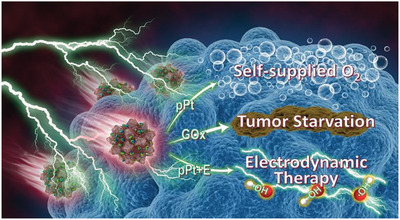
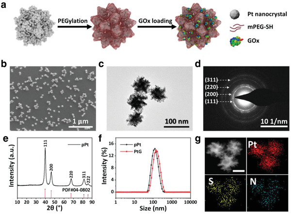
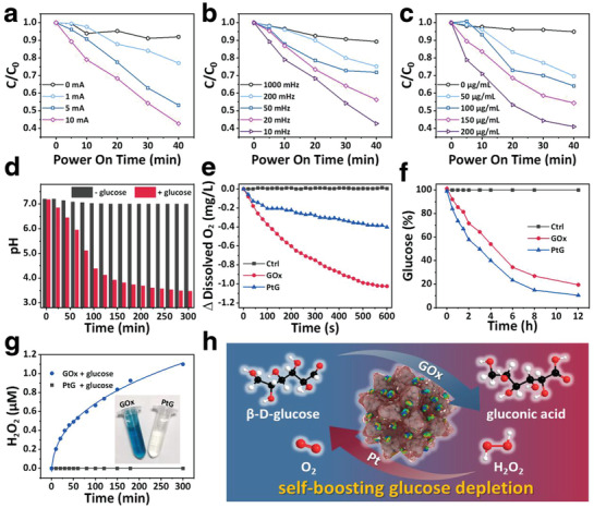
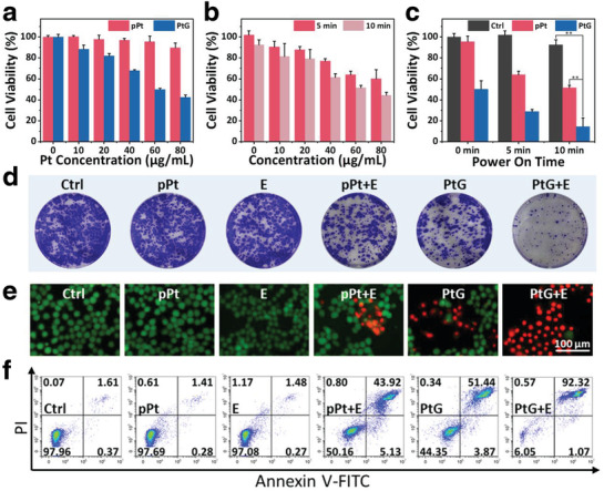
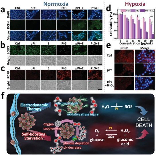
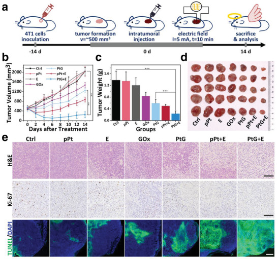
Glucose-oxidase (GOx)-mediated starvation by consuming intracellular glucose has aroused extensive exploration as an advanced approach for tumor treatment. However, this reaction of catalytic oxidation by GOx is highly dependent on the on-site oxygen content, and thus starvation therapy often suffers unexpected anticancer outcomes due to the intrinsic tumorous hypoxia. Herein, porous platinum nanospheres (pPts), incorporated with GOx molecules (PtGs), are synthesized to enable synergistic cancer therapy. In this system, GOx can effectively catalyze the oxidation of glucose to generate H2O2, while pPt triggers the decomposition of both endogenous and exogenous H2O2 to produce considerable content of O2 to facilitate the glucose consumption by GOx. Meanwhile, pPt induces remarkable content of intracellular reactive oxygen species (ROS) under an alternating electric field, leading to cellular oxidative stress injury and promotes apoptosis following the mechanism of electrodynamic therapy (EDT). In consequence, the PtG nanocomposite exhibits significant anticancer effect both in vitro and in vivo. This study has therefore demonstrated a fascinating therapeutic platform enabling oxygen-inductive starvation/EDT synergistic strategy for effective tumor treatment.

Keywords: electrodynamic therapy; oxygen‐inductive starvation; porous Pt nanospheres; synergistic tumor therapy.


Conflict of interest statement

The authors declare no conflict of interest.

Figures

Scheme 1
Scheme 1
Illustration of functioning mechanism of pPt@GOx (PtG).
Figure 1
Figure 1
a) Illustration of the synthetic procedure of PtGs. b) Scanning electron microscope (SEM) image of pPts. c) Typical transmission electron microscope (TEM) image and d) corresponding SAED pattern of pPts. e) XRD patterns of pPts. f) DLS analysis of pPts and PtGs in PBS. g) Energy dispersive spectroscopy (EDS) element mapping of PtGs (scale bar: 50 nm).
Figure 2
Figure 2
Degradation of MB in pPt solutions with varied a) current intensities, b) electric frequencies, and c) pPt concentrations. d) The variation of pH in PtG solutions with or without glucose. e) Oxygen and f) glucose consumption kinetics of GOx and PtGs solutions. g) H2O2 generation of PtGs and GOx in glucose solution (inset: photograph of solutions after chromogenic reaction). h) Schematic diagram of the self‐boosting glucose depletion of PtGs.
Figure 3
Figure 3
In vitro cytotoxicity assay. a) 4T1 cells viabilities treated with varied concentrations of pPts and PtGs for 24 h. b) Relative cell viabilities of 4T1 cells treated with pPts after agitated by a square‐wave current (5 mA, 10 mHz) for 5 or 10 min. c) Synergistic cell‐killing effect of PtGs with an electric field. p‐values: **p < 0.01. d–f) Corresponding colony formation, Live&Dead staining images, and quantitative flow cytometry analysis of cell apoptosis/necrosis of 4T1 cells treated with all seven groups.
Figure 4
Figure 4
Cellular abnormalities induced by electric field and starvation. Fluorescence images of a) intracellular ROS (scale bar: 100 µm), b) pH (scale bar: 200 µm), and c) oxygen (scale bar: 200 µm) in 4T1 cells cultured with different sample groups under a normoxia condition. d) Relative cell viabilities of 4T1 cells incubated with free GOx, PtGs, and PtGs+H2O2 under a hypoxic condition for 24 h. p‐values: ***p < 0.001. e) RDPP fluorescence in 4T1 cells treated with pPts in the absence and presence of H2O2 under a hypoxic condition (scale bar: 100 µm). f) Mechanism illustration of inhibition effect induced by PtGs.
Figure 5
Figure 5
In vivo antitumor efficacy. a) Demonstration of the treatment process. b) Variation of tumor volume after different treatments by all sample groups (n = 5). c) Average tumor weight on day 14 after various treatments. d) Photographs of tumors harvested from different treatment groups. e) H&E staining (scale bar: 100 µm), Ki‐67 staining (scale bar: 100 µm), and TUNEL staining (scale bar: 500 µm) images of tumor tissue collected from different mice groups 24 h after treatments. p‐values: ***p < 0.001, **p < 0.01.

References

    1. Yu S., Chen Z., Zeng X., Chen X., Gu Z., Theranostics 2019, 9, 8026. - PMC - PubMed
    1. a) Cairns R. A., Harris I. S., Mak T. W., Nat. Rev. Cancer 2011, 11, 85; - PubMed
    2. b) DeBerardinis R. J., Lum J. J., Hatzivassiliou G., Thompson C. B., Cell Metab. 2008, 7, 11. - PubMed
    1. a) Agemy L., Sugahara K. N., Kotamraju V. R., Gujraty K., Girard O. M., Kono Y., Mattrey R. F., Park J.‐H., Sailor M. J., Jimenez A. I., Cativiela C., Zanuy D., Sayago F. J., Aleman C., Nussinov R., Ruoslahti E., Blood 2010, 116, 2847; - PMC - PubMed
    2. b) Li S., Jiang Q., Liu S., Zhang Y., Tian Y., Song C., Wang J., Zou Y., Anderson G. J., Han J.‐Y., Chang Y., Liu Y., Zhang C., Chen L., Zhou G., Nie G., Yan H., Ding B., Zhao Y., Nat. Biotechnol. 2018, 36, 258; - PubMed
    3. c) Zhang C., Ni D., Liu Y., Yao H., Bu W., Shi J., Nat. Nanotechnol. 2017, 12, 378. - PubMed
    1. a) Fu L.‐H., Qi C., Lin J., Huang P., Chem. Soc. Rev. 2018, 47, 6454; - PubMed
    2. b) Hamanaka R. B., Chandel N. S., J. Exp. Med. 2012, 209, 211; - PMC - PubMed
    3. c) Maddocks O. D. K., Berkers C. R., Mason S. M., Zheng L., Blyth K., Gottlieb E., Vousden K. H., Nature 2013, 493, 542; - PMC - PubMed
    4. d) Selwan E. M., Finicle B. T., Kim S. M., Edinger A. L., FEBS Lett. 2016, 590, 885. - PMC - PubMed
    1. a) Bankar S. B., Bule M. V., Singhal R. S., Ananthanarayan L., Biotechnol. Adv. 2009, 27, 489; - PubMed
    2. b) Wilson R., Turner A. P. F., Biosens. Bioelectron. 1992, 7, 165;
    3. c) Wong C. M., Wong K. H., Chen X. D., Appl. Microbiol. Biotechnol. 2008, 78, 927. - PubMed

LinkOut - more resources