

A Lactose-Derived CRISPR/Cas9 Delivery System for Efficient Genome Editing In Vivo to Treat Orthotopic Hepatocellular Carcinoma
- PMID: 32995132
- PMCID: PMC7507475
- DOI: 10.1002/advs.202001424
A Lactose-Derived CRISPR/Cas9 Delivery System for Efficient Genome Editing In Vivo to Treat Orthotopic Hepatocellular Carcinoma
Abstract
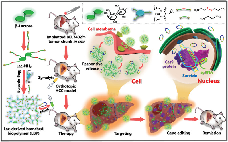
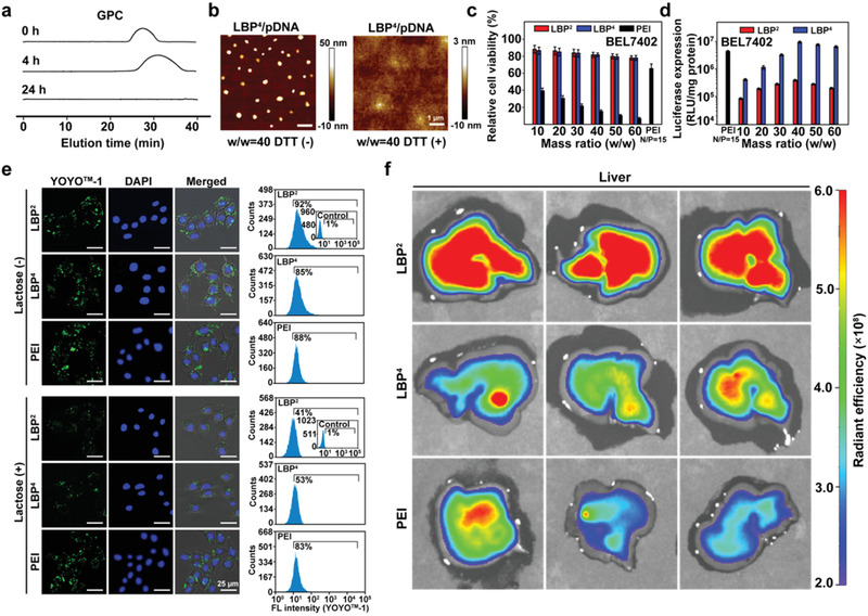
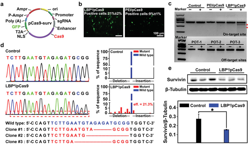
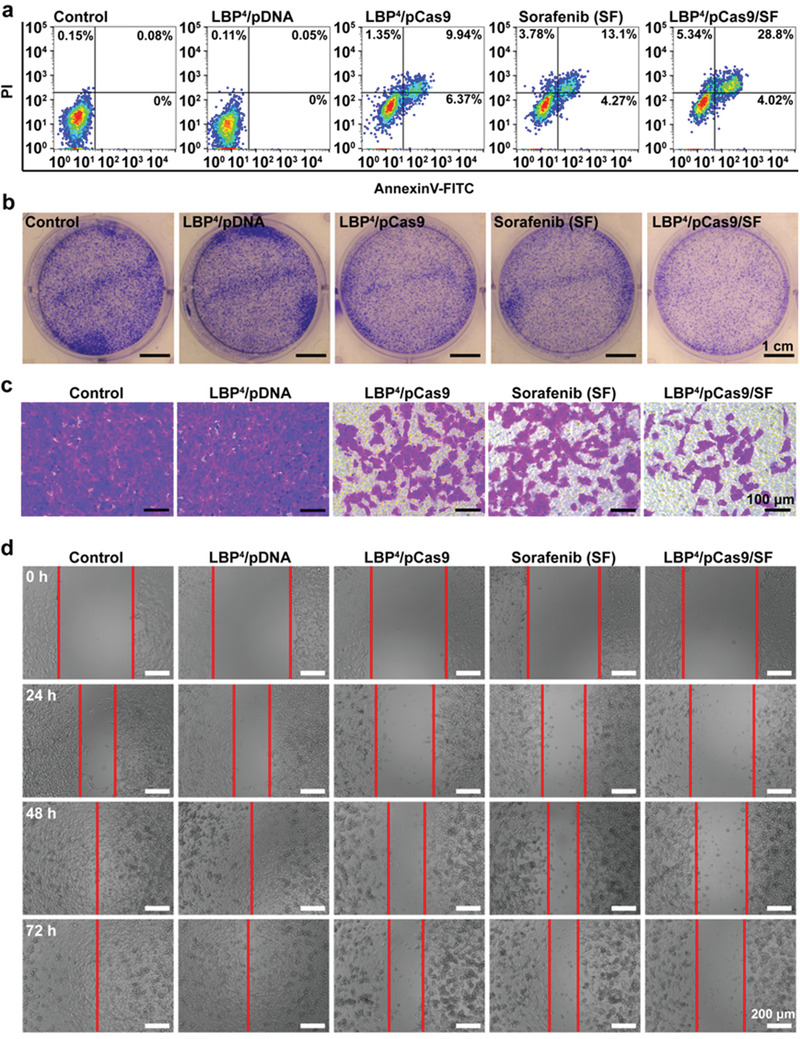
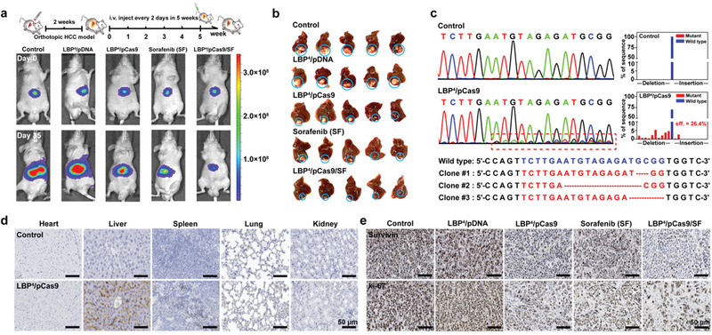
Gene editing is a crucial and effective strategy to treat genetic diseases. Safe and effective delivery vectors are specially required for efficient gene editing in vivo of CRISPR/Cas9 system. Interestingly, lactose, a natural saccharide, can specifically bind to asialoglycoprotein receptors, highly expressed on the surface of hepatocellular carcinoma (HCC) cells. Herein, a lactose-derived branched cationic biopolymer (LBP) with plentiful reducible disulfide linkages and hydroxyl groups is proposed as a potential delivery vector of CRISPR/Cas9 system for efficient genome editing in vivo to treat orthotopic HCC. LBP is synthesized via a facile one-pot ring-opening reaction. LBP possesses excellent compacting ability, degradability, biocompatibility, gene transfection performances, and HCC-targeting ability. LBP-mediated delivery of classical pCas9-survivin, which can target and knockout survivin oncogene, produces efficient gene editing performances, and superb anti-cancer activities in orthotopic HCC mouse models. This study provides an attractive and safe strategy for the rational design of CRISPR/Cas9 delivery system.
Keywords: CRISPR/Cas9; biopolymers; delivery vectors; lactose; orthotopic hepatocellular carcinoma.
© 2020 The Authors. Published by WILEY‐VCH Verlag GmbH & Co. KGaA, Weinheim.
Conflict of interest statement
The authors declare no conflict of interest.
Figures
Similar articles
-
Fluorinated Acid-Labile Branched Hydroxyl-Rich Nanosystems for Flexible and Robust Delivery of Plasmids.Small. 2018 Oct;14(42):e1803061. doi: 10.1002/smll.201803061. Epub 2018 Sep 20. Small. 2018. PMID: 30238691
-
Charge-reversal nanocomolexes-based CRISPR/Cas9 delivery system for loss-of-function oncogene editing in hepatocellular carcinoma.J Control Release. 2021 May 10;333:362-373. doi: 10.1016/j.jconrel.2021.03.030. Epub 2021 Mar 27. J Control Release. 2021. PMID: 33785418
-
A multifunctional non-viral vector for the delivery of MTH1-targeted CRISPR/Cas9 system for non-small cell lung cancer therapy.Acta Biomater. 2022 Nov;153:481-493. doi: 10.1016/j.actbio.2022.09.046. Epub 2022 Sep 24. Acta Biomater. 2022. PMID: 36162766
-
In vivo delivery of CRISPR-Cas9 genome editing components for therapeutic applications.Biomaterials. 2022 Dec;291:121876. doi: 10.1016/j.biomaterials.2022.121876. Epub 2022 Oct 28. Biomaterials. 2022. PMID: 36334354 Free PMC article. Review.
-
Recent advances in the delivery and applications of nonviral CRISPR/Cas9 gene editing.Drug Deliv Transl Res. 2023 May;13(5):1500-1519. doi: 10.1007/s13346-023-01320-z. Epub 2023 Mar 29. Drug Deliv Transl Res. 2023. PMID: 36988873 Free PMC article. Review.
Cited by
-
Biomaterials-mediated CRISPR/Cas9 delivery: recent challenges and opportunities in gene therapy.Front Chem. 2023 Sep 28;11:1259435. doi: 10.3389/fchem.2023.1259435. eCollection 2023. Front Chem. 2023. PMID: 37841202 Free PMC article. Review.
-
Biomaterial-Based CRISPR/Cas9 Delivery Systems for Tumor Treatment.Biomater Res. 2024 Apr 30;28:0023. doi: 10.34133/bmr.0023. eCollection 2024. Biomater Res. 2024. PMID: 38694229 Free PMC article.
-
CRISPR-Cas9 in basic and translational aspects of cancer therapy.Bioimpacts. 2024;14(6):30087. doi: 10.34172/bi.2024.30087. Epub 2024 Mar 10. Bioimpacts. 2024. PMID: 39493894 Free PMC article. Review.
-
Biomedical polymers: synthesis, properties, and applications.Sci China Chem. 2022;65(6):1010-1075. doi: 10.1007/s11426-022-1243-5. Epub 2022 Apr 24. Sci China Chem. 2022. PMID: 35505924 Free PMC article. Review.
-
In vivo targeted delivery of nucleic acids and CRISPR genome editors enabled by GSH-responsive silica nanoparticles.J Control Release. 2021 Aug 10;336:296-309. doi: 10.1016/j.jconrel.2021.06.030. Epub 2021 Jun 23. J Control Release. 2021. PMID: 34174352 Free PMC article.
References
-
- Ribeil J. A., Hacein‐Bey‐Abina S., Payen E., Magnani A., Semeraro M., Magrin E., Caccavelli L., Neven B., Bourget P., El N. W., N. Engl. J. Med. 2017, 376, 848. - PubMed
-
- Thompson A. A., Walters M. C., Kwiatkowski J., Jej R., Ribeil J. A., Hongeng S., Magrin E., Schiller G. J., Payen E., Semeraro M., N. Engl. J. Med. 2018, 378, 1479. - PubMed
-
- Martin J., Krzysztof C., Ines F., Michael H., Doudna J. A., Emmanuelle C., Science 2012, 337, 46. - PubMed
LinkOut - more resources
Full Text Sources
Miscellaneous