

Anatomic Characteristics of Tissues Attached to the Fifth Metatarsal Bone
- PMID: 32995346
- PMCID: PMC7503013
- DOI: 10.1177/2325967120947725
Anatomic Characteristics of Tissues Attached to the Fifth Metatarsal Bone
Abstract
Background: Two types of stress, bending stress and traction stress, have been reported to be involved in the mechanism of Jones fracture. However, little is known about the risk factors for traction stress.
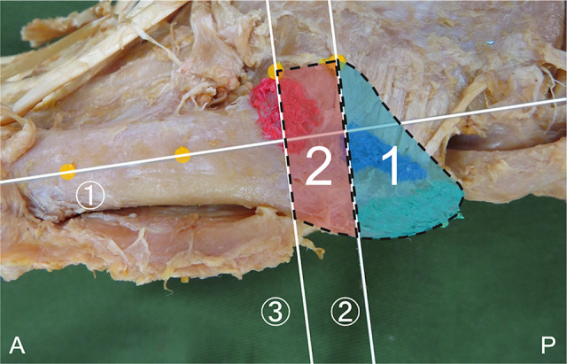
Purpose: To classify the attachment position of the peroneus brevis muscle (PB), peroneus tertius (PT), lateral band of the plantar aponeurosis (LB), and the long plantar ligament (LPL), focusing on the zone where a Jones fracture occurs (zone 2), and to compare the footprint area of each tissue type.
Study design: Descriptive laboratory study.
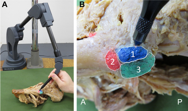
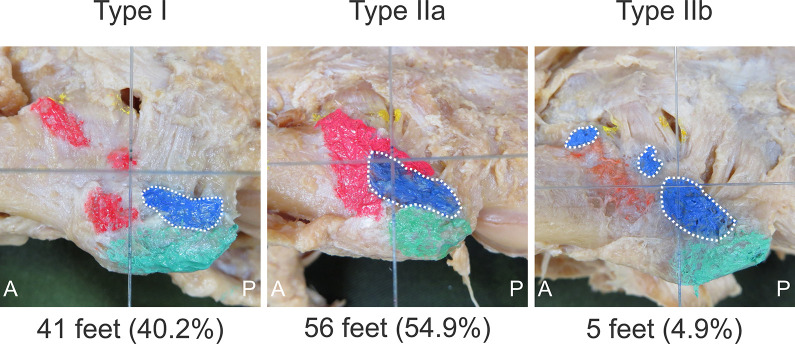
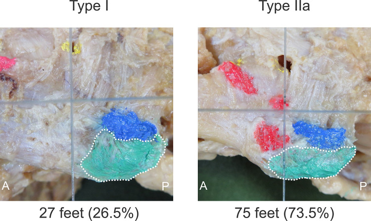
Methods: This study examined 102 legs from 55 Japanese cadavers. Type classification was performed by focusing on the positional relationship between each tissue attachment and the zone where Jones fracture occurs (zone 2). The classifications were as follows: type I, attached proximal to the border between zones 1 and 2; type IIa, attached to the border between zones 1 and 2 with one attached part; and type IIb, attached across the border between zones 1 and 2 with two or more attached parts. The footprint areas of the PB, PT, LB, and LPL were compared between tissue types and within each attachment classification.
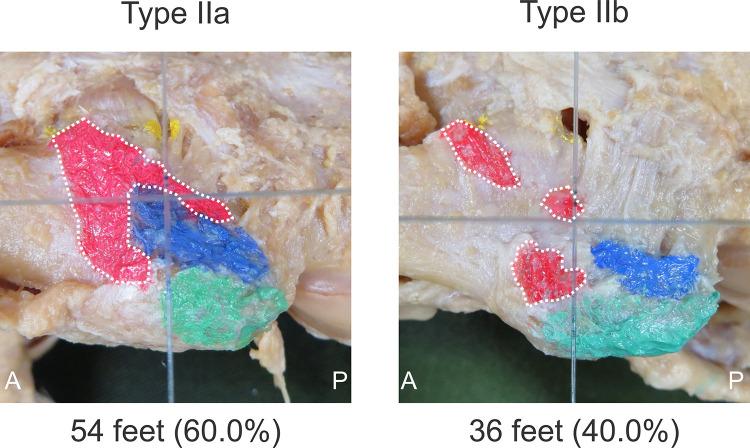
Results: The PB was recorded as type I in 41 feet (40.2%), type IIa in 56 feet (54.9%), and type IIb in 5 feet (4.9%); the PT was recorded as type IIa in 54 feet (60.0%) and type IIb in 36 feet (40.0%); and the LB was recorded as type I in 27 feet (26.5%) and type IIa in 75 feet (73.5%). The LPL did not attach to the fifth metatarsal bone. No significant difference was found in the footprint area between type I PB and type I LB.
Conclusion: The results indicate that type I, which attaches proximal to zone 2, occurs with PB and LB, and there was no significant difference in the footprint area between them. These findings suggest that type I is involved in traction stress. In the future, biomechanical research based on the results of this study will be necessary.
Clinical relevance: The results of this study provide basic research for investigating the mechanism of Jones fracture and the cause of delayed healing.
Keywords: Jones fracture; fifth metatarsal bone; gross anatomy; traction stress.
© The Author(s) 2020.
Conflict of interest statement
One or more of the authors has declared the following potential conflict of interest or source of funding: This study was supported by Japan Society for the Promotion of Science (JSPS) KAKENHI, grant JP19K11358, and a grant-in-aid program from Niigata University of Health and Welfare. AOSSM checks author disclosures against the Open Payments Database (OPD). AOSSM has not conducted an independent investigation on the OPD and disclaims any liability or responsibility relating thereto.
Figures
References
-
- Azevedo RR, da Rocha ES, Franco PS, Carpes FP. Plantar pressure asymmetry and risk of stress injuries in the foot of young soccer players. Phys Ther Sport. 2017;24:39–43. - PubMed
-
- Beaupied H, Lespessailles E, Benhamou CL. Evaluation of macrostructural bone biomechanics. Joint Bone Spine. 2007;74(3):233–239. - PubMed
-
- Dameron TB., Jr Fractures and anatomical variations of the proximal portion of the fifth metatarsal. J Bone Joint Surg Am. 1975;57(6):788–792. - PubMed
LinkOut - more resources
Full Text Sources
