. 2020 May 22:18:84-97.
doi: 10.1016/j.omtm.2020.05.021. eCollection 2020 Sep 11.

Systemic Delivery of AAV- Fdxr Mitigates the Phenotypes of Mitochondrial Disorders in Fdxr Mutant Mice

Affiliations

Systemic Delivery of AAV- Fdxr Mitigates the Phenotypes of Mitochondrial Disorders in Fdxr Mutant Mice

Li Yang et al. Mol Ther Methods Clin Dev. .

Abstract

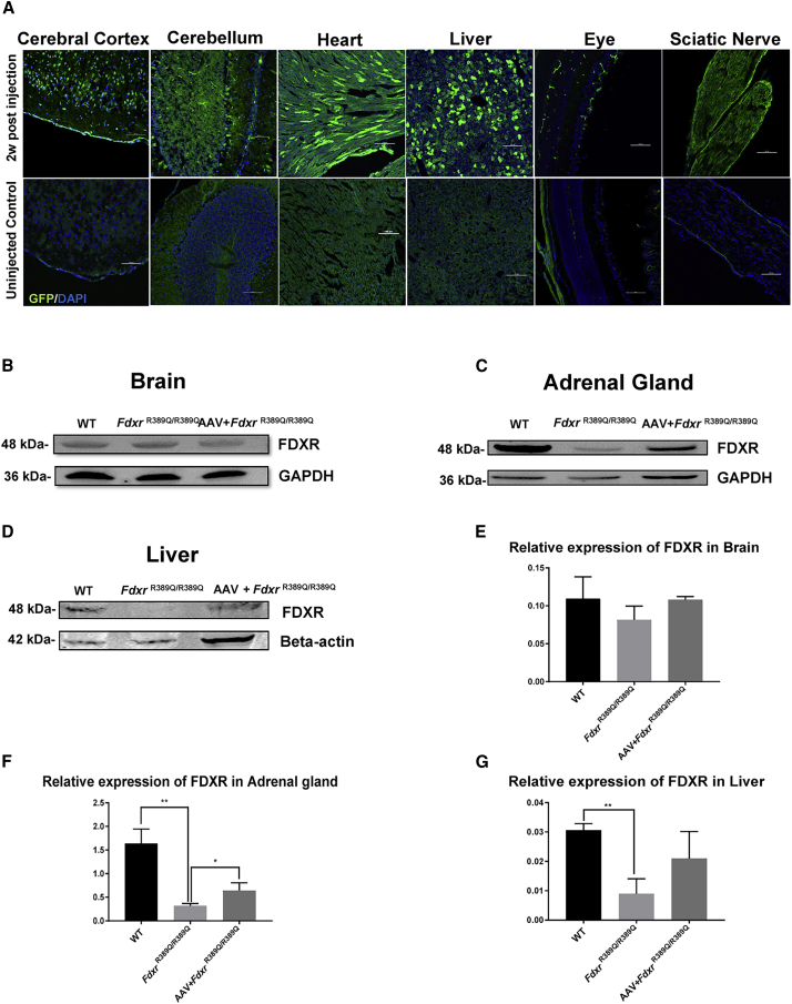
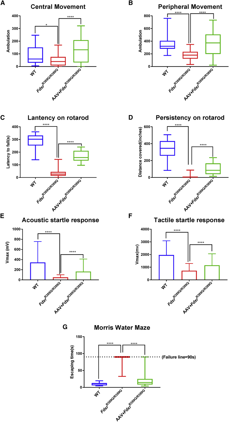
Gene therapy now provides a novel approach for treating inherited monogenetic disorders, including nuclear gene mutations associated with mitochondrial diseases. In this study, we have utilized a mouse model carrying a p.Arg389Gln mutation of the mitochondrial Ferredoxin Reductase gene (Fdxr) and treated them with neurotropic AAV-PHP.B vector loaded with the mouse Fdxr cDNA sequence. We then used immunofluorescence staining and western blot to test the transduction efficiency of this vector. Toluidine blue staining and electronic microscopy were also utilized to assess the morphology of optic and sciatic nerves, and the mitochondrial respiratory chain activity was determined as well. The AAV vector effectively transduced in the central nervous system and peripheral organs, and AAV-Fdxr treatment reversed almost all the symptoms of the mutants (Fdxr R389Q/R389Q ). This therapy also improved the electronic conductivity of the sciatic nerves, prevented optic atrophy, improved mobility, and restored mitochondrial complex function. Most notably, the sensory neuropathy, neurodegeneration, and chronic neuroinflammation in the brain were alleviated. Overall, we present the first demonstration of a potential definitive treatment that significantly improves optic and sciatic nerve atrophy, sensory neuropathy, and mitochondrial dysfunction in FDXR-related mitochondriopathy. Our study provides substantial support for the translation of AAV-based Fdxr gene therapy into clinical applications.


Figures

None
Graphical abstract
Figure 1
Figure 1
AAV-PHP.B Transduction in the Central Nervous System (CNS) and Peripheral Systems after Administration via Facial Vein Injection (A) Distribution of GFP reporter expression in the CNS and peripheral tissues. Scale bar, 100 μm. (B) FDXR expression in the brain lysate of 5-month-old mice. (C) FDXR expression in the adrenal gland of 5-month-old mice. (D) FDXR expression in the liver of 5-month-old mice. (E–G) Relative expression of FDXR in the brain (E), adrenal gland (F), and liver (G) lysates. Values are normalized to GAPDH (for brain and adrenal gland) or beta-actin (for liver) as internal controls.
Figure 2
Figure 2
The AAV-Fdxr Treatment Alleviates the Neuronal Gliosis and Neurodegeneration in the CNS of FdxrR389Q/R389Q Mutants (A) Immunofluorescence (IF) with GFAP staining in the mouse cerebellum. Scale bars, 100 μm. Nuclei were counterstained with 4′,6-diamidino-2-phenylindole (DAPI). (B) Fluoro-Jade C staining of Purkinje cells in the mouse brain. The green signal indicates degenerating neurons. Scale bars, 100 μm.
Figure 3
Figure 3
AAV-Fdxr Mitigates the Optic Atrophy of FdxrR389Q/R389Q Mutants (A) H&E staining of 4-month-old mouse retinas. Scale bar, 10 μm. Red arrows indicate cell loss in the retinal ganglion cell (RGC) layer of FdxrR389Q/R389Q mutants. (B) Brn3a IF staining of retinas from 4-month-old mice. Scale bar, 100 μm. The middle panel indicates the decrease in Brn3a positive signal in the RGC layer of the FdxrR389Q/R389Q mutant. The right panel shows that the reduction in Brn3a positive cells was reversed after AAV-Fdxr treatment. (C) Toluidine blue staining of semi-thin cross-sections of optic nerves from 4-month-old mice. Scale bar, 10 μm. (D) EM cross-sections of optic nerves from 4-month-old mice. Scale bar, 2 μm. Note the axon loss in the middle panel for the FdxrR389Q/R389Q mutant. (E) Cell counts in the RGC layer. (F) Quantification of the Brn3a–positive area in the retinas of 4 month-old mice. Brn3a signals were quantified from six retinal sections. (G) Quantification of myelinated axons from 4-month-old mice.
Figure 4
Figure 4
AAV-Fdxr Mitigates the Peripheral Neuropathy in the Hindlimbs of FdxrR389Q/R389Q Mutants (A) Schematic diagram of EMGs recorded from 4-month-old mice. Scales indicate volts and time. (B and C) The maximum amplitude (B) and conduction velocity (C) of 4-month-old mice were compared between each group. (D) Toluidine blue staining of cross-sections from 4-month-old mice. Orange arrows indicate axons undergo demyelination. Scale bar, 10 μm. (E) 1,000× magnification EM images of sciatic nerve cross-sections of 4-month-old mice. Note the demyelinating axons indicated by red arrows. (F) 2,000× magnification EM images of a cross-section of sciatic nerves from 4-month-old mice. Note the disintegrated axon in the middle panel. (G) Comparison of myelinated axons inside the sciatic nerves of each group. Values were obtained from 6 images from each group. (H) G-ratio of myelinated axons inside the sciatic nerves of each group. Values of G-ratio were obtained from 65 random axons from each group.
Figure 5
Figure 5
AAV-Fdxr Treatment Relieves the Dysfunction of the Mitochondrial Respiratory Chain (A) The complex I activities of various tissues. (B) The activities of complexes II+III. (C) Complex IV activities of various tissues. Mitochondria were isolated from different high energy consumption tissues (brain, heart, liver, and muscle) from 4-month-old male mice (n = 3 for each group).
Figure 6
Figure 6
The AAV-Fdxr Treatment Eliminates the Mitochondrial Iron Overload in Various Tissues of FdxrR389Q/R389Q Mutants Levels of mitochondrial iron content were measured using colorimetric methods. (A–D) Mitochondria were isolated from energy-intensive tissues such as the brain (A), heart (B), liver (C), and muscle (D) from 4-month-old male mice (n = 3 for each group).
Figure 7
Figure 7
AAV-Fdxr Recovered the Movement Disorder and Sensory Defect of FdxrR389Q/R389Q Mutants Behavioral tests of 5-month-old female mice. (A) Movement frequency in the central area of locomotor activity chambers for 5-month-old female mice. (B) Motion recording in the peripheral areas of the locomotor chambers. (C) Lantency on rotarod and (D) Persistency on rotarod for 5-month-old female mice (n = 5 for each group). (E) Acoustic and startle response and (F) tactile startle response of 5-month-old female mice. (n = 5 for each group). (G) MWM performance of 5-month-old mice. A total assay time over 90 s indicates a failure to escape from the water maze (n = 5 for each group).

References

    1. Hanukoglu I. Steroidogenic enzymes: structure, function, and role in regulation of steroid hormone biosynthesis. J. Steroid Biochem. Mol. Biol. 1992;43:779–804. - PubMed
    1. Shi Y., Ghosh M., Kovtunovych G., Crooks D.R., Rouault T.A. Both human ferredoxins 1 and 2 and ferredoxin reductase are important for iron-sulfur cluster biogenesis. Biochim. Biophys. Acta. 2012;1823:484–492. - PMC - PubMed
    1. Solish S.B., Picado-Leonard J., Morel Y., Kuhn R.W., Mohandas T.K., Hanukoglu I., Miller W.L. Human adrenodoxin reductase: two mRNAs encoded by a single gene on chromosome 17cen----q25 are expressed in steroidogenic tissues. Proc. Natl. Acad. Sci. USA. 1988;85:7104–7108. - PMC - PubMed
    1. Peng Y., Shinde D.N., Valencia C.A., Mo J.S., Rosenfeld J., Truitt Cho M., Chamberlin A., Li Z., Liu J., Gui B. Biallelic mutations in the ferredoxin reductase gene cause novel mitochondriopathy with optic atrophy. Hum. Mol. Genet. 2017;26:4937–4950. - PMC - PubMed
    1. Paul A., Drecourt A., Petit F., Deguine D.D., Vasnier C., Oufadem M., Masson C., Bonnet C., Masmoudi S., Mosnier I. FDXR Mutations Cause Sensorial Neuropathies and Expand the Spectrum of Mitochondrial Fe-S-Synthesis Diseases. Am. J. Hum. Genet. 2017;101:630–637. - PMC - PubMed