. 2020 Aug 21:18:556-572.
doi: 10.1016/j.omto.2020.08.008. eCollection 2020 Sep 25.

Doxorubicin Conjugation to Reovirus Improves Oncolytic Efficacy in Triple-Negative Breast Cancer

Affiliations

Doxorubicin Conjugation to Reovirus Improves Oncolytic Efficacy in Triple-Negative Breast Cancer

Jameson T L Berry et al. Mol Ther Oncolytics. .

Abstract

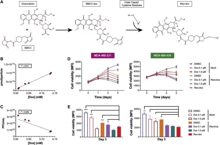
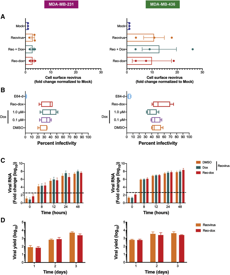
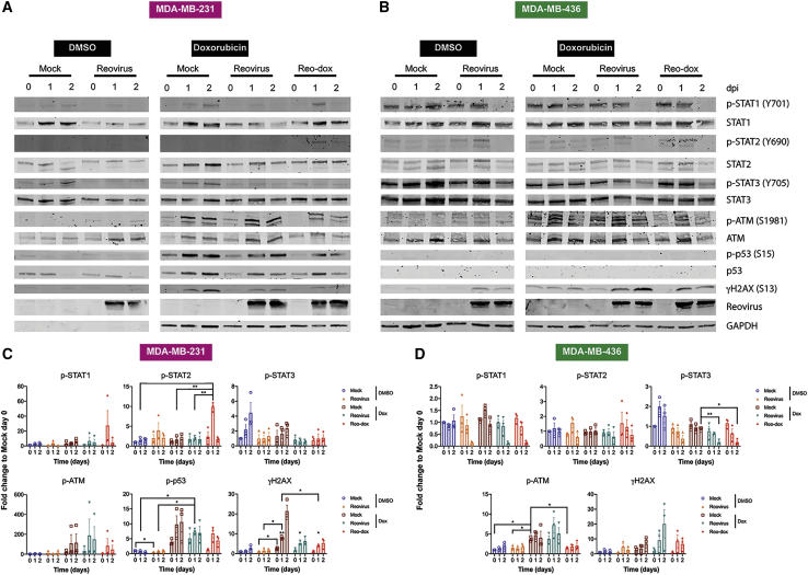
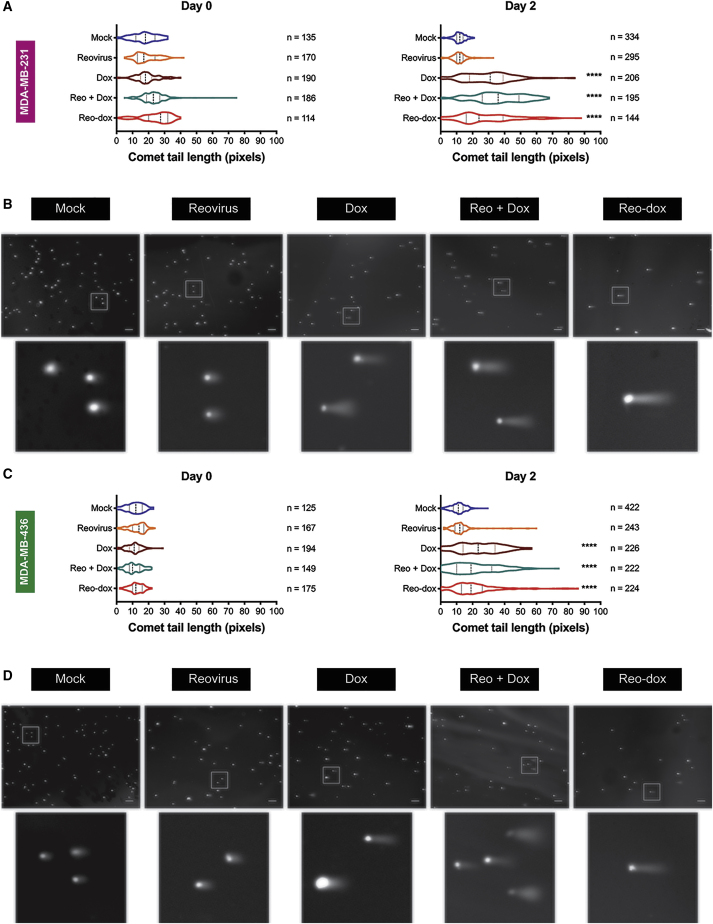
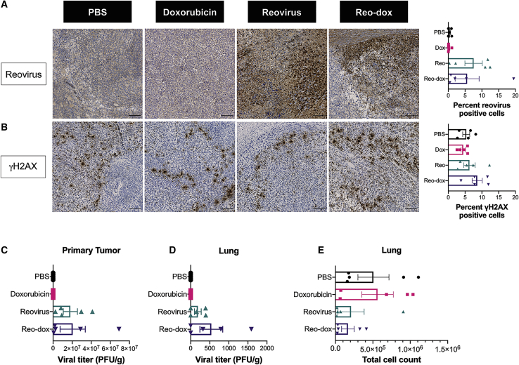
Breast cancer is the second leading cause of cancer-related deaths in women in the United States. The triple-negative breast cancer (TNBC) subtype associates with higher rates of relapse, shorter overall survival, and aggressive metastatic disease. Hormone therapy is ineffective against TNBC, leaving patients with limited therapeutic options. Mammalian orthoreovirus (reovirus) preferentially infects and kills transformed cells, and a genetically engineered reassortant reovirus infects and kills TNBC cells more efficiently than prototypical strains. Reovirus oncolytic efficacy is further augmented by combination with topoisomerase inhibitors, including the frontline chemotherapeutic doxorubicin. However, long-term doxorubicin use correlates with toxicity to healthy tissues. Here, we conjugated doxorubicin to reovirus (reo-dox) to control drug delivery and enhance reovirus-mediated oncolysis. Our data indicate that conjugation does not impair viral biology and enhances reovirus oncolytic capacity in TNBC cells. Reo-dox infection promotes innate immune activation, and crosslinked doxorubicin retains DNA-damaging properties within infected cells. Importantly, reovirus and reo-dox significantly reduce primary TNBC tumor burden in vivo, with greater reduction in metastatic burden after reo-dox inoculation. Together, these data demonstrate that crosslinking chemotherapeutic agents to oncolytic viruses facilitates functional drug delivery to cells targeted by the virus, making it a viable approach for combination therapy against TNBC.

Keywords: DNA damage; cell death; doxorubicin; drug delivery; oncolytics; reovirus; triple-negative breast cancer; virus; virus-host interactions.


Figures

None
Graphical abstract
Figure 1
Figure 1
Doxorubicin Conjugation to Reovirus Enhances Viral Cytotoxicity in TNBC Cells (A) Chemistry of doxorubicin conjugation to reovirus. The lone primary amine of doxorubicin reacts with the succinimide functional group of succinimidyl 4-(n-maleimidomethyl) cyclohexane-1-carboxylate (SMCC) to form SMCC-dox. Cysteine residues on viral capsid proteins (R1) react with the maleimide functional group of SMCC-dox, yielding a final crosslinked product or doxorubicin bound to reovirus (reo-dox). (B and C) UV-vis spectroscopy was performed on reo-dox preparations (Table S1). (B) Doxorubicin concentration was correlated with the amount of drug per reovirus particle and (C) viral titer. r2 values are presented for six independently labeled reo-dox preparations. (D and E) TNBC cells were pretreated with vehicle (DMSO) or doxorubicin. Cells were infected with mock, reovirus, or reo-dox at an MOI of 100 PFU/cell. (D) Cell viability was measured over 3 days post infection. (E) Cell viability at 3 dpi from (D). Data represent the mean of four independent experiments. Error bars, SEM. ∗p ≤ 0.05; ∗∗p ≤ 0.01; ∗∗∗p ≤ 0.001; ∗∗∗∗p ≤ 0.0001 by one-way ANOVA for reo-dox compared to all conditions.
Figure 2
Figure 2
Reo-dox Has Similar Attachment, Infectivity, and Replication Kinetics as Reovirus, But Enhanced Cytotoxicity in TNBC Cells Results for MDA-MB-231 cells are displayed on left and MDA-MB-436 cells are displayed on right for all panels. (A) TNBC cells were pretreated with vehicle (DMSO) or doxorubicin and adsorbed with mock, reovirus, or reo-dox at an MOI of 1 × 105 particles/cell for 1 h at 4°C. Cells were assessed for cell surface reovirus by flow cytometry using indirect immunofluorescence. (B) TNBC cells were pretreated with vehicle (DMSO), E64-d, or doxorubicin and infected with mock, reovirus, or reo-dox at an MOI of 100 PFU/cell for 18 h. Infectivity was assessed by indirect immunofluorescence. (C) TNBC cells were pretreated with vehicle (DMSO) or doxorubicin and infected with mock, reovirus, or reo-dox, and qPCR was performed to assess mRNA levels of reovirus S1 gene. Dashed line represents background baseline levels observed in mock. Data are shown as fold change normalized to a housekeeping gene. (D) TNBC cells were adsorbed with reovirus or reo-dox at an MOI of 10 PFU/cell over a 3 day time course. Viral titers were assessed by plaque assay on L929 mouse fibroblasts and viral yield was calculated as fold increase in titer compared to day 0. Data represent the means of three (A) or four (B–D) independent experiments. Error bars, SEM.
Figure 3
Figure 3
Reo-Dox Induces Type-III IFN in TNBC Cells (A) MDA-MB-231 and (B) MDA-MB-436 cells were pretreated with vehicle (DMSO) or doxorubicin and infected with mock, reovirus, or reo-dox. RNA was isolated from cells at times shown and qPCR was performed to assess mRNA levels of IFNL1, IFNB1, and IFNG. Data are shown as fold change to DMSO mock at 0 h for four independent experiments. Error bars, SEM. Levels of IFN-λ, IFN-β (MDA-MB-436 only), and IFN-γ in cell supernatants were detected by ELISA in (C) MDA-MB-231 and (D) MDA-MB-436 cells. Data are shown as pg/mL of IFN for four independent experiments. Error bars, SEM. ∗p ≤ 0.05; ∗∗p ≤ 0.01; ∗∗∗p ≤ 0.001; ∗∗∗∗p ≤ 0.0001 by two-way ANOVA. Open ended brackets indicate multiple comparisons to one condition, doubled-sided brackets indicate comparison between two conditions only.
Figure 4
Figure 4
Reo-Dox Activates DNA Damage Response Pathways and Modulates Innate Immune Activity in TNBC Cells (A and C) MDA-MB-231 and (B and D) MDA-MB-436 cells were treated with vehicle (DMSO) or doxorubicin and infected with reovirus or reo-dox at an MOI of 100 PFU/cell. (A and B) Whole cell lysates for 0, 1, and 2 dpi were resolved by SDS-PAGE and immunoblotted with antibodies specific for phosphorylated and total STAT1, STAT2, STAT3, ATM, p53, H2AX, reovirus, and GAPDH. Residues recognized by phosphorylation-specific antibodies are shown in parentheses. Blots are representative of three independent experiments. (C and D) Quantitative densitometry was performed on all phosphorylated and total proteins. Data represent means of three independent experiments normalized to respective mock day 0 values. Error bars, SEM. ∗p ≤ 0.05; ∗∗p ≤ 0.01 by two-way ANOVA. ∗ above γH2AX day 1 bar graphs in (C) indicate comparisons between reovirus plus dox or reo-dox and DMSO mock on the same day.
Figure 5
Figure 5
Reo-Dox Infection of TNBC Cells Induces DNA Double-Strand Breaks (A and B) MDA-MB-231 and (C and D) MDA-MB-436 cells were pretreated with vehicle (DMSO) or doxorubicin and infected with mock, reovirus, or reo-dox at an MOI of 100 PFU/cell. DNA double-strand break damage was assessed by single cell electrophoresis (comet assay) on 0 and 2 dpi. Chromatin was visualized by epifluorescence using DAPI staining. (A and C) Comet tail length was measured for imaged cells. Data represented as violin plots. Median and upper and lower quartiles are presented as dotted lines within violins. ∗∗∗∗p ≤ 0.0001 compared to mock and reovirus by one-way ANOVA for reo-dox compared to all conditions. (B and D) Representative images of comets on 2 dpi. Scale bar, 200 μm. Inset highlights boxed cells. Data are representative of two independent experiments.
Figure 6
Figure 6
Reo-Dox and Reovirus Infect and Kill 4T1 Cells In Vitro and In Vivo, and Reduce 4T1 Cell Metastatic Potential to the Lungs (A) 4T1 cells were pretreated with vehicle (DMSO) or doxorubicin and infected with mock, reovirus, or reo-dox. Infectivity was assessed after 20 h by indirect immunofluorescence using reovirus-specific antiserum. (B) 4T1 cells were treated with vehicle (DMSO) or doxorubicin, or they were infected with reovirus or reo-dox at increasing MOIs. Cell viability was assessed at times shown (left) and day 6 alone for statistical analysis (right). (A and B) Data represent mean of four independent experiments. Error bars, SEM. ∗p ≤ 0.05; ∗∗p ≤ 0.01; ∗∗∗p ≤ 0.001; ∗∗∗∗p ≤ 0.0001 by one-way ANOVA. ∗ above bar graphs in (B) indicate comparisons between reovirus and reo-dox at the same MOI. (C) Female, 8-week-old BALB/c mice were challenged with 5 × 104 4T1 cells via subcutaneous injection in the hind flank. At day 10 and 14 post challenge (arrows in D and E), mice were treated with PBS, 54.4 μg/mL doxorubicin, or 5 × 108 PFU of reovirus or reo-dox. The experimental endpoint was 21 days post tumor challenge. (D) Percent change in weight of mice was calculated as the weight on day of measurement normalized to weight at day 5. (E) Tumor area was measured at days indicated by data points. ∗∗p < 0.01, reovirus and reo-dox compared to PBS by two-way ANOVA. (C–E) n = 5 mice per treatment group. Error bars, SEM.
Figure 7
Figure 7
Reovirus Antigen and γH2AX Are Detected in 4T1 Tumors Infected with Reo-Dox Primary tumor tissues were assessed for (A) reovirus antigen and (B) γH2AX levels by indirect immunohistochemistry. (A and B) Inset images enlarged from representative whole tissue scans in Figure S4. Scale bars, 100 μm. Percent of cells in whole tissue scans positive for reovirus antigen and γH2AX presented on right. Bar graphs represent mean of representative tissue from each mouse. n = 5. Error bars, SEM (C) Titers for reovirus or reo-dox present in primary tumor tissue and (D) lungs were assessed by plaque assay on L929 mouse fibroblasts. (E) Total number of metastatic 4T1 cells in lungs were counted. (C–E) n = 5. Error bars, SEM.

References

    1. American Cancer Society . 2020. Cancer Facts & Figures 2020. (Atlanta: American Cancer Society) - PMC - PubMed
    1. American Cancer Society . 2019. Breast Cancer Facts & Figures 2019-2020. (Atlanta: American Cancer Society)
    1. NAACCR.org Available from: https://naaccr.org.
    1. Haffty B.G., Yang Q., Reiss M., Kearney T., Higgins S.A., Weidhaas J., Harris L., Hait W., Toppmeyer D. Locoregional relapse and distant metastasis in conservatively managed triple negative early-stage breast cancer. J. Clin. Oncol. 2006;24:5652–5657. - PubMed
    1. Dent R., Trudeau M., Pritchard K.I., Hanna W.M., Kahn H.K., Sawka C.A., Lickley L.A., Rawlinson E., Sun P., Narod S.A. Triple-negative breast cancer: clinical features and patterns of recurrence. Clin. Cancer Res. 2007;13:4429–4434. - PubMed

LinkOut - more resources