. 2020 Sep 16;5(1):e000545.
doi: 10.1136/bmjophth-2020-000545. eCollection 2020.

Povidone iodine treatment is deleterious to human ocular surface conjunctival cells in culture

Affiliations

Povidone iodine treatment is deleterious to human ocular surface conjunctival cells in culture

William Swift et al. BMJ Open Ophthalmol. .

Abstract

Objective: To determine the effect of povidone iodine (PI), an antiseptic commonly used prior to ocular surgery, on viability of mixed populations of conjunctival stratified squamous and goblet cells, purified conjunctival goblet cells and purified conjunctival stromal fibroblasts in primary culture.

Methods and analysis: Mixed population of epithelial cells (stratified squamous and goblet cells), goblet cells and fibroblasts were grown in culture from pieces of human conjunctiva using either supplemented DMEM/F12 or RPMI. Cell type was evaluated by immunofluorescence microscopy. Cells were treated for 5 min with phosphate-buffered saline (PBS); 0.25%, 2.5%, 5% or 10% PI in PBS; or a positive control of 30% H2O2. Cell viability was determined using Alamar Blue fluorescence and a live/dead kit using calcein/AM and ethidium homodimer-1 (EH-1).

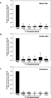
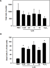
Results: Mixed populations of epithelial cells, goblet cells and fibroblasts were characterised by immunofluorescence microscopy. As determined with Alamar Blue fluorescence, all concentrations of PI significantly decreased the number of cells from all three preparation types compared with PBS. As determined by calcein/EH-1 viability test, mixed populations of cells and fibroblasts were less sensitive to PI treatment than goblet cells. All concentrations of PI, except for 0.25% used with goblet cells, substantially increased the number of dead cells for all cell populations. The H2O2 control also significantly decreased the number and viability of all three types of cells in both tests.

Conclusion: We conclude that PI, which is commonly used prior to ocular surgeries, is detrimental to human conjunctival stratified squamous cells, goblet cells and fibroblasts in culture.

Keywords: conjunctiva; ocular surface.


Conflict of interest statement

Competing interests: None declared.

Figures

Figure 1
Figure 1
Identification of cell types in cultured conjunctiva. Mixed populations of cells containing populations of stratified squamous cells (A) and goblet cells (B) were grown on coverslips, fixed and labelled against unique markers for each cell type and used to determine the fractional cell population in mixed preparations. Stratified squamous cells were identified from mixed populations using antibodies against cytokeratin 4 (red), Bandeiraea Simplicifolia lectin (green) and DAPI (blue). Goblet cells were identified from mixed populations of cells (B) or purified goblet cell cultures (C) using antibodies against cytokeratin 7 (red), and Ulex Europeaus Agglutin 1 (green). Fibroblasts (D) were identified in purified cultures using antibodies against vimentin (red), and DAPI (blue). Representative stratified squamous and goblet cells are indicated with arrows in A and B. Cells were analysed from three individuals in A and B and four individuals in C and D. Cell types were counted and percentage of cell type is shown in E. Data are expressed as mean±SEM. DAPI, 4′,6-diamino-2-phenylindole.
Figure 2
Figure 2
Viability of cells after treatment with PI. Cultured mixed cells (A), purified goblet cells (B) and purified fibroblasts (C) were treated with PBS, 0.25%–10% PI or 30% H2O2 for 5 min. Relative fluorescence intensity of Alamar Blue is expressed as fold change when intensity of PBS was set to 1. Data are mean±SEM from three individuals. Asterisk (*) indicates significance from PBS. PBS, phosphate-buffered saline; PI, povidone iodine.
Figure 3
Figure 3
Viability of cultured mixed epithelial cells after treatment with PI. Cultured mixed epithelial cells were treated with PBS, 0.25%–10% PI or 30% H2O2 for 5 min. Calcein staining (green) indicates live cells, and EH-1 (red) indicates dead cells (A). The total number of cells counted for each group was recorded and analysed (B), and the percentage of dead cells as a per cent of total cells is indicated on the y-axis (C). Data are mean±SEM from three individuals. Asterisk (*) indicates significance from PBS. EH-1, ethidium homodimer-1; PBS, phosphate-buffered saline; PI, povidone iodine.
Figure 4
Figure 4
Viability of cultured goblet cells after treatment with PI. Cultured goblet cells were treated with PBS, 0.25%–10% PI or 30% H2O2 for 5 min. The total number of cells counted for each group was recorded and analysed (A), and the percentage of dead cells as a per cent of total cells is indicated on the y-axis (B). Data are mean±SEM from three individuals. Asterisk (*) indicates significance from PBS. PBS, phosphate-buffered saline; PI, povidone iodine.
Figure 5
Figure 5
Viability of cultured fibroblasts after treatment with PI. Cultured fibroblasts were treated with PBS, 0.25%–10% PI, or 30% H2O2 for 5 min. The total number of cells counted for each group was recorded and analysed (A), and the percentage of dead cells as a per cent of total cells is indicated on the y-axis (B). Data are mean±SEM from three individuals. Asterisk (*) indicates significance from PBS. PBS, phosphate-buffered saline; PI, povidone iodine.

References

    1. Dartt DA. Regulation of mucin and fluid secretion by conjunctival epithelial cells. Prog Retin Eye Res 2002;21:555–76. 10.1016/S1350-9462(02)00038-1 - DOI - PubMed
    1. Uchino Y, Woodward AM, Argüeso P. Differential effect of rebamipide on transmembrane mucin biosynthesis in stratified ocular surface epithelial cells. Exp Eye Res 2016;153:1–7. 10.1016/j.exer.2016.10.007 - DOI - PubMed
    1. Gipson I. Anatomy of the Conjunctiva, Cornea, and Limbus : Smolin G, Thoft R, The Cornea. Boston: Little, Brown, and Company, 1994: 3–24.
    1. Diebold Y, Ríos JD, Hodges RR, et al. . Presence of nerves and their receptors in mouse and human conjunctival goblet cells. Invest Ophthalmol Vis Sci 2001;42:2270–82. - PubMed
    1. McGilligan VE, Gregory-Ksander MS, Li D, et al. . Staphylococcus aureus activates the NLRP3 inflammasome in human and rat conjunctival goblet cells. PLoS One 2013;8:e74010. 10.1371/journal.pone.0074010 - DOI - PMC - PubMed