Review
. 2020 Sep 2:6:100044.
doi: 10.1016/j.tcsw.2020.100044. eCollection 2020 Dec.

Antibiotics and resistance: the two-sided coin of the mycobacterial cell wall

Affiliations
Review

Antibiotics and resistance: the two-sided coin of the mycobacterial cell wall

Sarah M Batt et al. Cell Surf. .

Abstract

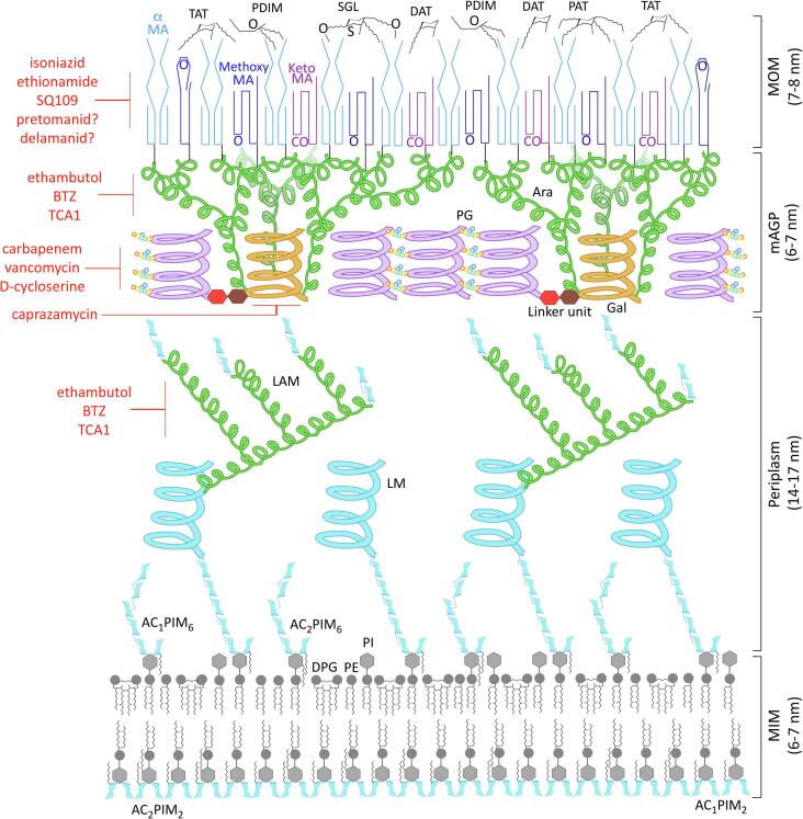
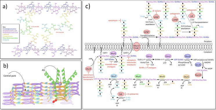
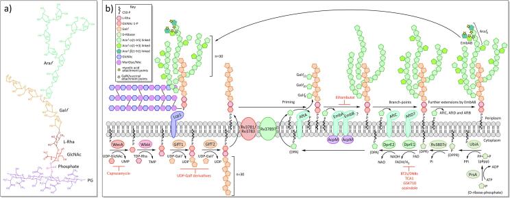
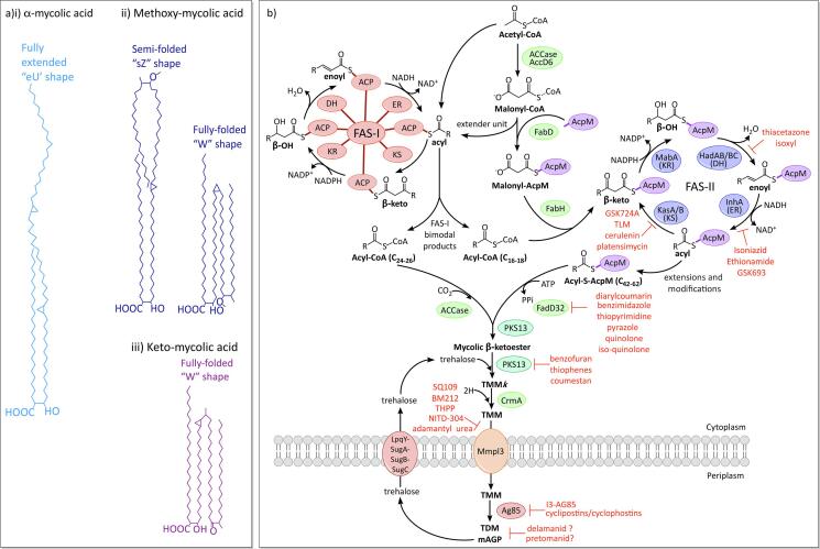
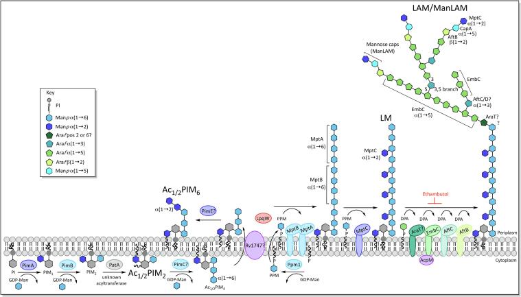
Mycobacterium tuberculosis, the bacterium responsible for tuberculosis, is the global leading cause of mortality from an infectious agent. Part of this success relies on the unique cell wall, which consists of a thick waxy coat with tightly packed layers of complexed sugars, lipids and peptides. This coat provides a protective hydrophobic barrier to antibiotics and the host's defences, while enabling the bacterium to spread efficiently through sputum to infect and survive within the macrophages of new hosts. However, part of this success comes at a cost, with many of the current first- and second-line drugs targeting the enzymes involved in cell wall biosynthesis. The flip side of this coin is that resistance to these drugs develops either in the target enzymes or the activation pathways of the drugs, paving the way for new resistant clinical strains. This review provides a synopsis of the structure and synthesis of the cell wall and the major current drugs and targets, along with any mechanisms of resistance.

Keywords: Arabinogalactan; Cell wall; Lipoarabinomannan; Mycobacterium tuberculosis; Mycolic acids.


Conflict of interest statement

The authors declare that they have no known competing financial interests or personal relationships that could have appeared to influence the work reported in this paper.

Figures

Fig. 1
Fig. 1
The cell wall of Mycobacterium tuberculosis. The inner leaflet of the plasma membrane contains a high quantity of Ac1/Ac2PIM2 (tri- and tetra-acylated phosphatidyl-myo-inositol-dimannoside), while the outer membrane has Ac1/Ac2PIM6 (tri- and tetra-acylated phosphatidyl-myo-inositol-hexamannoside), along with the more usual phospholipids, DPG (diphosphatidylglycerol), PE (phosphatidylethanolamine) and PI (phosphatidylinositol); the methyl groups of the unique tuberculostearic acids of mycobacteria are depicted here (Minnikin et al., 2015). Also anchored into the plasma membrane are LM (lipomannan) and LAM (lipoarabinomannan), which project out into the periplasm; the mannose sugars and mannan domains are coloured light blue and the branched arabinan is green. According to the ‘scaffold model’, the glycan back bone (purple) of the PG (peptidoglycan) forms a matrix of helices orientated perpendicular to the plasma membrane (Dmitriev et al., 2000). These surround the AG (arabinogalactan) and LAM (lipoarabinomannan) and are connected by the peptide cross-links (coloured circles: orange = L-alanine, yellow = D-isoglutamine, green = meso-diaminopimelate and blue = D-alanine). The PG is connected to the base of the Gal (galactan; orange) via a unique rhamnose-N-acetylglucosamine linker. Three highly branched Ara (arabinan; green) domains project from the base of the Gal towards the MA layer (mycolic acids; dark blues and purples), which is covalently attached to most of the non-reducing ends of the Ara and forms the inner layer of the MOM (mycobacterial outer membrane). The PG, AG and MA make up the mycolylarabinogalactan-peptidoglycan complex (mAGP). The free lipids of the outer leaflet consist of PDIM (phthiocerol dimycocerosates); DAT, TAT, PAT and SGL (di-, tri- and penta-acyl trehalose and sulfated trehalose glycolipids) (Jankute et al., 2015, Minnikin et al., 2015). The diagram is roughly to scale using dimensions obtained from cryo-electron microscopy (Zuber et al., 2008). The main current and pipe-line drugs targeting the biosynthesis/transport pathways of the cell wall are shown in red. (For interpretation of the references to colour in this figure legend, the reader is referred to the web version of this article.)
Fig. 2
Fig. 2
Structural features of peptidoglycan and biosynthesis A) Chemical structure of peptidoglycan (see key for details). B) Diagram of the ‘scaffold model’ of peptidoglycan. The glycan (purple) of the peptidoglycan forms a matrix of helices orientated perpendicular to the plasma membrane, joined together by peptide cross-links and forming central pores to fit other structures, such as arabinogalactan (AG; orange and green helices) (Dmitriev et al., 2000). C) Biosynthesis of peptidoglycan. (For interpretation of the references to colour in this figure legend, the reader is referred to the web version of this article.)
Fig. 3
Fig. 3
Structural features of arabinogalactan and biosynthesis A) Chemical structure of arabinogalactan. B) Biosynthesis of arabinogalactan and the rhamnose-N-acetylglucosamine linker unit.
Fig. 4
Fig. 4
Chemical features of mycolic acids and biosynthesis A) Structures and common conformations of the three classes of MAs: i) α-, ii) Methoxy- and iii) Keto- (Minnikin et al., 2015). B) Mycolic acid biosynthesis.
Fig. 5
Fig. 5
Biosynthesis of phosphatidyl-inositol-mannosides (PIMs), lipomannan (LM) and lipoarabinomannan (LAM).

References

    1. Abrahams K.A., Chung C.-W., Ghidelli-Disse S., Rullas J., Rebollo-López M.J., Gurcha S.S., Cox J.A.G., Mendoza A., Jiménez-Navarro E., Martínez-Martínez M.S., Neu M., Shillings A., Homes P., Argyrou A., Casanueva R., Loman N.J., Moynihan P.J., Lelièvre J., Selenski C., Axtman M., Kremer L., Bantscheff M., Angulo-Barturen I., Izquierdo M.C., Cammack N.C., Drewes G., Ballell L., Barros D., Besra G.S., Bates R.H. Identification of KasA as the cellular target of an anti-tubercular scaffold. Nat. Commun. 2016;7:12581. doi: 10.1038/ncomms12581. - DOI - PMC - PubMed
    1. Aggarwal A., Parai M.K., Shetty N., Wallis D., Woolhiser L., Hastings C., Dutta N.K., Galaviz S., Dhakal R.C., Shrestha R., Wakabayashi S., Walpole C., Matthews D., Floyd D., Scullion P., Riley J., Epemolu O., Norval S., Snavely T., Robertson G.T., Rubin E.J., Ioerger T.R., Sirgel F.A., van der Merwe R., van Helden P.D., Keller P., Böttger E.C., Karakousis P.C., Lenaerts A.J., Sacchettini J.C. Development of a novel lead that targets m. tuberculosis polyketide synthase 13. Cell. 2017;170:249–259.e25. doi: 10.1016/j.cell.2017.06.025. - DOI - PMC - PubMed
    1. Alderwick L.J., Radmacher E., Seidel M., Gande R., Hitchen P.G., Morris H.R., Dell A., Sahm H., Eggeling L., Besra G.S. Deletion of Cg-emb in corynebacterianeae leads to a novel truncated cell wall arabinogalactan, whereas inactivation of Cg-ubiA results in an Arabinan-deficient mutant with a cell wall galactan core. J. Biol. Chem. 2005;280:32362–32371. doi: 10.1074/jbc.M506339200. - DOI - PubMed
    1. Alderwick L.J., Seidel M., Sahm H., Besra G.S., Eggeling L. Identification of a novel arabinofuranosyltransferase (AftA) involved in cell wall arabinan biosynthesis in Mycobacterium tuberculosis. J. Biol. Chem. 2006;281:15653–15661. doi: 10.1074/jbc.M600045200. - DOI - PubMed
    1. Alderwick L.J., Dover L.G., Veerapen N., Gurcha S.S., Kremer L., Roper D.L., Pathak A.K., Reynolds R.C., Besra G.S. Expression, purification and characterisation of soluble GlfT and the identification of a novel galactofuranosyltransferase Rv3782 involved in priming GlfT-mediated galactan polymerisation in Mycobacterium tuberculosis. Protein Expr. Purif. 2008;58:332–341. doi: 10.1016/j.pep.2007.11.012. - DOI - PubMed

LinkOut - more resources