Review
. 2020 Jul 21;2(6):100149.
doi: 10.1016/j.jhepr.2020.100149. eCollection 2020 Dec.

Approach to the patient with acute severe autoimmune hepatitis

Affiliations
Review

Approach to the patient with acute severe autoimmune hepatitis

Mussarat N Rahim et al. JHEP Rep. .

Abstract

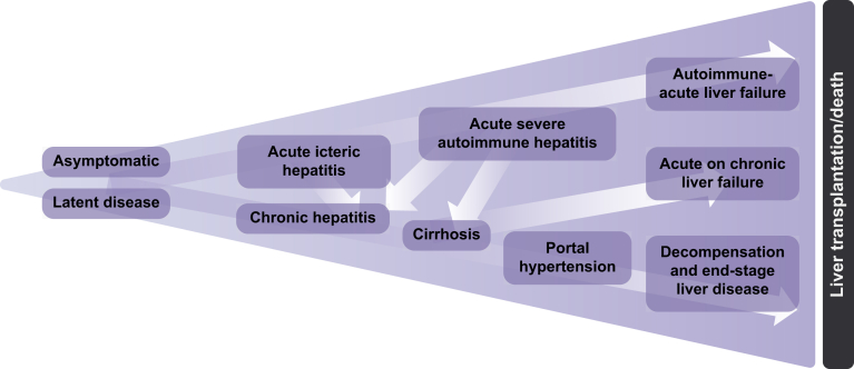
Autoimmune hepatitis is associated with varied clinical presentations and natural history, as well as somewhat unpredictable treatment responses. Understanding how to stratify patients who require further escalation of therapy will help clinicians manage these patients. The presentation of acute severe autoimmune hepatitis (AS-AIH) is relatively uncommon, although its prevalence is potentially greater than currently perceived. Previous studies consist of small retrospective single-centre series and are not directly comparable due to the diversity of presentations, disease definitions and non-standardised treatment regimens. We define AS-AIH as those who present acutely with AIH and are icteric with an international normalised ratio ≥1.5 and no evidence of hepatic encephalopathy. Those with hepatic encephalopathy should be defined as having AS-AIH with acute liver failure. In this review, we provide a structured practical approach for diagnosing and managing this unique group of patients.

Keywords: ACLF, acute-on-chronic liver failure; AIH, autoimmune hepatitis; ALF, acute liver failure; ALI, acute liver injury; ALT, alanine aminotransferase; ANA, anti-nuclear antibody; AS-AIH, acute severe autoimmune hepatitis; ASMA, anti-smooth muscle antibody; AST, aspartate aminotransferase; AUROC, analysis of area under the receiver operator characteristic curve; Acute liver failure; Acute severe presentation; Autoimmune hepatitis; CT, computed tomography; Corticosteroids; DILI, drug-induced liver injury; EBV, Epstein-Barr virus; HE, hepatic encephalopathy; HLA, human leukocyte antigen; IAIHG, International Autoimmune Hepatitis Group; INR, international normalised ratio; LT, liver transplantation; Liver transplantation; MELD, model for end-stage liver disease; MELD-Na, model for end-stage liver disease-sodium; MHN, massive hepatic necrosis; NAC, N-acetylcysteine; PT, prothrombin time; UKELD, United Kingdom end-stage liver disease; USALF, United States Acute Liver Failure; anti-LC-1, anti-liver cytosol-1; anti-LKM, anti-liver kidney microsomal; anti-SLA/LP, anti-soluble liver antigen/liver pancreas.


Conflict of interest statement

The authors declare no conflicts of interest that pertain to this work. Please refer to the accompanying ICMJE disclosure forms for further details.

Figures

Fig. 1
Fig. 1
The spectrum of presentations in AIH. AIH, autoimmune hepatitis.
Fig. 4
Fig. 4
Autoimmune-like acute hepatitis in a patient on nitrofurantoin. (A) Severe liver damage with areas of recent bridging necrosis and post-necrotic collapse (H&E, 40×). (B) The hepatic parenchyma shows nodular configuration secondary to post-necrosis collapse, with no obvious established fibrosis or very early collagen deposition (Picrosirius red, 40×). (C) Lobular disarray with multiple necro-inflammatory foci and reactive changes of the liver cell plates with regenerative rosettes (H&E, 200×). (D) Detail of an inflammatory area with clusters of plasma cells (arrowheads), hepatocyte reactive changes with cell ballooning and focal emperipolesis (arrow) (H&E, 400×).
Fig. 2
Fig. 2
Schematic diagram of a hepatic lobule and the potential histological changes in acute presentations of AIH. AIH, autoimmune hepatitis; AIH-ALF, autoimmune hepatitis-acute liver failure; AS-AIH, acute severe autoimmune hepatitis.
Fig. 3
Fig. 3
Typical histological features of autoimmune hepatitis. (A) Portal inflammation with interface activity (H&E, 100×). (B) Detail of the interface between portal tract and hepatic parenchyma, with moderate activity associated with conspicuous plasma cell component (H&E, 200×). (C) Lobular hepatitis with disarray of the liver cell plates. Diffuse necro-inflammatory activity and regenerative hepatocellular resetting (H&E, 200×). (D) Occasional emperipolesis activity. Intracytoplasmic lymphocytes are spotted in some hepatocytes (arrows) (H&E, 400×).
Fig. 6
Fig. 6
Comparing the diagnostic sensitivity of the IAIHG criteria in acute-onset AIH. AIH, autoimmune hepatitis; IAIHG, International Autoimmune Hepatitis Group. Reproduced from Fujiwara et al.
Fig. 5
Fig. 5
Histopathological spectrum of liver damage at the time of transplantation. (A) Chronic autoimmune hepatitis in stage of established cirrhosis, with no inflammatory activity, in a 22-year-old female, diagnosed with autoimmune hepatitis aged 12. (A1) Macroscopy of the explanted liver, with macronodular cirrhosis predominantly. (A2) Macroscopy of a liver section with macro-regenerative nodules up to 15 mm in greatest dimension. (A3) On microscopy, regenerative nodules show well-demarcated outlines, with predominantly thin fibrous septa (arrows) (Picrosirius red, 20×). (A4) No residual significant inflammation is of note at the porto-septal areas. No interface activity is present (arrows), (H&E, 200×). (B) Chronic autoimmune hepatitis in stage of cirrhosis, with persistent inflammatory activity, in a male patient aged 53, diagnosed with AIH 13 years ago. He developed portal hypertension and suffered episodes of variceal bleeding, ascites and encephalopathy. (B1) Macroscopy of the explanted liver (1,299 grams). Of note is the heterogeneous capsular surface, with whitish areas of capsular depression. An incidental hepatocellular carcinoma (inset) was found at the explant, 18 mm, moderately differentiated. (B2) Detail of the liver capsule surface, with micro-macronodular cirrhosis and a large area of fibrous scarring (∗) secondary to previous multiacinar necrosis. B3, Fibrous septa and regenerative nodules show variable appearance. Some of the regenerative nodules show well-defined outline (thin arrows) while blurred interface is observed in many areas (thick arrows) (Picrosirius red, 20×). (B4) Detail of the blurred interface area in image B3, corresponding with marked interface activity (H&E, 200×). (C) Small explanted liver (857 grams) in the clinical context of subacute liver failure secondary to AIH in a 21-year-old male with no underlying chronic liver disease. (B1) Notice the smooth capsular surface in comparison with the liver explants in figure A1 and B1. (C2) Macroscopic appearance of liver sections, showing map-like areas of residual cholestatic (green) parenchyma∗, alternating with areas of parenchymal extinction where massive hepatocellular loss has occurred∗∗. (C3) Microscopy of the Glisson's capsule (thin and smooth) and subcapsular parenchyma (area ∗ of figure C2). The hepatic parenchyma shows vague nodularity secondary to bridging necrosis but no evidence of fibrosis, indicating the acute/subacute damage (Picrosirius red, 20×). (C4) Microscopy of the area of collapse (area ∗∗ of figure C2), with complete hepatocellular loss in areas of multiacinar necrosis and secondary micro-haemorrhage in the centrilobular area, centred by a terminal hepatic venule (CV). Portal tracts (PT) at the periphery of this area show mild inflammatory component and some degree of ductular reaction (H&E, 200×).

References

    1. Lohse A.W., Chazouillères O., Dalekos G., Drenth J., Heneghan M., Hofer H. EASL clinical practice guidelines: autoimmune hepatitis. J Hepatol. 2015;63(4):971–1004. - PubMed
    1. Mack C.L., Adams D., Assis D.N., Kerkar N., Manns M.P., Mayo M.J. Diagnosis and management of autoimmune hepatitis in adults and children: 2019 practice guidance and guidelines from the American Association for the Study of Liver Diseases. Hepatology. 2019;72(2):671–722. - PubMed
    1. Lee W.M., Stravitz R.T., Larson A.M. Introduction to the revised American Association for the Study of Liver Diseases position paper on acute liver failure 2011. Hepatology. 2012;55(3):965–967. - PMC - PubMed
    1. Trey C., Davidson C.S. The management of fulminant hepatic failure. Prog Liver Dis. 1970;3:282–298. - PubMed
    1. Williams R., Schalm S.W., O'Grady J.G. Acute liver failure: redefining the syndromes. Lancet. 1993;342(8866):273–275. - PubMed