. 2020 Oct 20;1(7):100123.
doi: 10.1016/j.xcrm.2020.100123. Epub 2020 Sep 24.

ReScan, a Multiplex Diagnostic Pipeline, Pans Human Sera for SARS-CoV-2 Antigens

Affiliations

ReScan, a Multiplex Diagnostic Pipeline, Pans Human Sera for SARS-CoV-2 Antigens

Colin R Zamecnik et al. Cell Rep Med. .

Abstract

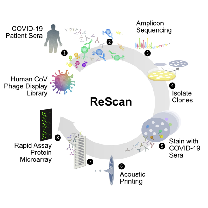
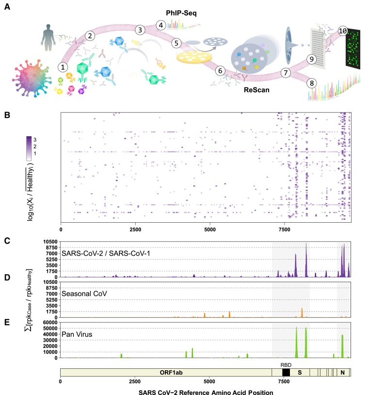
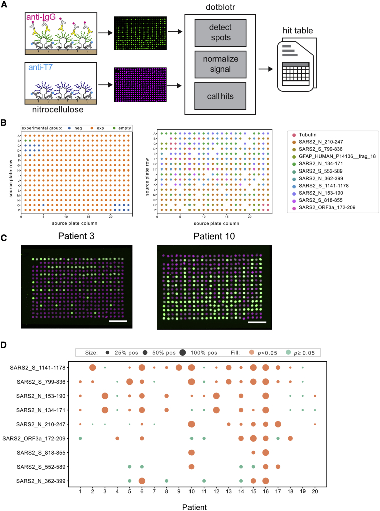
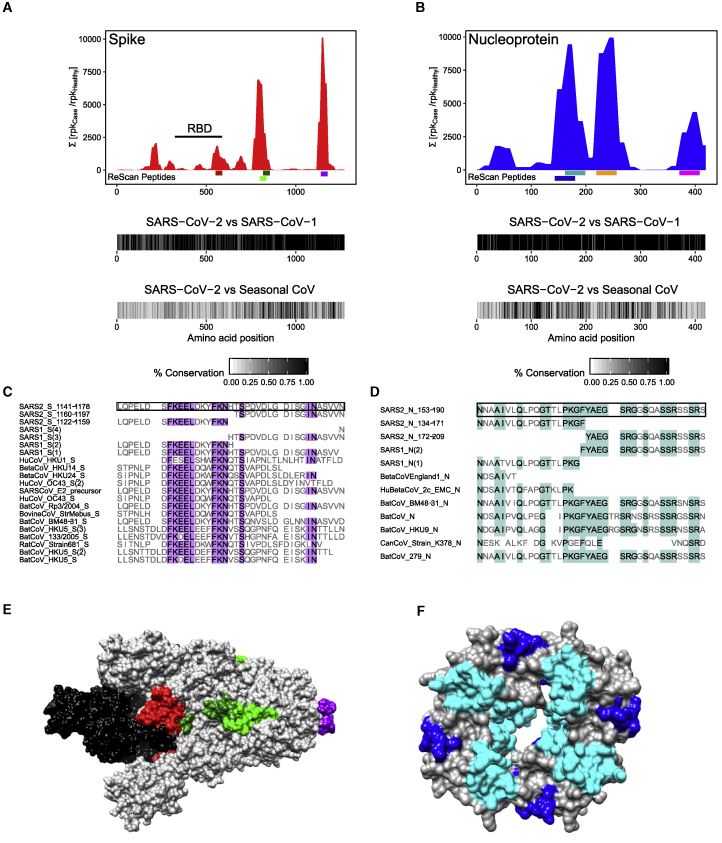
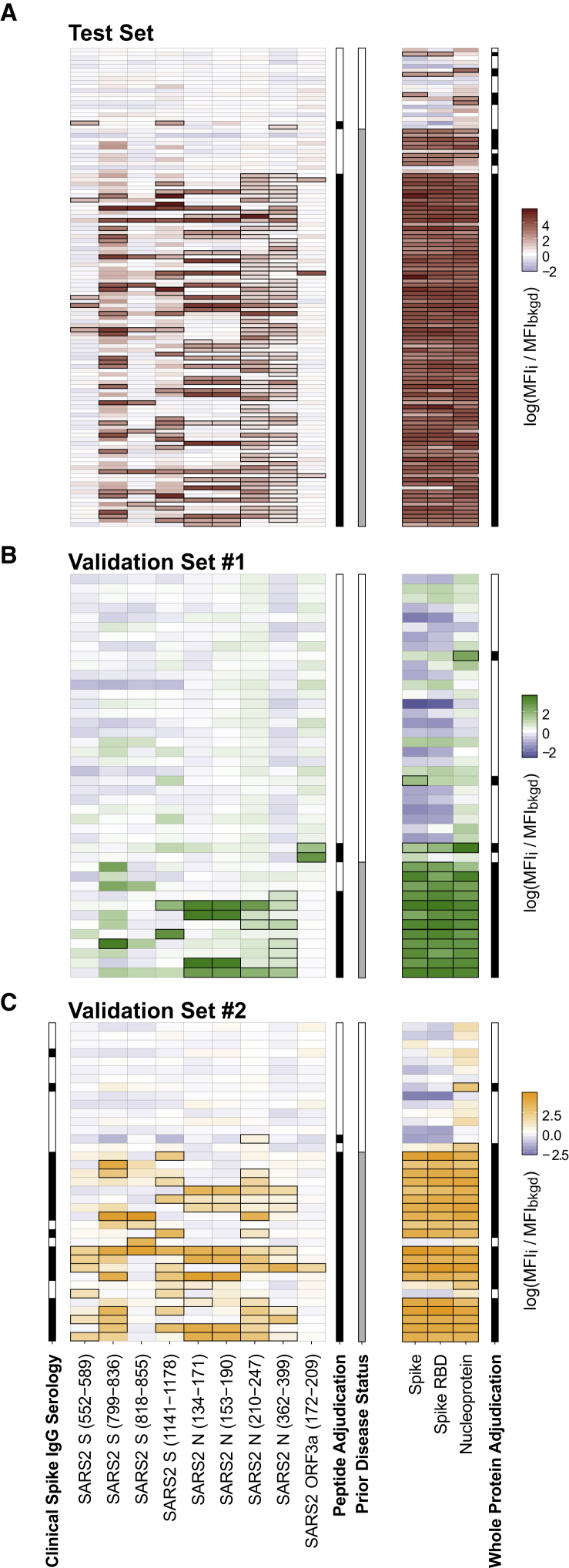
Comprehensive understanding of the serological response to SARS-CoV-2 infection is important for both pathophysiologic insight and diagnostic development. Here, we generate a pan-human coronavirus programmable phage display assay to perform proteome-wide profiling of coronavirus antigens enriched by 98 COVID-19 patient sera. Next, we use ReScan, a method to efficiently sequester phage expressing the most immunogenic peptides and print them onto paper-based microarrays using acoustic liquid handling, which isolates and identifies nine candidate antigens, eight of which are derived from the two proteins used for SARS-CoV-2 serologic assays: spike and nucleocapsid proteins. After deployment in a high-throughput assay amenable to clinical lab settings, these antigens show improved specificity over a whole protein panel. This proof-of-concept study demonstrates that ReScan will have broad applicability for other emerging infectious diseases or autoimmune diseases that lack a valid biomarker, enabling a seamless pipeline from antigen discovery to diagnostic using one recombinant protein source.

Keywords: COVID-19; SARS-CoV-2; assay development; diagnostics; phage display; serology.


Conflict of interest statement

The authors declare no competing interests.

Figures

None
Graphical abstract
Figure 1
Figure 1
General Workflow for ReScan and Epitope Mapping SARS-CoV-2 Using PhIP-Seq (A) VirScan T7 phage display system with SARS-CoV-2 antigens in overlapping 38-aa fragments (1) are incubated with antibodies (2) from patient sera and undergo 2 rounds of amplification (3). Amplified lysates are either analyzed by phage immunoprecipitation sequencing (PhIP-seq), or are lifted, stained, and isolated for ReScan (4 and 5). Microarrays of isolated clonal phage populations are printed via acoustic liquid transfer (6), stained with patient sera, and analyzed for identity (7 and 8) and commonality (9) via dotblotr. (B and C) Heatmap displaying results from individual patients (B) and (C) cumulative fold enrichment of significant (p < 0.01) SARS-CoV peptides from HuCoV library relative to pre-pandemic controls aligned to the SARS-CoV-2 genome. (D) Cumulative fold enrichment of significant other seasonal CoV peptides in the HuCoV library relative to pre-pandemic controls aligned to the SARS-CoV-2 genome. (E) Cumulative enrichment of significant pan-viral library peptides relative to pre-pandemic controls aligned to the SARS-CoV-2 genome. One technical replicate for each library.
Figure 2
Figure 2
ReScan Analysis Using SARS-CoV-2 Phage Display Library (A) Schematic of microarray design. (B) ReScan assay configuration layout (left) and map of each clonal phage population on plate (right). (C) Examples of staining patterns on patients positive for nucleocapsid and spike peptides; scale bar, 5 mm. Anti-T7 tag signal is magenta, anti-IgG signal is green. (D) SARS-CoV-2 peptide analysis from ReScan microarrays; dot size is proportional to the fraction of dots for a given peptide that stained positive by a given patient sample. One technical replicate per serum sample.
Figure 3
Figure 3
Similarity of Enriched ReScan and VirScan SARS-CoV-2 Peptides to Other CoVs (A and B) Comparing underlined SARS-CoV-2 spike (A) and nucleocapsid (B) regions found by ReScan with other CoV sequences that were enriched over healthy controls in the HuCoV library (S552−589 = red, S799−836 = light green, S818−855 = dark green, S1,141−1,178 = purple; N134−171 = blue, N153−190 = teal, N210−247 = orange, N362−399 = pink). Homology maps with SARS-CoV-1 and seasonal coronaviruses in the HuCoV libraries are seen below. (C and D) Diverse CoV peptide sequences with partial similarity to the regions spanning (1,1411,178) and (153190) of the S and N proteins that were enriched in both the HuCoV and pan-viral libraries. (E and F) CryoEM structures of prefusion S glycoprotein (6VSB) and RNA-binding domain of N protein (6WKP) with color-coded ReScan peptides overlaid in colors corresponding to those in the coverage maps, and the S glycoprotein receptor-binding domain (RBD) highlighted in black.
Figure 4
Figure 4
ReScan Peptides as High-Throughput Serology Assay for COVID-19 (A) Test set of patients who were analyzed by either VirScan and ReScan, with each column representing either a ReScan-identified peptide or a whole-protein antigen. The outline around a result indicates a significant (i.e., exceeds Z score of 3) signal established by the background population of pre-pandemic controls. Adjudication is made from either a single peptide or a single protein determined to be positive within their respective panels by exceeding 3 standard deviations from the mean of pre-pandemic control distribution. The prior disease status columns represent patients either with a historically positive qPCR swab or neutralizing antibodies against SARS-CoV-2. (B) Validation set of new patient sera from blinded plate. (C) Second blinded validation set of new patient samples with an accompanying clinical serology IgG result measuring anti-Spike antibodies. All of the data are represented as the log of secondary fluorescence signal on each antigen normalized to 2 separate negative controls in each well. In all of the panels, each row represents a unique patient. n = 2 technical replicates averaged for each data point.

Similar articles

Cited by

References

    1. Zhou P., Yang X.L., Wang X.G., Hu B., Zhang L., Zhang W., Si H.R., Zhu Y., Li B., Huang C.L. A pneumonia outbreak associated with a new coronavirus of probable bat origin. Nature. 2020;579:270–273. - PMC - PubMed
    1. Wu F., Zhao S., Yu B., Chen Y.M., Wang W., Song Z.G., Hu Y., Tao Z.W., Tian J.H., Pei Y.Y. A new coronavirus associated with human respiratory disease in China. Nature. 2020;579:265–269. - PMC - PubMed
    1. Zhu N., Zhang D., Wang W., Li X., Yang B., Song J., Zhao X., Huang B., Shi W., Lu R., China Novel Coronavirus Investigating and Research Team A Novel Coronavirus from Patients with Pneumonia in China, 2019. N. Engl. J. Med. 2020;382:727–733. - PMC - PubMed
    1. Wolfel R., Corman V.M., Guggemos W., Seilmaier M., Zange S., Muller M.A., Niemeyer D., Jones T.C., Vollmar P., Rothe C. Virological assessment of hospitalized patients with COVID-2019. Nature. 2020;581:465–469. - PubMed
    1. Petherick A. Developing antibody tests for SARS-CoV-2. Lancet. 2020;395:1101–1102. - PMC - PubMed

Publication types