. 2020 Sep;10(5):e182.
doi: 10.1002/ctm2.182.

In vivo and in vitro activation of dormant primordial follicles by EGF treatment in mouse and human

Affiliations

In vivo and in vitro activation of dormant primordial follicles by EGF treatment in mouse and human

Jiawei Zhang et al. Clin Transl Med. 2020 Sep.

Abstract

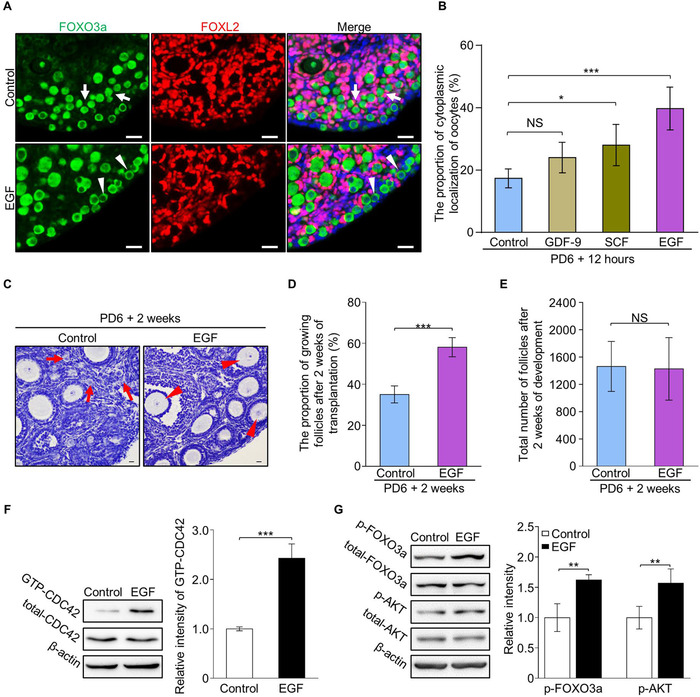
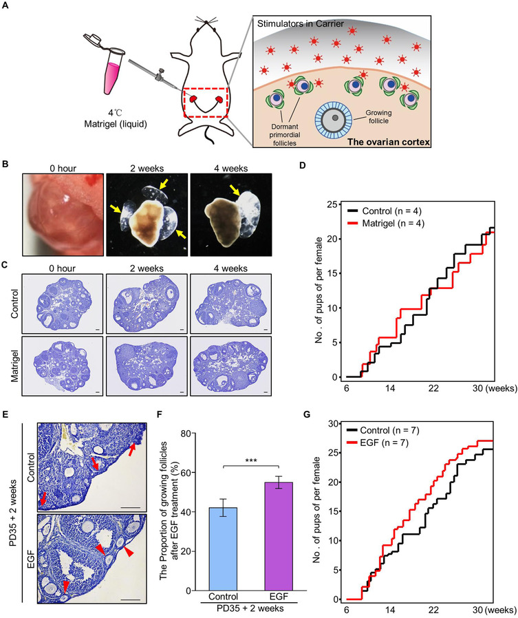
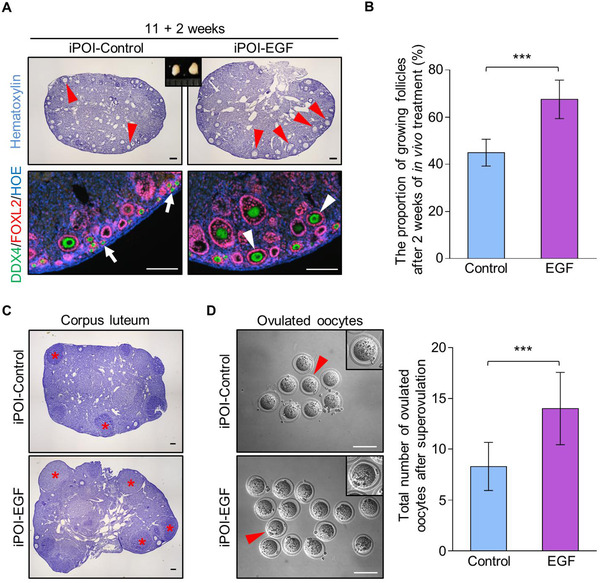
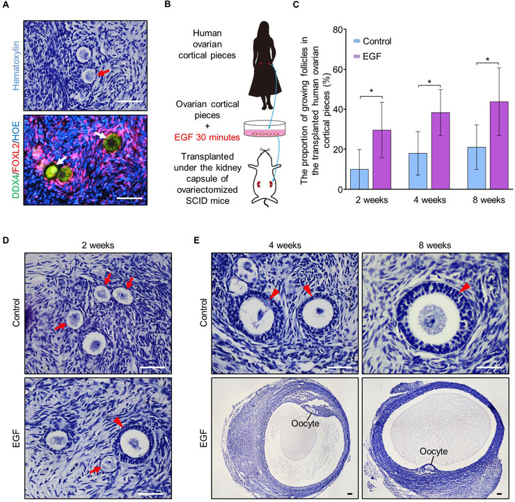
In the mammalian ovaries, dormant primordial follicles represent the reproductive reserve of individual females. Recently, stimulating the activation of primordial follicles in vitro has been practiced, making the utilization of those dormant follicles to treat female infertility possible. However, there are still lacks of effective upstream molecule and strategy to elevate follicle activation in vivo. In the current study, we revealed that growth factor EGF improved a transiently primordial follicle activation in mice by elevating the CDC42-PI3K signaling activity, and EGF treatment also improved the activation and development of human follicles in ovarian cortical pieces. Using a liquid-solid phase transition bio-gel as a carrier, an efficient in vivo activation system was established by ovarian topical EGF administration to living mice. We found that EGF treatment led to an increase of primordial follicle activation in short time but had no effect on long-term fertility in females. By establishing an inducible premature ovarian insufficiency (POI) mouse model through selectively ablating growing follicles in Zp3-Cre;iDTR mice, we further revealed that our in vivo EGF treatment system improved primordial follicle activation and ovulation of POI ovaries significantly. Taken together, our results revealed that in situ ovarian EGF administration could improve the activation of primordial follicles in living animals, and manipulating activation and development of primordial follicles in vivo might be an efficient approach to improve reproductive health in women.

Keywords: EGF; in vivo activation; noninvasive administration; premature ovarian insufficiency; primordial follicle activation.


Conflict of interest statement

Jiawei Zhang, Lei Yan, Yibo Wang, Shuo Zhang, Xueqiang Xu, Yanli Dai, Shidou Zhao, Zhen Li, Yan Zhang, Guoliang Xia, Yingying Qin, and Hua Zhang declare that they have no conflict of interest.

Figures

FIGURE 1
FIGURE 1
EGF treatment improves the activation of primordial follicles by elevating CDC42‐PI3K signaling in mouse ovaries. (A) FOXO3a (green) shuttled from the nuclei (arrows) to the cytoplasm (arrowheads) with oocyte activation. EGF treatment for 30 minutes markedly increased cytoplasmic localization of FOXO3a (arrowheads) in oocytes compared to controls. Granulosa cells were dyed with FOXL2 antibody (purple) to show the follicles and nuclei were dyed with a Hoechst counterstain (blue). (n = 6). (B) Quantification results revealed a significantly increased ratio of cytoplasm localization of FOXO3a in the oocytes of EGF (40 ± 6%), GDF‐9 (24 ± 4%) and SCF (28 ± 6%) treated ovaries compared to vehicle (17 ± 3%) treated ovaries (n > 3). (C) After 2 weeks of transplantation, primordial follicles in clusters (arrows) were detected in the control ovaries (n = 5) whereas an increased number of growing follicles (arrowheads) was observed in the cortical region of EGF‐treated ovaries (n = 5). (D) Follicle quantification showed a significantly increased percentage of growing follicles in the EGF group compared to controls (58 ± 4% vs 35 ± 4%) (n = 5). (E) EGF treatment had no effect on follicle survival (1428 ± 410 vs 1464 ± 372) (n = 5). (F) The CDC42‐GTP pull‐down assay showed that CDC42‐GTP levels were significantly increased in EGF‐treated ovaries compared to the controls. (G) Signaling studies in EGF‐treated and control ovaries, showing an enhanced level of p‐AKT and p‐FOXO3a in the EGF group compared to control ovaries. Levels of total FOXO3a, AKT, and β‐actin were used as internal controls. The experiments were repeated at least three times. In B, D, E, F, and G, data represent the mean ± SD of biological triplicate experiments. NS, > .05, *P < .05, **P < .01, and ***P < .001, by two‐tailed unpaired Student's t test. Scale bars: 25 µm
FIGURE 2
FIGURE 2
In vivo stimulation of the activation of primordial follicles by ovarian topical administration of EGF in PD35 mice. (A) Schema for the in vivo stimulator administration system to activate dormant primordial follicles in the ovaries of live mice. Using a liquid‐solid phase transition bio‐gel (Matrigel) as a carrier, the primordial follicle stimulator was delivered to the ovarian bursa and covered the ovarian surface in which the dormant primordial follicles localized. (B‐D) Validation of the system of ovarian topical Matrigel administration. (B) The Matrigel became solidification in ovarian bursa immediately after injection, and was existing on the ovarian surface at 2 and 4 weeks after injection (n = 4). (C) A normal follicular distribution was observed in the ovaries after 2 and 4 weeks of Matrigel treatment (n = 4) compared to control (n = 4). (D) Fertility check showed that Matrigel injection had no effect on female reproduction (n = 4). (E‐G) Ovarian topical administration of Matrigel‐carried EGF improved the proportion of growing follicles in the ovaries of adult mice. (E) Two weeks after in vivo EGF treatment, histological analysis showed that normal distribution of primordial follicles (arrows) was observed in the control ovaries (n = 3) whereas an increased number of growing follicles (arrowheads) was detected in the cortical region of EGF‐treated ovaries (n = 3). (F) Follicle counting revealed a significantly increased proportion of growing follicles in EGF‐treated ovaries compared to the proportion in controls (55 ± 3% vs 42 ± 4%) (n = 3). (G) EGF treatment had no dramatic effect on female fertility compared to that of the controls (n = 7). Data represent the mean ± SD of biological triplicate experiments. ***P < .001, by two‐tailed unpaired Student's t test. Scale bars: 100 µm
FIGURE 3
FIGURE 3
Establishing an inducible premature ovarian insufficiency mouse model by selective ablation of growing follicles in Zp3‐Cre;iDTR ovary. (A) Illustration of the inducible premature ovarian insufficiency (iPOI) model by DT‐induced ablation of growing follicles in Zp3‐Cre;iDTR ovaries. (B) Upon continuous DT administration, the growing follicles that contain Zp3‐expressing oocytes were selectively ablated to eliminate the follicle reserve in the ovary with no side effect on dormant primordial follicles and other ovarian cells. (C) DT was given to Zp3‐Cre;iDTR or NoCre;iDTR females for 10 consecutive weeks from 1 week after labor (10 µg/kg, one injection per week), and ovaries were examined at PD21 and 11‐weeks. Histological analysis showed a dramatic decrease in ovarian size with DT treatment in Zp3‐Cre;iDTR females. (D) Immunofluorescent labeling of follicles revealed that few growing follicles and a number of primordial follicles were found in the Zp3‐Cre;iDTR females after DT administration (green: DDX4, purple: FOXL2, blue: Hoechst, n = 3). (E) Primordial follicle counting showed a significantly decreased number of follicle reserve in ovaries of iPOI mice compared to the number in controls after DT treatment (n = 3 per group). (F) ELISA analysis showed that no change of serum FSH levels at 3 weeks old iPOI females after DT treatment (0.47 ± 0.21 vs 0.33 ± 0.09 ng/mL), but significantly elevated serum FSH levels were observed at PD35 (2.03 ± 0.49 vs 0.39 ± 0.18 ng/mL) and 11‐week old iPOI females (2.50 ± 0.36 vs 0.47 ± 0.22 ng/mL) compared to levels in controls (n = 12 per group). Data represent the mean ± SD of biological triplicate experiments. NS, > .05, **P < .01 and ***P < .001, by two‐tailed unpaired Student's t test. Scale bars: 100 µm
FIGURE 4
FIGURE 4
In vivo EGF administration improves primordial follicle activation and the oocyte retrieval rate from iPOI ovaries. (A) The bilateral ovaries from each iPOI female were covered by Matrigel with or without EGF through ovarian bursa injection. After 2 weeks of treatment, a markedly increased number of growing follicles (arrowheads) was observed in the cortical regions of EGF‐treated ovaries compared to that of the control ovaries with majority of primordial follicles (arrows) (green: DDX4, purple: FOXL2, blue: Hoechst, n = 3). (B) Follicle counting results showed a significant increase in growing follicles in EGF‐treated ovaries compared to that of the controls (68 ± 7% vs 45 ± 5%) (n = 6). (C) Three weeks after EGF treatment, superovulation was performed to detect the oocyte retrieval rate in iPOI females. Histological analysis showed a markedly increased number of corpus luteum (asterisks) in EGF‐treated ovaries compared to controls (n = 13). (D) A significantly increased number of ovulated oocytes after superovulation was collected from EGF‐treated ovaries compared to that of controls (14 ± 4 vs 8 ± 2) (n = 13). In B and D, data represent the mean ± SD of biological triplicate experiments. ***P < .001, by two‐tailed unpaired Student's t test. Scale bars: 100 µm
FIGURE 5
FIGURE 5
EGF treatment improves the activation and development of human follicles. (A) Histological and immunostaining detection to test the existence of primordial follicles in fresh human ovarian pieces before transplantation (green: DDX4, purple: FOXL2, blue: Hoechst, n = 5). (B) Detecting the effect of EGF on stimulating human dormant follicle activation. Human ovarian cortical pieces which contained a large number of primordial follicles were cut into small cubes (1 mm3) and incubated with or without EGF for 30 minutes in dishes, then the treated tissues were xenografted into the kidney capsule of ovariectomized SCID mice for further development. (C) After 2 weeks (30 ± 12% vs 10 ± 9%), 4 weeks (38 ± 10% vs 18 ± 10%) and 8 weeks (44 ± 15% vs 21 ± 10%) of in vivo development under kidney capsule, follicle counting showed a significantly increased proportion of growing follicles in EGF‐treated human ovarian pieces compared to that of the controls (n = 5 per group). (D) After 2 weeks of transplantation, secondary follicles with multilayer granulosa cells (arrowhead) were observed in EGF‐treated ovarian pieces, and only primary follicles were found in the control group (n = 5). (E) Preovulatory follicles were found in EGF‐treated ovarian pieces after 4 and 8 weeks of transplantation, whereas only the secondary follicles were found in the control group (n = 5). Data represent the mean ± SD of biological triplicate experiments. *P < .05, by two‐tailed unpaired Student's t test. Scale bars: 50 µm

References

    1. Eppig JJ. Intercommunication between mammalian oocytes and companion somatic cells. Bioessays. 1991;13:569‐574. - PubMed
    1. Adhikari D, Liu K. Molecular Mechanisms Underlying the Activation of Mammalian Primordial Follicles. Endocr Rev. 2009;30:438‐464. - PubMed
    1. Pedersen T, Peters H. Proposal for a classification of oocytes and follicles in the mouse ovary. J Reprod Fertil. 1968;17:555‐557. - PubMed
    1. Gougeon A. Regulation of Ovarian Follicular Development in Primates: facts and Hypotheses. Endocr Rev. 1996;17:121‐155. - PubMed
    1. Mcgee EA, Hsueh AJ. Initial and cyclic recruitment of ovarian follicles. Endocr Rev. 2000;21:200‐214. - PubMed