Review
. 2020 Feb 13;217(3):e20190103.
doi: 10.1084/jem.20190103. Print 2020 Mar 2.

Transforming growth factor-β in tissue fibrosis

Affiliations
Review

Transforming growth factor-β in tissue fibrosis

Nikolaos Frangogiannis. J Exp Med. .

Abstract

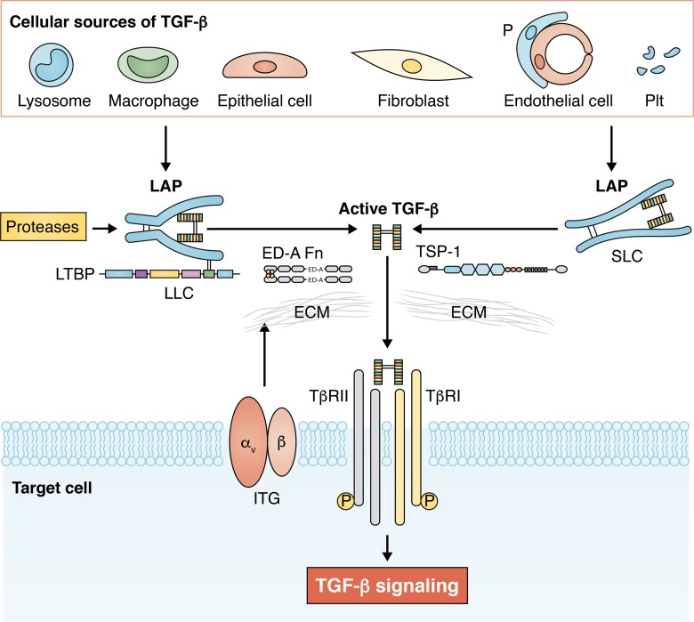
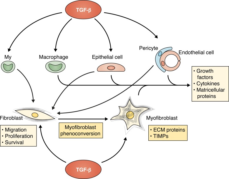
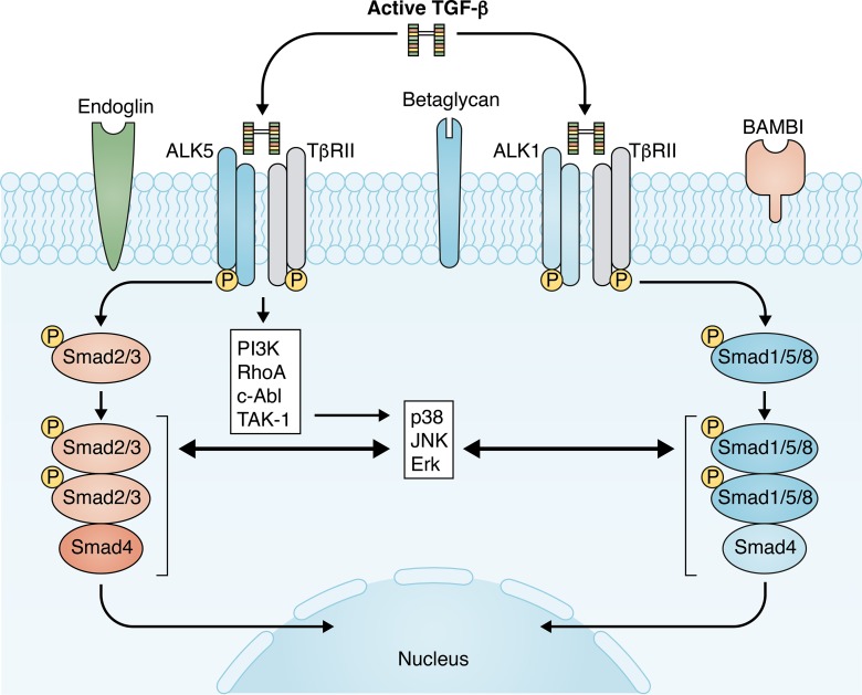
TGF-β is extensively implicated in the pathogenesis of fibrosis. In fibrotic lesions, spatially restricted generation of bioactive TGF-β from latent stores requires the cooperation of proteases, integrins, and specialized extracellular matrix molecules. Although fibroblasts are major targets of TGF-β, some fibrogenic actions may reflect activation of other cell types, including macrophages, epithelial cells, and vascular cells. TGF-β-driven fibrosis is mediated through Smad-dependent or non-Smad pathways and is modulated by coreceptors and by interacting networks. This review discusses the role of TGF-β in fibrosis, highlighting mechanisms of TGF-β activation and signaling, the cellular targets of TGF-β actions, and the challenges of therapeutic translation.


Conflict of interest statement

Disclosures: The author declares no competing interests exist.

Figures

Figure 1.
Figure 1.
TGF-β secretion and activation in fibrotic tissues. Many different cell types, including macrophages, lymphocytes, epithelial cells, fibroblasts, pericytes (P), endothelial cells, and platelets (Plt), can produce and secrete TGF-β isoforms in sites of injury. The specific cellular origins of TGF-βs are dependent on the type of injury and on the cellular composition of the affected organ. TGF-βs are secreted in a latent form, typically as the tripartite LLC, comprised of mature TGF-β, its propeptide called LAP, and LTBP. Liberation of mature TGF-β from the latent form involves effects of proteases, integrin (ITG)-mediated actions, and contributions of specialized ECM proteins that may serve to localize the activation process or interact with LAP to release the active molecule. Active TGF-β binds to its receptors on the surface of the target cell, initiating signaling responses that promote fibrosis. SLC, small latent complex.
Figure 2.
Figure 2.
The cellular targets of TGF-βs in tissue fibrosis. Although resident fibroblasts are major cellular targets of TGF-βs in fibrotic conditions, some TGF-β–mediated fibrogenic effects may involve actions on other cell types, including myeloid fibroblast progenitors (My), macrophages, epithelial cells, pericytes, and endothelial cells. TGF-β is a central mediator in fibroblast-to-myofibroblast conversion and promotes a matrix-preserving phenotype, associated with secretion of ECM proteins and tissue inhibitors of metalloproteinases (TIMPs). Some studies have suggested that expansion of myofibroblasts in fibrotic tissues may also involve TGF-β–mediated conversion of circulating progenitors, epithelial cells, pericytes, and endothelial cells into fibroblasts. TGF-β may also exert indirect activating effects on fibroblasts by promoting secretion of fibrogenic cytokines, growth factors, and matricellular proteins by macrophages, vascular cells, and epithelial cells.
Figure 3.
Figure 3.
TGF-β signaling pathways in tissue fibrosis. TGF-β stimulates a matrix-preserving transcriptional program in fibroblasts by activating Smad-dependent and non-Smad pathways. TGF-βs bind to TβR complexes, comprising a single type II receptor (TβRII) and two forms of type I receptors (ALK5 or ALK1). ALK5 activates Smad2/3, whereas ALK1 activates Smad1/5/8. Although the role of the TβRII–ALK5–Smad2/3 axis in fibrosis is relatively well established, the potential significance of ALK1-Smad1/5/8 remains unclear. TGF-β–mediated activation of non-Smad cascades may also contribute to the fibrotic response. R-Smads exhibit an extensive network of interactions with non-Smad pathways. Fibrogenic TGF-β signaling is also modulated through accessory receptors, such as endoglin, betaglycan, and BAMBI. The complexity of TGF-β signaling pathways, the extensive cross-talk with other signaling networks, and the variable expression of coreceptors depending on the differentiation state of the cells and the microenvironment may explain the context-specific in vivo actions of TGF-βs.

References

    1. Abe R., Donnelly S.C., Peng T., Bucala R., and Metz C.N.. 2001. Peripheral blood fibrocytes: differentiation pathway and migration to wound sites. J. Immunol. 166:7556–7562. 10.4049/jimmunol.166.12.7556 - DOI - PubMed
    1. Abreu J.G., Ketpura N.I., Reversade B., and De Robertis E.M.. 2002. Connective-tissue growth factor (CTGF) modulates cell signalling by BMP and TGF-beta. Nat. Cell Biol. 4:599–604. 10.1038/ncb826 - DOI - PMC - PubMed
    1. Accornero F., van Berlo J.H., Correll R.N., Elrod J.W., Sargent M.A., York A., Rabinowitz J.E., Leask A., and Molkentin J.D.. 2015. Genetic Analysis of Connective Tissue Growth Factor as an Effector of Transforming Growth Factor β Signaling and Cardiac Remodeling. Mol. Cell. Biol. 35:2154–2164. 10.1128/MCB.00199-15 - DOI - PMC - PubMed
    1. Ahn J.Y., Park S., Yun Y.S., and Song J.Y.. 2010. Inhibition of type III TGF-β receptor aggravates lung fibrotic process. Biomed. Pharmacother. 64:472–476. 10.1016/j.biopha.2010.01.006 - DOI - PubMed
    1. Akhurst R.J. 2017. Targeting TGF-β Signaling for Therapeutic Gain. Cold Spring Harb. Perspect. Biol. 9:a022301 10.1101/cshperspect.a022301 - DOI - PMC - PubMed

Publication types

Substances