Comment
. 2021 Jan 15;203(2):263-265.
doi: 10.1164/rccm.202008-3186LE.

Positive Bubble Study in Severe COVID-19 Indicates the Development of Anatomical Intrapulmonary Shunts in Response to Microvascular Occlusion

Affiliations
Comment

Positive Bubble Study in Severe COVID-19 Indicates the Development of Anatomical Intrapulmonary Shunts in Response to Microvascular Occlusion

Robin Cherian et al. Am J Respir Crit Care Med. .
No abstract available


Figures

Figure 1.
Figure 1.
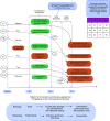
Schematic representation of the mechanisms of hypoxemia in coronavirus disease (COVID-19) lung injury and clinical implications. Pulmonary in situ thrombosis results in hypoxemia owing to flow diversion and overperfusion of other areas with intact perfusion, leading to reduced V./Q. ratio in these segments. The dead spaces that occur because of loss of perfusion do not participate in gas exchange and are not relevant for oxygenation. Extensive perfusion loss is required before becoming clinically apparent owing to its moderate effect on effective V./Q. ratio, nonlinear relationship between V./Q. ratio and PaO2 (7, 8), and the presence of early compensatory mechanisms. However, when these mechanisms are exhausted, clinical deterioration is quick, aggravated by right heart compromise owing to acute pressure overload and reduced mixed venous saturations. Silent hypoxemia can be explained by a state of increased tissue extraction maintaining tissue oxygenation, at the expense of low arterial and venous oxygen saturations. This is facilitated by the subacute nature of the disease process and the lack of significant parenchymal injury at this stage; the work of breathing is not significantly increased, and dyspnea is minimal. CO = cardiac output; critical Do2 = critical oxygen delivery; O2 = oxygen; RV = right ventricular; SILI = self-induced lung injury; Sv-O2 = mixed venous oxygen saturation; TRBC = red blood cell transit time in the alveoli.

Comment in

Comment on

References

    1. Reynolds AS, Lee AG, Renz J, DeSantis K, Liang J, Powell CA, et al. Pulmonary vascular dilatation detected by automated transcranial Doppler in COVID-19 pneumonia [letter] Am J Respir Crit Care Med. [online ahead of print] 6 Aug 2020; DOI: 10.1164/rccm.202006-2219LE. - PMC - PubMed
    1. Krowka MJ. Hepatopulmonary syndromes. Gut. 2000;46:1–4. - PMC - PubMed
    1. Stickland MK, Welsh RC, Haykowsky MJ, Petersen SR, Anderson WD, Taylor DA, et al. Intra-pulmonary shunt and pulmonary gas exchange during exercise in humans. J Physiol. 2004;561:321–329. - PMC - PubMed
    1. Patel BV, Arachchillage DJ, Ridge CA, Bianchi P, Doyle JF, Garfield B, et al. Pulmonary angiopathy in severe COVID-19: physiologic, imaging, and hematologic observations. Am J Respir Crit Care Med. 2020;202:690–699. - PMC - PubMed
    1. Lins M, Vandevenne J, Thillai M, Lavon BR, Lanclus M, Bonte S, et al. Assessment of small pulmonary blood vessels in COVID-19 patients using HRCT. Acad Radiol. [online ahead of print] 25 Jul 2020; DOI: 10.1016/j.acra.2020.07.019. - PMC - PubMed

LinkOut - more resources