

PHLPPing the Script: Emerging Roles of PHLPP Phosphatases in Cell Signaling
- PMID: 32997603
- PMCID: PMC11003498
- DOI: 10.1146/annurev-pharmtox-031820-122108
PHLPPing the Script: Emerging Roles of PHLPP Phosphatases in Cell Signaling
Abstract
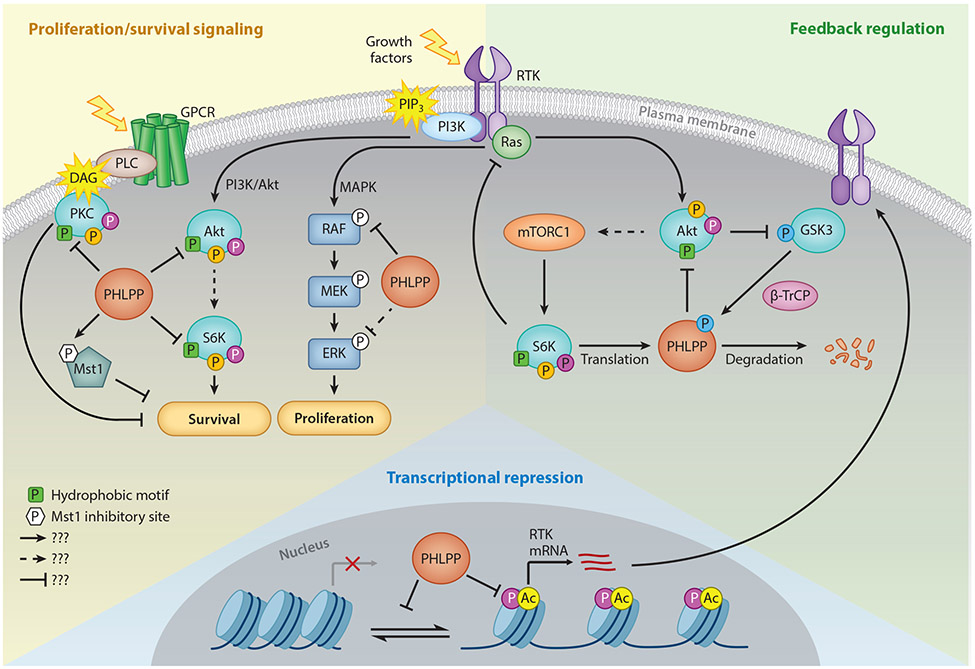
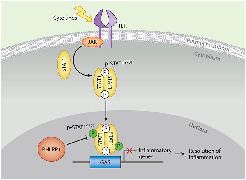
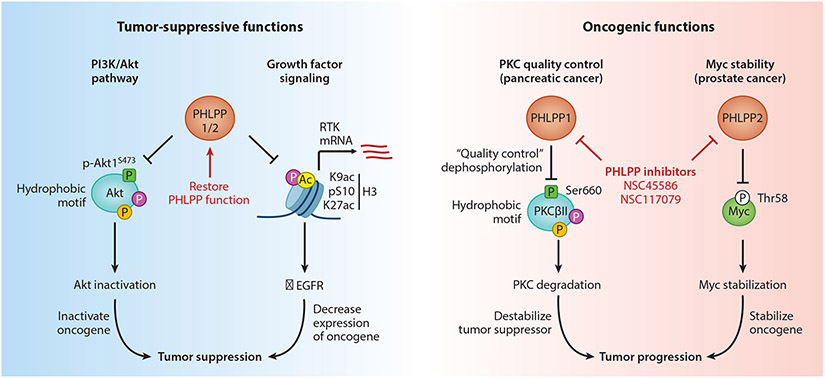
Whereas protein kinases have been successfully targeted for a variety of diseases, protein phosphatases remain an underutilized therapeutic target, in part because of incomplete characterization of their effects on signaling networks. The pleckstrin homology domain leucine-rich repeat protein phosphatase (PHLPP) is a relatively new player in the cell signaling field, and new roles in controlling the balance among cell survival, proliferation, and apoptosis are being increasingly identified. Originally characterized for its tumor-suppressive function in deactivating the prosurvival kinase Akt, PHLPP may have an opposing role in promoting survival, as recent evidence suggests. Additionally, identification of the transcription factor STAT1 as a substrate unveils a role for PHLPP as a critical mediator of transcriptional programs in cancer and the inflammatory response. This review summarizes the current knowledge of PHLPP as both a tumor suppressor and an oncogene and highlights emerging functions in regulating gene expression and the immune system. Understanding the context-dependent functions of PHLPP is essential for appropriate therapeutic intervention.
Keywords: Akt; PHLPP; PKC; cancer; inflammation; phosphatase; phosphorylation; transcription.
Figures

Similar articles
-
Pleckstrin homology domain leucine-rich repeat protein phosphatase (PHLPP): a new player in cell signaling.J Biol Chem. 2012 Feb 3;287(6):3610-6. doi: 10.1074/jbc.R111.318675. Epub 2011 Dec 5. J Biol Chem. 2012. PMID: 22144674 Free PMC article.
-
PHLPPing through history: a decade in the life of PHLPP phosphatases.Biochem Soc Trans. 2016 Dec 15;44(6):1675-1682. doi: 10.1042/BST20160170. Biochem Soc Trans. 2016. PMID: 27913677 Free PMC article. Review.
-
Suppression of survival signalling pathways by the phosphatase PHLPP.FEBS J. 2013 Jan;280(2):572-83. doi: 10.1111/j.1742-4658.2012.08537.x. Epub 2012 Mar 16. FEBS J. 2013. PMID: 22340730 Free PMC article.
-
Turning off AKT: PHLPP as a drug target.Annu Rev Pharmacol Toxicol. 2014;54:537-58. doi: 10.1146/annurev-pharmtox-011112-140338. Annu Rev Pharmacol Toxicol. 2014. PMID: 24392697 Free PMC article. Review.
-
The deubiquitination enzyme USP46 functions as a tumor suppressor by controlling PHLPP-dependent attenuation of Akt signaling in colon cancer.Oncogene. 2013 Jan 24;32(4):471-8. doi: 10.1038/onc.2012.66. Epub 2012 Mar 5. Oncogene. 2013. PMID: 22391563 Free PMC article.
Cited by
-
RNF149 confers cisplatin resistance in esophageal squamous cell carcinoma via destabilization of PHLPP2 and activating PI3K/AKT signalling.Med Oncol. 2023 Sep 2;40(10):290. doi: 10.1007/s12032-023-02137-z. Med Oncol. 2023. PMID: 37658961
-
On the PHLPPside: Emerging roles of PHLPP phosphatases in the heart.Cell Signal. 2021 Oct;86:110097. doi: 10.1016/j.cellsig.2021.110097. Epub 2021 Jul 25. Cell Signal. 2021. PMID: 34320369 Free PMC article. Review.
-
Postnatal deletion of Phlpp1 in chondrocytes delays post-traumatic osteoarthritis in male mice.Osteoarthr Cartil Open. 2024 Sep 27;7(1):100525. doi: 10.1016/j.ocarto.2024.100525. eCollection 2025 Mar. Osteoarthr Cartil Open. 2024. PMID: 39811690 Free PMC article.
-
Unraveling the Mystery of Insulin Resistance: From Principle Mechanistic Insights and Consequences to Therapeutic Interventions.Int J Mol Sci. 2025 Mar 19;26(6):2770. doi: 10.3390/ijms26062770. Int J Mol Sci. 2025. PMID: 40141412 Free PMC article. Review.
-
The role of serine/threonine phosphatases in human development: Evidence from congenital disorders.Front Cell Dev Biol. 2022 Oct 13;10:1030119. doi: 10.3389/fcell.2022.1030119. eCollection 2022. Front Cell Dev Biol. 2022. PMID: 36313552 Free PMC article. Review.
References
-
- Chen MJ, Dixon JE, and Manning G, Genomics and evolution of protein phosphatases. Sci Signal, 2017. 10(474). - PubMed
-
- Gao T, Furnari F, and Newton AC, PHLPP: a phosphatase that directly dephosphorylates Akt, promotes apoptosis, and suppresses tumor growth. Mol Cell, 2005. 18(1): p. 13–24. - PubMed
-
- Shi Y, Serine/threonine phosphatases: mechanism through structure. Cell, 2009. 139(3): p. 468–84. - PubMed
Publication types
MeSH terms
Substances
Grants and funding
LinkOut - more resources
Full Text Sources
Other Literature Sources
Medical
Research Materials
Miscellaneous
