. 2020 Sep 30;18(9):e3000827.
doi: 10.1371/journal.pbio.3000827. eCollection 2020 Sep.

Full-length three-dimensional structure of the influenza A virus M1 protein and its organization into a matrix layer

Affiliations

Full-length three-dimensional structure of the influenza A virus M1 protein and its organization into a matrix layer

Lisa Selzer et al. PLoS Biol. .

Abstract

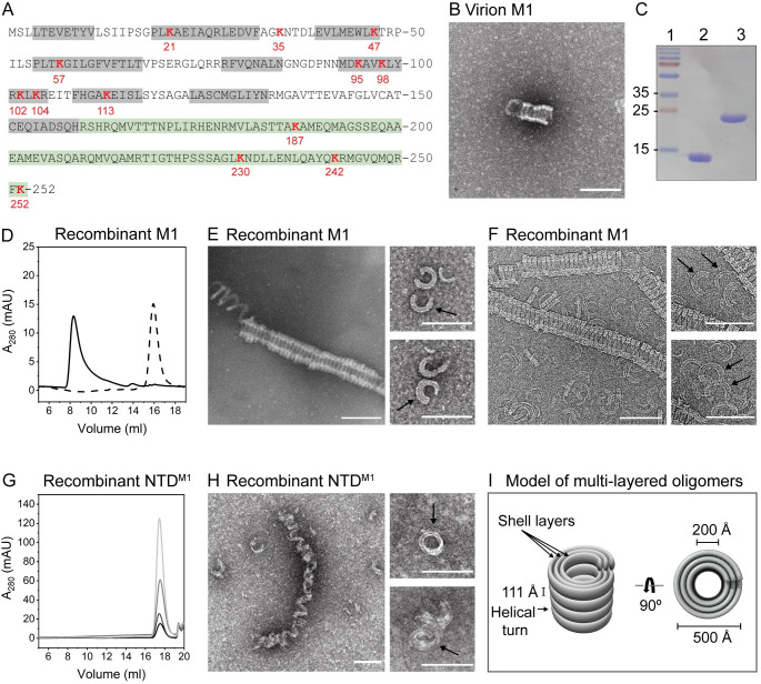
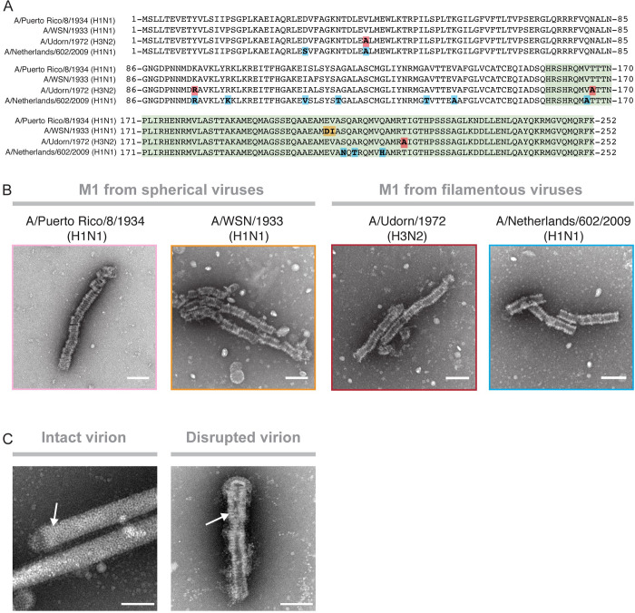
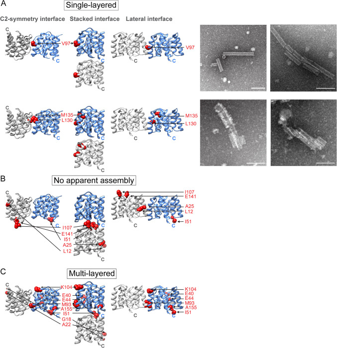
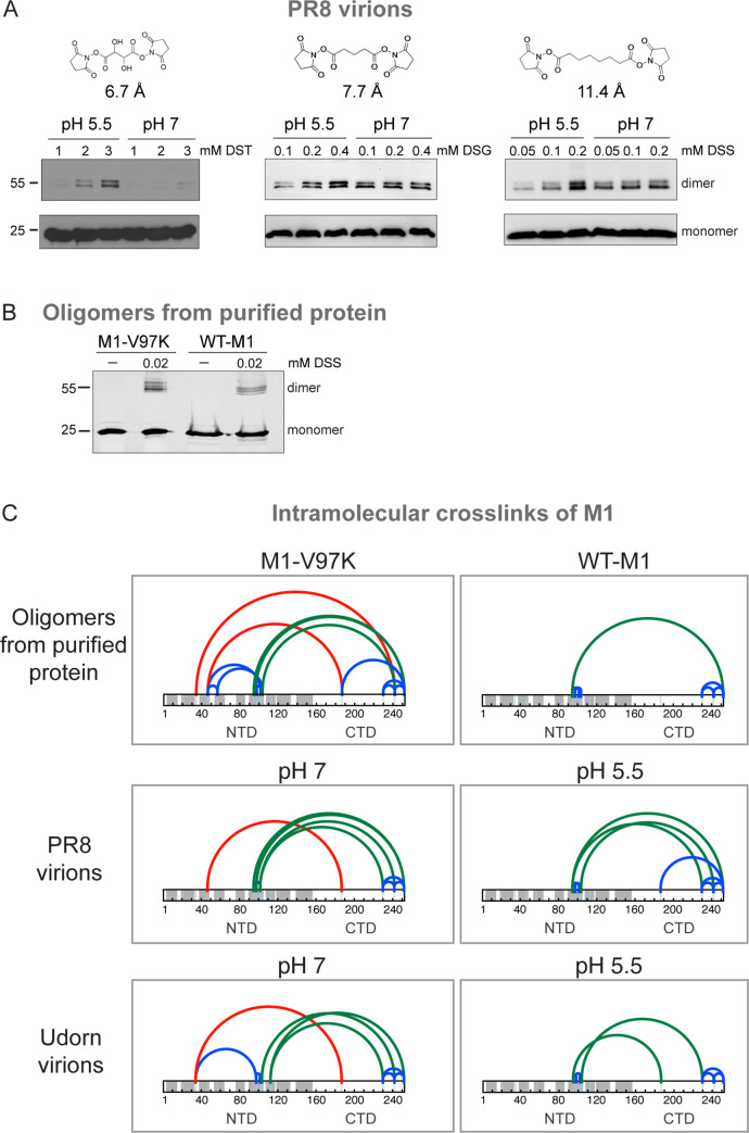
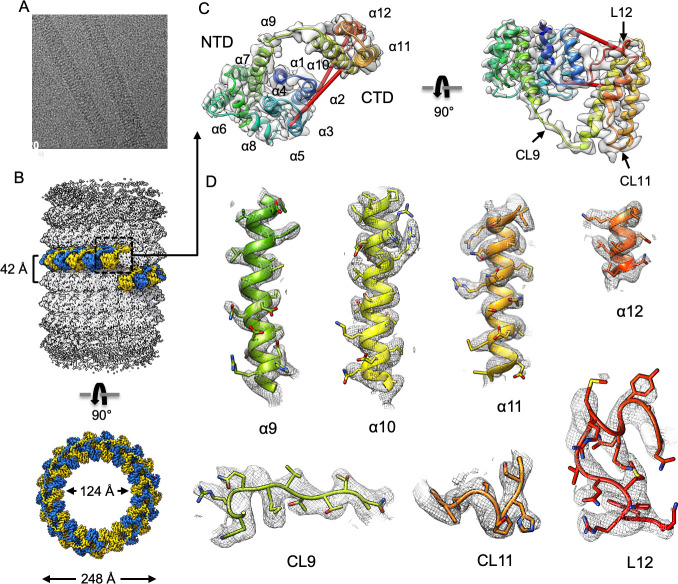
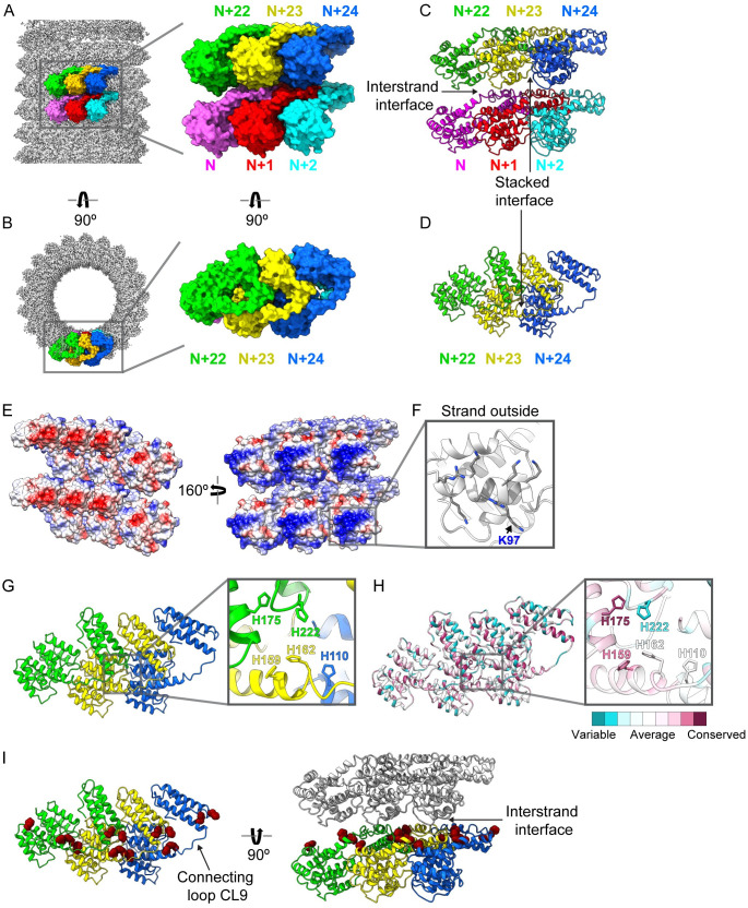
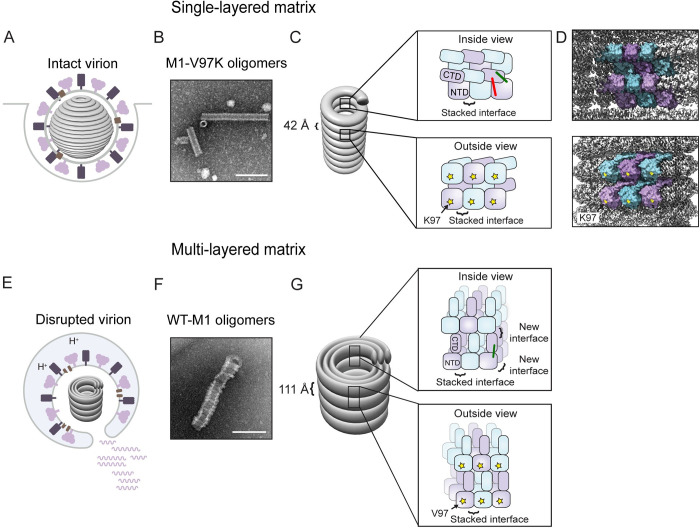
Matrix proteins are encoded by many enveloped viruses, including influenza viruses, herpes viruses, and coronaviruses. Underneath the viral envelope of influenza virus, matrix protein 1 (M1) forms an oligomeric layer critical for particle stability and pH-dependent RNA genome release. However, high-resolution structures of full-length monomeric M1 and the matrix layer have not been available, impeding antiviral targeting and understanding of the pH-dependent transitions involved in cell entry. Here, purification and extensive mutagenesis revealed protein-protein interfaces required for the formation of multilayered helical M1 oligomers similar to those observed in virions exposed to the low pH of cell entry. However, single-layered helical oligomers with biochemical and ultrastructural similarity to those found in infectious virions before cell entry were observed upon mutation of a single amino acid. The highly ordered structure of the single-layered oligomers and their likeness to the matrix layer of intact virions prompted structural analysis by cryo-electron microscopy (cryo-EM). The resulting 3.4-Å-resolution structure revealed the molecular details of M1 folding and its organization within the single-shelled matrix. The solution of the full-length M1 structure, the identification of critical assembly interfaces, and the development of M1 assembly assays with purified proteins are crucial advances for antiviral targeting of influenza viruses.


Conflict of interest statement

The authors have declared that no competing interests exist.

Figures

Fig 1
Fig 1. Full-length influenza M1 assembles into helical multilayered oligomers.
(A) Amino acid sequence of full-length M1, with α-helices in the NTD shown in gray and the CTD in green. All lysine residues are shown in red. (B) Negative-stain EM of M1 purified from virions after incubation of 1 μM protein in the presence of 2 M NaCl. (C) SDS-PAGE analysis of purified recombinant NTDM1 (lane 2) and full-length M1 (lane 3). (D) Size-exclusion chromatograms of full-length M1 following incubation of 10 μM in the presence (solid line) and absence (dotted line) of 2 M NaCl. (E) Negative-stain and (F) cryo-EM of full-length M1 after incubation of 2 μM in the presence of 2 M NaCl. Black arrows point to multilayered rings. (G) Size-exclusion chromatograms of NTDM1 after incubation of 10–80 μM in the presence of 2 M NaCl. Increasing concentrations of protein are represented as lighter shades of gray. (H) Negative-stain EM of NTDM1 incubated at 170 μM in the presence of 2 M NaCl. (I) Model of multilayered oligomers. Scale bar = 1,000 Å. See also S2 and S3 Figs. cryo-EM, cryo-electron microscopy; CTD, C-terminal domain; EM, electron microscopy; mAU, milli-absorbance units; M1, matrix protein 1; NTD, N-terminal domain.
Fig 2
Fig 2. Formation of multilayered oligomers is independent of virus morphology.
(A) Amino-acid–sequence alignment of M1 sequences derived from 2 spherical (PR8, A/WSN/1933[H1N1]) and 2 filamentous (Udorn, A/Netherlands/602/2009[H1N1]) virus strains. Amino acid changes compared with the PR8 reference sequence are highlighted. (B) Negative-stain electron micrographs of assembled M1 derived from i) PR8, ii) A/WSN/1933(H1N1), iii) Udorn, and iv) A/Netherlands/602/2009(H1N1). (C) Negative-stain electron micrographs showing the matrix layer inside Udorn virions with an intact (left panel) and a disrupted envelope (right panel). White arrows point to the matrix layers. Scale bar = 1,000 Å. M1, matrix protein 1; PR8, A/Puerto Rico/8/1934(H1N1); Udorn, A/Udorn/1972(H3N2) filamentous virus.
Fig 3
Fig 3. Mutations that alter assembly.
Mutated amino acid residues are displayed as red spheres and are shown in the C2-symmetry, stacked, and lateral NTDM1 dimer orientation. Shown are mutations that resulted in (A) formation of completely and partially assembled single-layered helical oligomers, (B) no assembly, or (C) multilayered helical oligomers. Panels in (A) show negative-stain electron micrographs of oligomers formed by the V97K (top) and the L130Q/M135Q (bottom) mutant. Scale bar = 1,000 Å. See also S1 Fig and S2 Table. M1, matrix protein 1; NTD, N-terminal domain.
Fig 4
Fig 4. Crosslinking and MS analysis of virions and oligomers formed from purified M1.
(A) SDS-PAGE analysis of M1 crosslinked within intact PR8 virions at increasing concentrations of DST, DSG, and DSS, with crosslinker distances of 6.7 Å, 7.7 Å, and 11.4 Å, after incubation at pH 5.5 or pH 7. M1 was visualized by immunoblotting using an anti-M1 antibody. (B) SDS-PAGE analysis of crosslinked M1-V97K and WT-M1 oligomers using 0.02 mM DSS stained with SYPRO Ruby. (C) Identified intramolecular DSS crosslinks of M1 within oligomers from purified WT-M1 and M1-V97K proteins are shown in the upper panels. Middle panes show DSS crosslinks of M1 within spherical PR8 virions. Lower panels show DSS crosslinks within filamentous Udorn virions. Crosslinks within the N- or CTD are shown as blue arches, crosslinks between helix α6 of the NTD and the CTD are shown as green arches, and crosslinks between helix α2 or α3 of the NTD and the CTD are displayed as red arches. See also S1–S6 Data. CTD, C-terminal domain; DSG, disuccinimidyl glutarate; DSS, disuccinimidyl suberate; DST, disuccinimidyl tartrate; M1, matrix protein 1; NTD, N-terminal domain; PR8, A/Puerto Rico/8/1934(H1N1); Udorn, A/Udorn/1972(H3N2) filamentous virus; WT-M1, full-length PR8 M1.
Fig 5
Fig 5. Cryo-EM helical reconstruction of M1-V97K filament.
(A) Representative micrograph. (B) Helical reconstruction of M1-V97K oligomers reveals a hollow tube with inner and outer diameters of 124 Å and 248 Å. One helical turn comprises approximately 21 asymmetric units, highlighted in alternating blue and yellow colors. (C) Ribbon representation of one M1-V97K asymmetric unit, which includes the NTD helices α1–9, the newly resolved CTD helices α10–12, and connecting loops CL9–11 and terminal loop L12. Identified red crosslinks are shown as red rods. (D) Cryo-EM maps and models of M1-V97K helices α9–12 and connecting loops CL9–11 and terminal loop L12. Most side chains are clearly resolved in the cryo-EM map at 3.4 Å resolution. See also S4 and S5 Figs and S3 Table. cryo-EM, cryo-electron microscopy; CTD, C-terminal domain; M1, matrix protein 1; NTD, N-terminal domain.
Fig 6
Fig 6. M1-V97K cryo-EM structure reveals molecular interactions between adjacent protein subunits.
Here, we define horizontal subunits in the diagram as forming a strand and the vertical interactions between subunits as “interstrand” interactions. A group of 6 protein subunits are highlighted in (A) a side view and (B) a top view of the oligomer with the lower strand consisting of N (pink), N + 1 (red), and N + 2 (cyan) and the upper strand consisting of N + 22 (green), N + 23 (yellow), and N + 24 (blue) because of the 21-subunit structure of the helix. (C, D) Stacked interactions that form the strand and interstrand contacts are shown. (E) Surface representation of 6 adjacent subunits are shown in 2 orientations colored by electrostatic potential, with positive residues in blue and negative residues in red. (F) A detailed view of the tube outside highlighting residues that form the many of positive charges, including mutated residue K97. (G) Top view of 3 adjacent protein subunits reveals a cluster of histidine residues at the 3-subunit contact point. (H) Top view of 3 adjacent protein subunits and the histidine cluster, with each residue colored by its conservation score, ranging from least conserved in dark cyan to most conserved in dark pink. (I) Top and side view of 3 adjacent protein subunits showing all histidine residues displayed as dark red spheres. Gray protein subunits are shown to display the interstrand interface. See also S6–S9 Figs and S1 Movie. CL, connecting loop; cryo-EM, cryo-electron microscopy; M1, matrix protein 1.
Fig 7
Fig 7. Summary and model.
(A) During virion formation, M1 assembles into a metastable matrix layer that is attached to the viral envelope. (B) We hypothesize that M1-V97K oligomers mimic the matrix layer observed in intact virions. (C, D) Within the M1-V97K oligomer, matrix proteins interact via the stacked dimer interface while the CTDs and NTDs are in close contact, shown by red and green crosslinks. K97 residues, shown as yellow stars, are facing the outside of the tube. The V97K mutation within M1-V97K oligomers and interactions with the viral membrane in intact virions prevent the addition of new M1 layers. (E) Within the endosome, a trigger converts the matrix layer into the multilayered matrix. (F) We hypothesize that multilayered oligomers assembled from WT-M1 mimic the oligomers found in disrupted virions. (G) Within WT-M1 oligomers, an increase in helical pitch to 111 Å could be caused by conformational changes of the CTD, indicated by the loss of the red crosslinks. New interfaces at the interstrand region and through disruption of interactions with the viral envelope allow the addition of new layers of matrix protein. CTD, C-terminal domain; M1, matrix protein 1; NTD, N-terminal domain; WT-M1, full-length PR8 M1.

Similar articles

Cited by

References

    1. Kirkegaard K, van Buuren NJ, Mateo R. My Cousin, My Enemy: quasispecies suppression of drug resistance. Curr Opin Virol. 2016;20: 106–111. 10.1016/j.coviro.2016.09.011 - DOI - PMC - PubMed
    1. Arzt S, Baudin F, Barge A, Timmins P, Burmeister WP, Ruigrok RW. Combined results from solution studies on intact influenza virus M1 protein and from a new crystal form of its N-terminal domain show that M1 is an elongated monomer. Virology. 2001;279: 439–446. 10.1006/viro.2000.0727 - DOI - PubMed
    1. Bui M, Whittaker G, Helenius A. Effect of M1 protein and low pH on nuclear transport of influenza virus ribonucleoproteins. J Virol. 1996;70: 8391–8401. Available from: https://www.ncbi.nlm.nih.gov/pmc/articles/PMC190928/pdf/708391.pdf 10.1128/JVI.70.12.8391-8401.1996 - DOI - PMC - PubMed
    1. Zhang J, Lamb RA. Characterization of the Membrane Association of the Influenza Virus Matrix Protein in Living Cells. Virology. 1996;225: 255–266. 10.1006/viro.1996.0599 - DOI - PubMed
    1. Martin K, Helenius A, Heleniust A, Helenius A. Nuclear transport of influenza virus ribonucleoproteins: the viral matrix protein (M1) promotes export and inhibits import. Cell. 1991;67: 117–130. 10.1016/0092-8674(91)90576-k - DOI - PubMed

Publication types

MeSH terms