. 2020 Sep 30;16(9):e1008819.
doi: 10.1371/journal.pgen.1008819. eCollection 2020 Sep.

The STRIPAK signaling complex regulates dephosphorylation of GUL1, an RNA-binding protein that shuttles on endosomes

Affiliations

The STRIPAK signaling complex regulates dephosphorylation of GUL1, an RNA-binding protein that shuttles on endosomes

Valentina Stein et al. PLoS Genet. .

Abstract

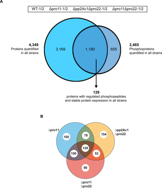
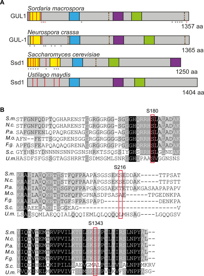
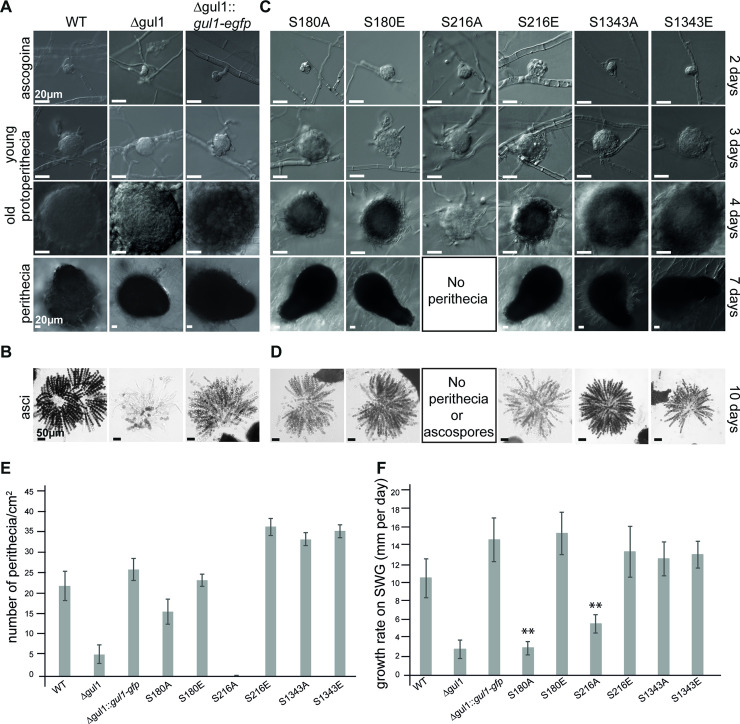
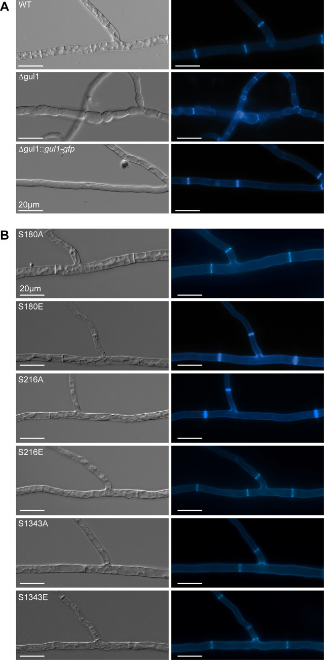
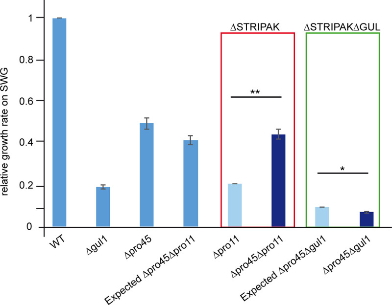
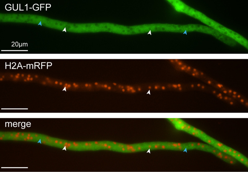
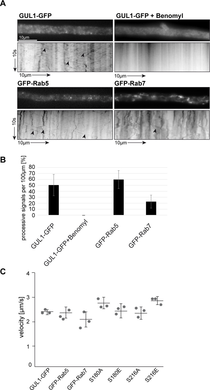
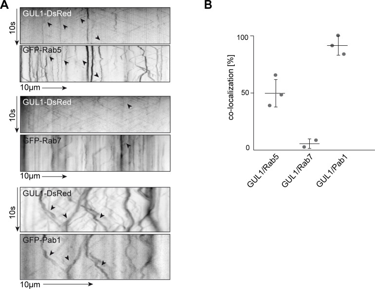
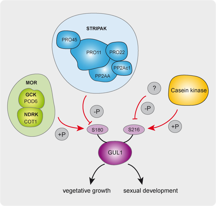
The striatin-interacting phosphatase and kinase (STRIPAK) multi-subunit signaling complex is highly conserved within eukaryotes. In fungi, STRIPAK controls multicellular development, morphogenesis, pathogenicity, and cell-cell recognition, while in humans, certain diseases are related to this signaling complex. To date, phosphorylation and dephosphorylation targets of STRIPAK are still widely unknown in microbial as well as animal systems. Here, we provide an extended global proteome and phosphoproteome study using the wild type as well as STRIPAK single and double deletion mutants (Δpro11, Δpro11Δpro22, Δpp2Ac1Δpro22) from the filamentous fungus Sordaria macrospora. Notably, in the deletion mutants, we identified the differential phosphorylation of 129 proteins, of which 70 phosphorylation sites were previously unknown. Included in the list of STRIPAK targets are eight proteins with RNA recognition motifs (RRMs) including GUL1. Knockout mutants and complemented transformants clearly show that GUL1 affects hyphal growth and sexual development. To assess the role of GUL1 phosphorylation on fungal development, we constructed phospho-mimetic and -deficient mutants of GUL1 residues. While S180 was dephosphorylated in a STRIPAK-dependent manner, S216, and S1343 served as non-regulated phosphorylation sites. While the S1343 mutants were indistinguishable from wild type, phospho-deficiency of S180 and S216 resulted in a drastic reduction in hyphal growth, and phospho-deficiency of S216 also affects sexual fertility. These results thus suggest that differential phosphorylation of GUL1 regulates developmental processes such as fruiting body maturation and hyphal morphogenesis. Moreover, genetic interaction studies provide strong evidence that GUL1 is not an integral subunit of STRIPAK. Finally, fluorescence microscopy revealed that GUL1 co-localizes with endosomal marker proteins and shuttles on endosomes. Here, we provide a new mechanistic model that explains how STRIPAK-dependent and -independent phosphorylation of GUL1 regulates sexual development and asexual growth.


Conflict of interest statement

The authors have declared that no competing interests exist.

Figures

Fig 1
Fig 1. Proteins and phosphoproteins found in the wild type and three STRIPAK deletion strains.
(A) We analysed the proteome and the phosphoproteome of the wild type, Δpro11, Δpp2Ac1Δpro22 and Δpro11Δpro22. In total, we identified 4,349 proteins in all strains and 2,465 phosphoproteins. The intersection of the Venn diagram gives the number of proteins found in both, the protein and the phosphoprotein analyses (1,180). Moreover, the number of regulated phosphoproteins from all strains are given that were identified with similar abundances in the global proteome. (B) Venn diagram of 129 phosphoproteins with regulated phosphorylation sites in STRIPAK deletion strains. Given is the total number of 129 phosphoproteins in the intersection of the Venn diagram which are differentially phosphorylated in Δpro11, Δpp2Ac1Δpro22, Δpro11Δpro22. Some phosphoproteins are given in more than one intersection because they exhibit multiple regulated phosphorylation sites (see also S1 and S2 Datasets).
Fig 2
Fig 2. Primary structure and amino acid sequence of GUL1 and its homologues.
(A) Identical protein domains in S. macrospora GUL1 and its homologue Ssd1 in Neurospora crassa, Saccharomyces cerevisiae and Ustilago maydis. Domains were analysed with ELM and have the following designation: yellow, Prion-like domain; red, LATS/NDR kinase recognition sites; blue, Nuclear localization signal; green, RNA binding domain; purple, nuclear export signal; brown dashed lines, PP2A-binding sites. Asterisks indicate phosphorylation sites, red asteriks in GUL1 were further investigated in this study (S180, S216, S1343). Yeast Ssd1 phosphorylation sites were adopted from Kurischko and Broach (2017). (B) Alignment of specific regions of the GUL1 protein from S. macrospora sequence with homologues from N. crassa (N.c., NCU01197), P. anserina (P.a., PODANS_2_6040), M. oryzae (M.o., MGG_08084), F. graminearum (F.g., FG05_07009), S. cerevisiae (S.c., SCY_1179) and U. maydis (U.m., UMAG_01220). Phosphorylation sites S180, S216 and S1343 are framed in red. S180, S216, and S1343 were investigated in the phosphorylation analysis.
Fig 3
Fig 3. Phenotypic analysis of wild type, Δgul1, a complemented Δgul1 strain (Δgul1::gul1-gfp), and phospho-mimetic and–deficient GUL1 strains.
(A, B) Sexual development. Ascogonia, young and old protoperithecia, as well as perithecia were examined after 2, 3, 4, and 7 days of growth BMM-slides. Samples were grown on BMM-medium. All bars represent 20 μm. (C, D) Wild type and the Δgul1::gul1-gfp complete ascus rosettes, while the gul1 deletion strain forms only a few ascospores. Phospho-deficient GUL1 strain S180A and both phospho-mimetic GUL1 strains S180E and S216E show complete ascus rosettes. Phospho-deficient GUL1 strain S216A does not form any spores. Phospho-deficient GUL1 strain S1343A and phospho-mimetic GUL1 strains S1343E show a wild-type like fertility. Bar represent 50 μm. (E) Quantification of perithecia per square centimetre on solid BMM-medium after 10 days (n = 9). (F) Growth rate of GUL1 phospho-mutants compared to Δgul1::gul1-gfp on SWG. Asterisks indicate significant differences compared to the complemented strain. Error bars in E and F indicate the standard deviation.
Fig 4
Fig 4. Septation and hyphal morphology of the wt, Δgul1 and the complemented Δgul1-strain Δgul1::gul1-gfp compared to phospho-mimetic and–deficient gul1 strains.
(A) The septation of hyphae in the wild type as reference, as well as in the complemented strain is regular and hyphal compartments are straight. In the gul1 deletion strain hyphae are hyperseptate and the compartments appear in a bubble-like structure. Hyphae of wild type are highly vacuolated due to the scarce nutrient supply in minimal medium. The slow-growing hyphae of Δgul1 have a reduced requirement of nutrients and therefore are not highly vacuolated. (B) Phospho-mimetic and–deficient GUL1 strains show no difference compared to wild type. Strains were grown on MMS for 2 days and stained with Calcofluor White M2R. Bars: 20 μm.
Fig 5
Fig 5. Analysis of the genetic interaction between gul1 and the slmap homologue pro45.
Genetic interaction was evaluated by comparing the daily vegetative growth rates of the indicated strains. The evaluation is based on the phenotype of the double mutant Δgul1/Δpro45 compared to the single mutants. The double mutant Δpro45/Δpro11 served as a control. Dark blue bars indicate experimentally generated values for single and double mutants, while light blue bars represent expected values for the double mutants based on multiplication of the values of the single mutants. The value of the wild type (wt) was set to 1 and all other values are given in relation to the wt. Absolute and relative values can be found in S2 Table. Error bars indicate standard deviations. Significant differences were evaluated by paired one-tailed Student´s t-test and are shown by lines * P ≤ 0.05; ** P ≤ 0.01. (n = 9 see Strains and growth conditions for details).
Fig 6
Fig 6. Localization of GUL1-GFP and H2A-mRFP in hyphae of Δgul1.
GUL1 localizes in dot-like structures within the cytoplasm (blue arrows). White arrows indicate a localization of GUL1 close to the nucleus (see also S1 Movie).
Fig 7
Fig 7. Shuttling of GUL1, Rab5 and Rab7.
(A) Kymographs comparing hyphae expressing GUL1-GFP in the gul1 deletion strain, GFP-Rab5 and GFP-Rab7 in the wild type. Processive signals are marked by black arrowheads; arrow length on the left and bottom indicates time and distance, 10 s and 10 μm, respectively; see also S2–S5 Movies). GUL1-GFP shuttling was inhibited by treatment with the microtubule inhibitor benomyl. (B) Amount of processive signals of GUL1-GFP, GUL1-GFP with benomyl, GFP-Rab5 and GFP-Rab7. Given are the processive signals (moving distance ≥5 μm) in percent per 100 μm hyphal length (7 to 18 hyphae per strain in three independent experiments). Error bars indicate standard deviation (S3 data set). (C) The average velocity of fluorescent signals per kymograph is indicated for all strains. Data points represent three means out of seven to 21 independent hyphae. At least 10 signals/hypha were analysed in all strains (S4 Dataset).
Fig 8
Fig 8. Kymographical analysis of co-localization between GUL1-DsRed and GFP-Rab5 and GFP-Rab7.
(A) Example of kymographs, used for the analysis of co-localization of GUL1-DsRed and GFP-Rab5 and GFP-Rab7. Fluorescence signals were detected simultaneously using dual-view technology. Kymographs were generated for a distance of at least 50 μm. Processive co-localizing signals are marked by black arrowheads. (B) Quantification of co-localization in percent. The shuttling of GUL1-DsRed and GFP-Rab5 was measured in 11 different hyphae per strain. The shuttling of GUL1-DsRed and GFP-Rab7 was measured in 4 different hyphae per strain. Analysis of Rab5 was done in triplicates. Analysis of Rab7 was done in duplicates. Mean is indicated by a horizontal line, dots show means of each experiment, error bars indicate standard deviation (S6–S8 Movies, S5 Dataset).
Fig 9
Fig 9. Schematic overview of phosphorylation dependent GUL1 function in sexual and asexual development.
S180 is a molecular switch for hyphal growth and morphology, which is targeted by COT1 and STRIPAK. In contrast, S216 is probably targeted by casein kinase and a so far unknown phosphatase. Abbreviations: MOR = morphogenesis orb6 network; STRIPAK: striatin-interacting phosphatase and kinase; GCK = germinal centre kinase; NDRK = nuclear dbf2-related kinase.

References

    1. Rogers S, McCloy R, Watkins DN, Burgess A. Mechanisms regulating phosphatase specificity and the removal of individual phosphorylation sites during mitotic exit. Bioessays. 2016;38 Suppl 1:S24–32. - PubMed
    1. Schuhmacher D, Sontag JM, Sontag E. Protein phosphatase 2A: More than a passenger in the regulation of epithelial cell-cell junctions. Front Cell Dev Biol. 2019;7:30 10.3389/fcell.2019.00030 - DOI - PMC - PubMed
    1. Goudreault M, D'Ambrosio LM, Kean MJ, Mullin MJ, Larsen BG, Sanchez A, et al. A PP2A phosphatase high density interaction network identifies a novel striatin-interacting phosphatase and kinase complex linked to the cerebral cavernous malformation 3 (CCM3) protein. Mol Cell Proteomics. 2009;8(1):157–71. 10.1074/mcp.M800266-MCP200 - DOI - PMC - PubMed
    1. Castets F, Bartoli M, Barnier JV, Baillat G, Salin P, Moqrich A, et al. A novel calmodulin-binding protein, belonging to the WD-repeat family, is localized in dendrites of a subset of CNS neurons. J Cell Biol. 1996;134(4):1051–62. 10.1083/jcb.134.4.1051 - DOI - PMC - PubMed
    1. Pöggeler S, Kück U. A WD40 repeat protein regulates fungal cell differentiation and can be replaced functionally by the mammalian homologue striatin. Eukaryot Cell. 2004;3(1):232–40. 10.1128/ec.3.1.232-240.2004 - DOI - PMC - PubMed

Publication types

MeSH terms