. 2020 Nov 2;219(11):e202006094.
doi: 10.1083/jcb.202006094.

Spatial and temporal control of targeting Polo-like kinase during meiotic prophase

Affiliations

Spatial and temporal control of targeting Polo-like kinase during meiotic prophase

James N Brandt et al. J Cell Biol. .

Abstract

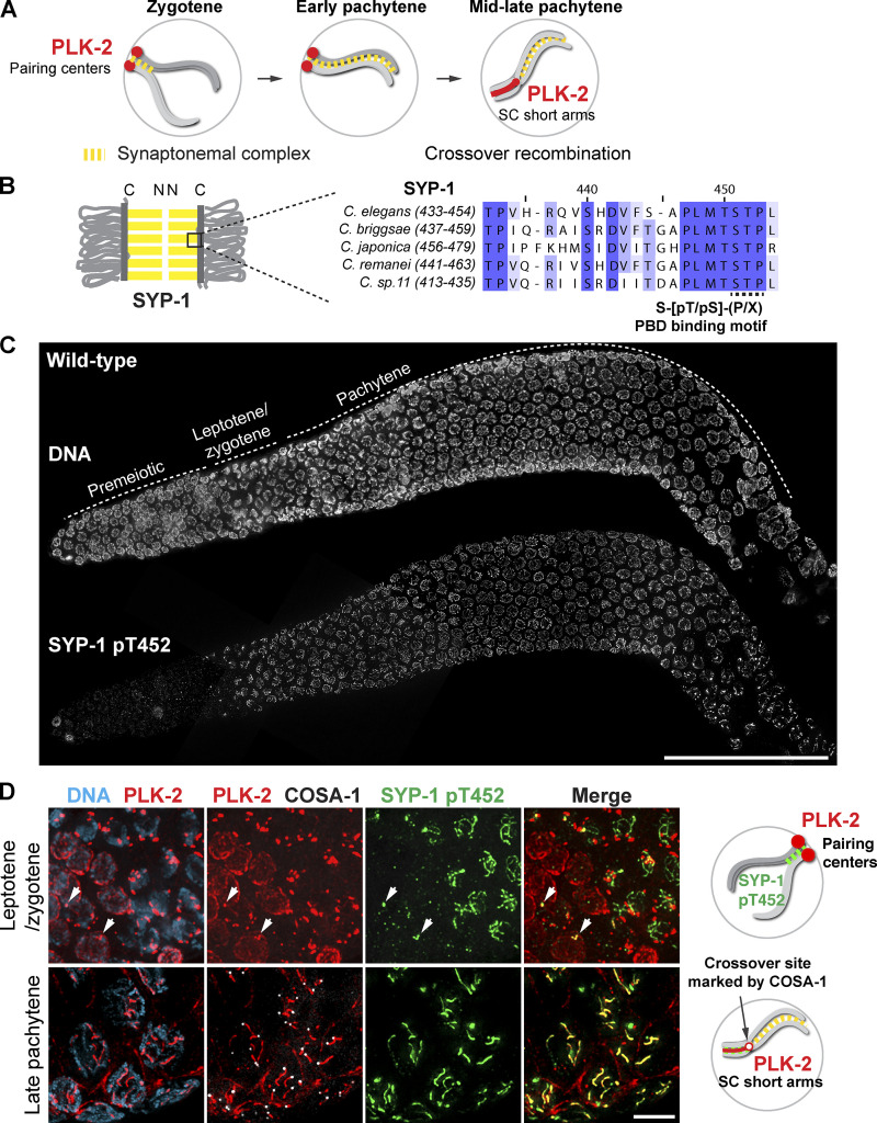
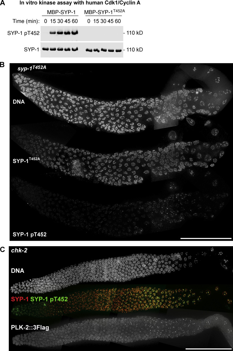
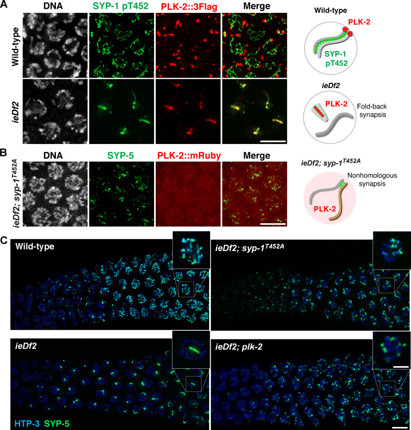
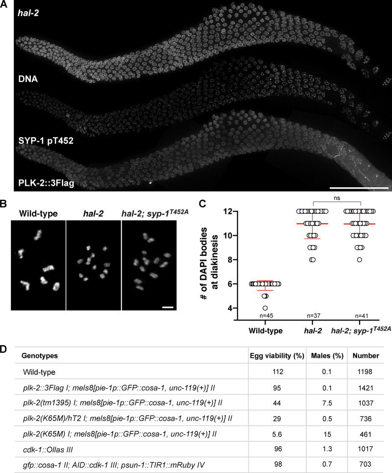
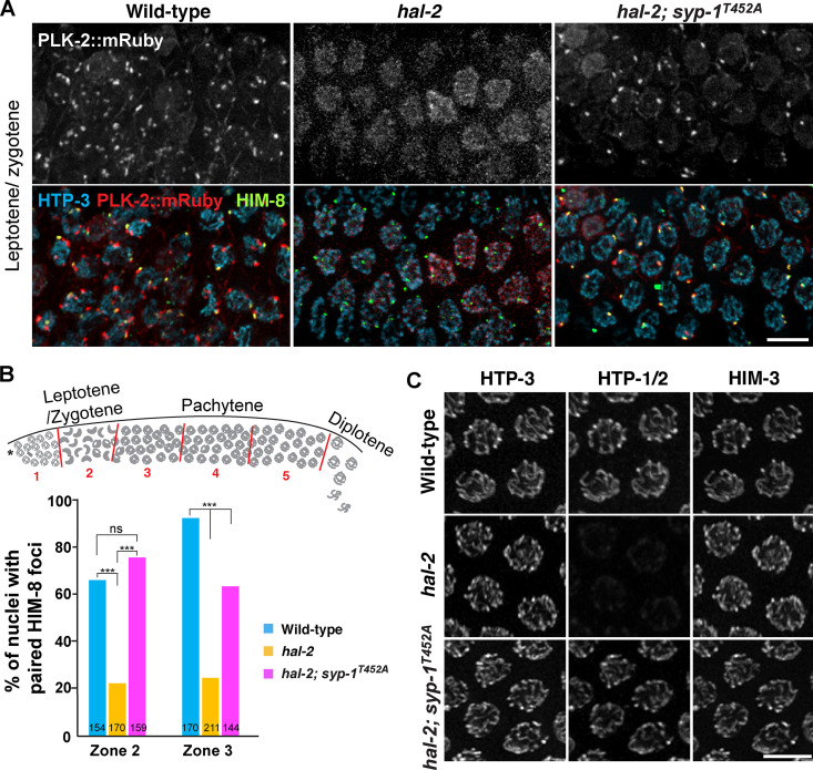
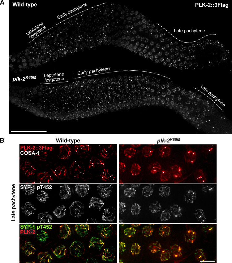
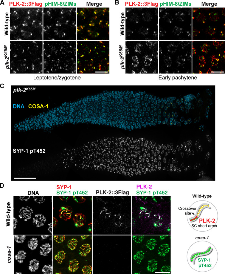
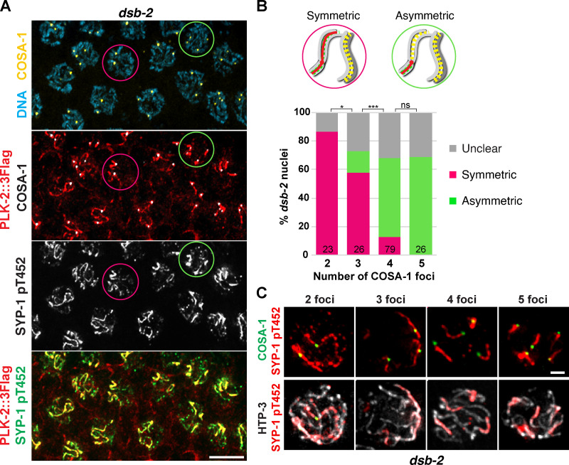
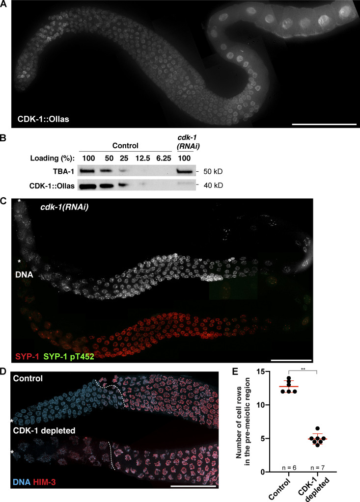
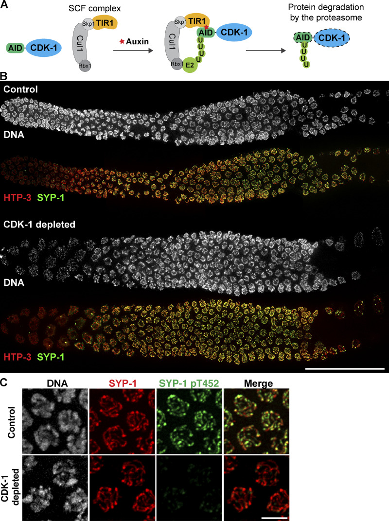
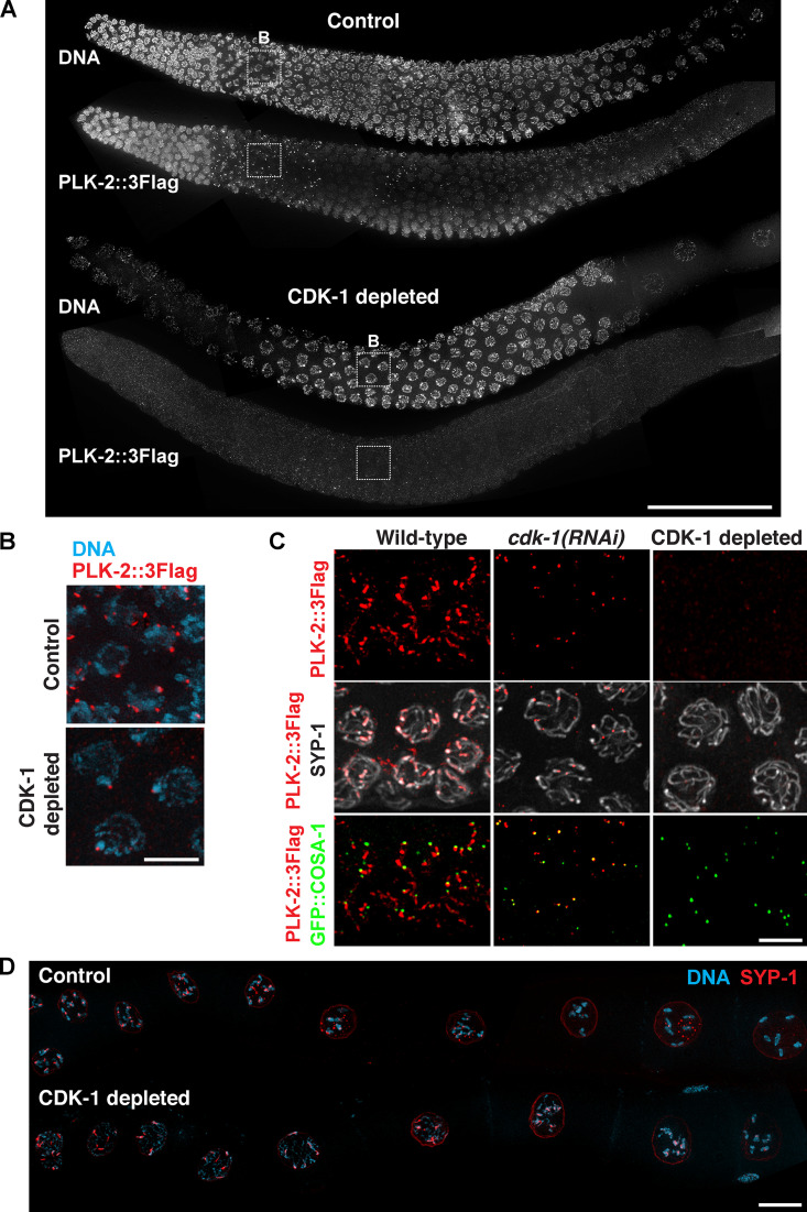
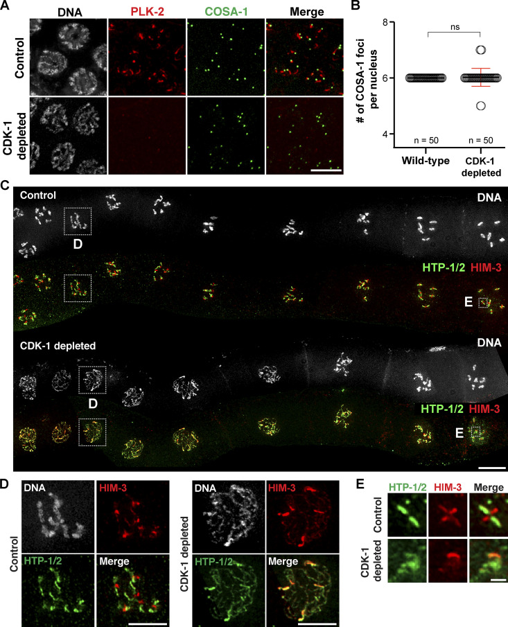
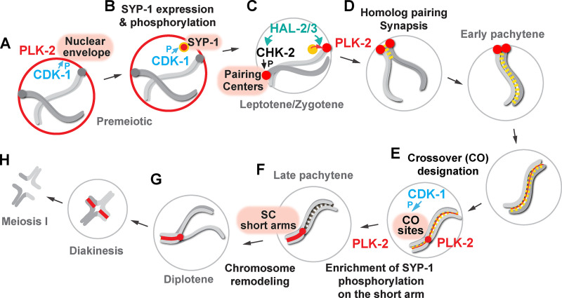
Polo-like kinases (PLKs) play widely conserved roles in orchestrating meiotic chromosome dynamics. However, how PLKs are targeted to distinct subcellular localizations during meiotic progression remains poorly understood. Here, we demonstrate that the cyclin-dependent kinase CDK-1 primes the recruitment of PLK-2 to the synaptonemal complex (SC) through phosphorylation of SYP-1 in C. elegans. SYP-1 phosphorylation by CDK-1 occurs just before meiotic onset. However, PLK-2 docking to the SC is prevented by the nucleoplasmic HAL-2/3 complex until crossover designation, which constrains PLK-2 to special chromosomal regions known as pairing centers to ensure proper homologue pairing and synapsis. PLK-2 is targeted to crossover sites primed by CDK-1 and spreads along the SC by reinforcing SYP-1 phosphorylation on one side of each crossover only when threshold levels of crossovers are generated. Thus, the integration of chromosome-autonomous signaling and a nucleus-wide crossover-counting mechanism partitions holocentric chromosomes relative to the crossover site, which ultimately defines the pattern of chromosome segregation during meiosis I.


Figures

Figure 1.
Figure 1.
PLK-2 is preferentially targeted to pairing centers in early meiotic prophase despite the presence of SYP-1 T452 phosphorylation in the C. elegans germline. (A) A schematic illustrating the dynamic localization of PLK-2 during meiotic prophase in C. elegans. (B) A schematic and sequence alignment showing the conserved PBD-binding motif within the C-terminal region of SYP-1. C, C-terminus. N, N-terminus. (C) Composite projection images of a whole gonad dissected from wild-type hermaphrodite and stained for DNA and SYP-1 pT452. Scale bar, 50 µm. (D) Immunofluorescence images of leptotene/zygotene nuclei (upper panels) and late pachytene nuclei (lower panels) showing DNA (blue), PLK-2 (red), COSA-1 (white), and SYP-1 pT452 (green). Arrows indicate the localization of PLK-2 to polycomplexes in early meiotic prophase. Scale bar, 5 µm. Diagrams illustrating the results are shown on the right.
Figure S1.
Figure S1.
Phospho-specificity of the SYP-1 pT452 antibody used in this study, and the analysis of SYP-1 T452 phosphorylation and PLK-2 localization in chk-2 mutants. (A) In vitro kinase assays using human CDK-1/cyclin A to phosphorylate MBP–SYP-1 with or without the T452A mutation. Western blot using antibodies against SYP-1 and phosphorylated SYP-1 at T452 is shown. (B) Composite immunofluorescence images of a whole gonad dissected from syp-1T452A mutants showing DNA, SYP-1, and SYP-1 pT452 staining. Scale bar, 50 µm. (C) Composite immunofluorescence images of a whole CHK-2–depleted gonad showing DNA (white), SYP-1 (red), SYP-1 pT452 (green), and PLK-2::3Flag staining. Scale bar, 50 µm.
Figure 2.
Figure 2.
Constrained PLK-2 activity at the pairing centers is essential for proper synapsis. (A) Immunofluorescence images of early pachytene nuclei from wild-type and ieDf2 mutants stained for DNA (white), SYP-1 pT452 (green), and PLK-2::3Flag (red). Scale bar, 5 µm. (B) Immunofluorescence images of mid-pachytene nuclei from ieDf2; syp-1T452A mutants stained for DNA (white), SYP-1 (green), and PLK-2::mRuby (red). Scale bar, 5 µm. Diagrams illustrating the results are shown on the right. (C) Composite projection images of gonad sections from indicated genotypes, spanning from meiotic entry to mid-pachytene, showing HTP-3 (blue) and SYP-5 (green). Scale bar, 5 µm. Insets show zoomed-in view of a representative nucleus from the boxed regions. Scale bar, 2 µm.
Figure S2.
Figure S2.
The SYP-1 T452A mutation does not rescue the failure to form crossovers in hal-2 mutants. (A) Composite immunofluorescence images of a whole gonad from hal-2 mutants showing DNA, SYP-1 pT452, and PLK-2::3Flag staining. Scale bar, 50 µm. (B) Oocyte nuclei at diakinesis from indicated genotypes were stained with DAPI. Scale bar, 3 µm. (C) Graph showing the number of DAPI-staining bodies in oocytes at diakinesis (mean ± SD). Numbers of nuclei scored are indicated (n = 45 for wild type; n = 37 for hal-2; n = 41 for hal-2; syp-1T452A). ns, not significant (P > 0.05) by two-tailed Mann-Whitney test. (D) Table showing the percent viable and male progeny from C. elegans hermaphrodites of indicated genotypes.
Figure 3.
Figure 3.
The SYP-1 T452A mutation partially rescues targeting of PLK-2 to pairing centers in hal-2 mutants. (A) Immunofluorescence images of leptotene/zygotene nuclei from wild-type, hal-2, and hal-2; syp-1T452A mutants showing PLK-2::mRuby (white or red), HTP-3 (blue), and HIM-8 (green). Scale bar, 5 µm. (B) Graph showing the percentage of nuclei with paired HIM-8 foci from the indicated genotypes. To score pairing of the X chromosome pairing center, the germline was divided into five zones as shown in the diagram. Zone 2 contains leptotene/zygotene nuclei (n = 154 for wild type; n = 170 for hal-2; n = 159 for hal-2; syp-1T452A), and zone 3 contains nuclei in early pachytene (n = 170 for wild type; n = 211 for hal-2; n = 144 for hal-2; syp-1T452A). ***, P < 0.0001; ns, not significant (P > 0.05) by χ2 analysis. (C) Immunofluorescence images of pachytene nuclei from wild-type, hal-2, and hal-2; syp-1T452A mutants showing the staining for HTP-3, HTP-1/2, and HIM-3. Scale bar, 5 µm.
Figure 4.
Figure 4.
The kinase activity of PLK-2 is required for enrichment of SYP-1 T452 phosphorylation along chromosomes that have designated crossovers. (A) Composite immunofluorescence images of whole gonads dissected from wild-type and plk-2K65M hermaphrodites and stained for PLK-2::3Flag. Scale bar, 50 µm. (B) Immunofluorescence images of late pachytene nuclei from wild-type and plk-2K65M mutants showing PLK-2::3Flag (red), COSA-1 (white), and SYP-1 pT452 (green) staining. Scale bar, 5 µm.
Figure S3.
Figure S3.
Enrichment of SYP-1 phosphorylation on the short arm requires PLK-2 kinase activity and crossover formation. (A and B) Zoomed-in images of nuclei from leptotene/zygotene (A) and early pachytene (B) showing PLK-2::3Flag (red) and phospho-HIM-8/ZIMs (green) staining. Scale bars, 5 µm. (C) Composite immunofluorescence images of a whole gonad dissected from the plk-2K65M (kinase dead) homozygote animal showing DNA (blue), COSA-1 (yellow), and SYP-1 pT452 staining (white). Scale bar, 50 µm. (D) Late-pachytene nuclei from wild-type and cosa-1(tm3298) mutants were stained for DNA, SYP-1 (red), SYP-1 pT452 (green), and PLK-2 (magenta). Scale bar, 5 µm. Diagrams illustrating the results are shown on the right.
Figure 5.
Figure 5.
Threshold levels of crossovers are required for asymmetric enrichment of SYP-1 T452 phosphorylation. (A) Immunofluorescence images of late pachytene nuclei from wild-type and dsb-2 mutants showing DNA (blue), COSA-1 (yellow), PLK-2 (red), and SYP-1 pT452 (green) staining. Circles represent examples of nuclei showing either symmetric (magenta) or asymmetric (green) SYP-1 pT452/PLK-2 localization relative to COSA-1. Scale bar, 5 µm. (B) Diagrams illustrating nuclei with the symmetric versus asymmetric SYP-1 pT452 staining and a graph showing the distribution of SYP-1 pT452 staining in dsb-2 nuclei with two COSA-1 foci (n = 23), three COSA-1 foci (n = 26), four COSA-1 foci (n = 79), and five COSA-1 foci (n = 26). *, P < 0.05; ***, P < 0.0001; ns, not significant (P > 0.05) by Fisher’s exact test. (C) Late pachytene nuclei from dsb-2 mutants were stained for SYP-1 pT452 (red), COSA-1 (green), and HTP-3 (white) and categorized by the number of COSA-1 foci. Scale bar, 1 µm.
Figure S4.
Figure S4.
Knockdown of CDK-1 by RNAi abolishes the phosphorylation of SYP-1 at T452 and abrogates PLK-2 targeting to the SC. (A) Composite immunofluorescence image of a full-length gonad dissected from a worm strain expressing CDK-1::Ollas. Scale bar, 50 µm. (B) Western blots showing the knockdown of CDK-1 by RNAi. The level of CDK-1::Ollas in cdk-1(RNAi) worms was compared with serially diluted worm lysates from control animals. Tubulin (TBA-1) was used as a loading control. Molecular weights for both proteins are indicated on the right. (C) Composite immunofluorescence images of a full-length gonad from a cdk-1 RNAi–treated worm showing DNA (white), SYP-1 (red), and SYP-1 pT452 staining (green). Asterisks indicate the distal tip of the germline. Scale bar, 50 µm. (D) Immunofluorescence images of distal germlines from control and CDK-1–depleted animals showing DNA (blue) and HIM-3 (red) staining. Asterisks indicate the distal tip of the germline, and dotted lines indicate the meiotic entry. Scale bar, 50 µm. (E) Graph showing the number of cell rows in the premeiotic region in control versus CDK-1–depleted germline. Mean ± SD is shown. Numbers of gonads scored are indicated on the bottom (n = 6 for control; n = 7 for CDK-1 depleted). **, P < 0.01 by two-tailed Mann-Whitney test.
Figure 6.
Figure 6.
CDK-1 is required for chromosome remodeling after crossover formation. (A) A schematic illustrating the strategy to deplete CDK-1 in the adult germline using the AID system. U, ubiquitination. (B) Whole gonads dissected from control and CDK-1–depleted animals were stained for DNA (white), HTP-3 (red), and SYP-1 (green). Composite immunofluorescence images are shown. Scale bar, 50 µm. (C) Immunofluorescence of mid-pachytene nuclei from control and CDK-1–depleted animals showing DNA (white), SYP-1 (red), and SYP-1 pT452 (green). Scale bar, 5 µm.
Figure S5.
Figure S5.
Depletion of CDK-1 in the germline by AID system leads to failures in targeting PLK-2 to distinct subnuclear structures. (A) Full-length gonads dissected from control versus CDK-1–depleted animals were stained for DNA and PLK-2::3Flag. Scale bar, 50 µm. (B) Zoomed-in images of the boxed regions in A showing DNA (blue) and PLK-2 (red) staining from control and CDK-1–depleted germlines during leptotene/zygotene. Scale bar, 5 µm. (C) Immunofluorescence of late-pachytene nuclei from wild-type, cdk-1(RNAi), and CDK-1–depleted worms showing SYP-1, PLK-2 (red), and COSA-1 (green). Scale bar, 5 µm. (D) Composite immunofluorescence images of diakinesis nuclei from control versus CDK-1–depleted germline showing DNA (blue) and SYP-1 (red). Scale bar, 5 µm.
Figure 7.
Figure 7.
CDK-1 is required for chromosome remodeling after crossover formation. (A) Immunofluorescence images of late-pachytene nuclei from control and CDK-1–depleted animals showing DNA (white), PLK-2 (red), and COSA-1 (green) staining. Scale bar, 5 µm. (B) Graph showing the quantification of COSA-1 number per nucleus in wild-type (n = 50) and CDK-1–depleted animals (n = 50). Mean ± SD is shown. ns, not significant (P > 0.05) by two-tailed Mann-Whitney test. (C) Composite immunofluorescence images of diakinesis nuclei from control and CDK-1–depleted animals showing DNA (white), HTP-1/2 (green), and HIM-3 (red) staining. Scale bar, 10 µm. (D) Zoomed-in images of a diplotene nucleus from control and CDK-1–depleted germlines as indicated in C. Scale bars, 5 µm. (E) Zoomed-in images of an individual chromosome in diakinesis from control and CDK-1–depleted germlines as indicated in C. Scale bar, 1 µm.
Figure 8.
Figure 8.
Model for targeting PLK-2 to distinct subnuclear structures during meiotic prophase in C. elegans. (A) In the premeiotic region of the germline, CDK-1 primes the localization of PLK-2 to the nuclear envelope. P, phosphorylation. (B) CDK-1 phosphorylates newly expressed SYP-1 at T452 just before the meiotic onset, and a pool of PLK-2 can localize to SC polycomplexes. (C) Upon meiotic entry, CHK-2 becomes active and phosphorylates the pairing center proteins (Kim et al., 2015), which serve as the preferred docking sites for PLK-2. The nucleoplasmic HAL-2/3 complex ensures the PLK-2 localization to pairing centers by promoting CHK-2 activity and by preventing premature association of PLK-2 to SYP proteins. (D) Constrained PLK-2 activity at the pairing centers is essential for proper axis assembly, homologue pairing, and synapsis. (E) CDK-1 is also responsible for targeting PLK-2 to the crossover-designated sites. (F) SYP-1 phosphorylation and PLK-2 are enriched on the SC short arm relative to the crossover site, and this requires PLK-2 kinase activity and a threshold level of crossover within the nucleus. (G) PLK-2 drives the asymmetric SC disassembly. (H) The short arm becomes the site of cohesion loss during meiosis I.

References

    1. Altendorfer E., Láscarez-Lagunas L.I., Nadarajan S., Mathieson I., and Colaiácovo M.P.. 2020. Crossover Position Drives Chromosome Remodeling for Accurate Meiotic Chromosome Segregation. Curr. Biol. 30:1329–1338.e7: E7. 10.1016/j.cub.2020.01.079 - DOI - PMC - PubMed
    1. Archambault V., and Glover D.M.. 2009. Polo-like kinases: conservation and divergence in their functions and regulation. Nat. Rev. Mol. Cell Biol. 10:265–275. 10.1038/nrm2653 - DOI - PubMed
    1. Argunhan B., Leung W.-K., Afshar N., Terentyev Y., Subramanian V.V., Murayama Y., Hochwagen A., Iwasaki H., Tsubouchi T., and Tsubouchi H.. 2017. Fundamental cell cycle kinases collaborate to ensure timely destruction of the synaptonemal complex during meiosis. EMBO J. 36:2488–2509. 10.15252/embj.201695895 - DOI - PMC - PubMed
    1. Attner M.A., Miller M.P., Ee L., Elkin S.K., and Amon A.. 2013. Polo kinase Cdc5 is a central regulator of meiosis I. Proc. Natl. Acad. Sci. USA. 110:14278–14283. 10.1073/pnas.1311845110 - DOI - PMC - PubMed
    1. Bazile F., St-Pierre J., and D’Amours D.. 2010. Three-step model for condensin activation during mitotic chromosome condensation. Cell Cycle. 9:3263–3275. 10.4161/cc.9.16.12620 - DOI - PubMed

Publication types

MeSH terms

Substances