. 2021 Mar;76(3):853-865.
doi: 10.1111/all.14608. Epub 2020 Nov 13.

Accuracy of serological testing for SARS-CoV-2 antibodies: First results of a large mixed-method evaluation study

Affiliations

Accuracy of serological testing for SARS-CoV-2 antibodies: First results of a large mixed-method evaluation study

Daniel Brigger et al. Allergy. 2021 Mar.

Abstract

Background: Serological immunoassays that can identify protective immunity against SARS-CoV-2 are needed to adapt quarantine measures, assess vaccination responses, and evaluate donor plasma. To date, however, the utility of such immunoassays remains unclear. In a mixed-design evaluation study, we compared the diagnostic accuracy of serological immunoassays that are based on various SARS-CoV-2 proteins and assessed the neutralizing activity of antibodies in patient sera.

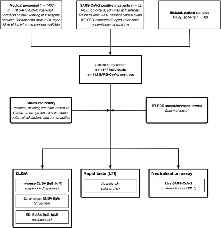
Methods: Consecutive patients admitted with confirmed SARS-CoV-2 infection were prospectively followed alongside medical staff and biobank samples from winter 2018/2019. An in-house enzyme-linked immunosorbent assay utilizing recombinant receptor-binding domain (RBD) of the SARS-CoV-2 spike protein was developed and compared to three commercially available enzyme-linked immunosorbent assays (ELISAs) targeting the nucleoprotein (N), the S1 domain of the spike protein (S1), and a lateral flow immunoassay (LFI) based on full-length spike protein. Neutralization assays with live SARS-CoV-2 were performed.

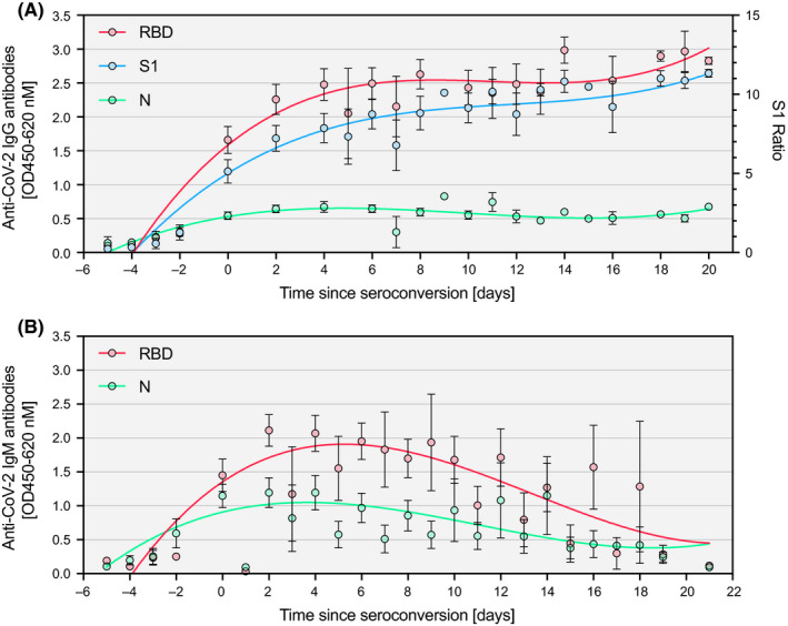
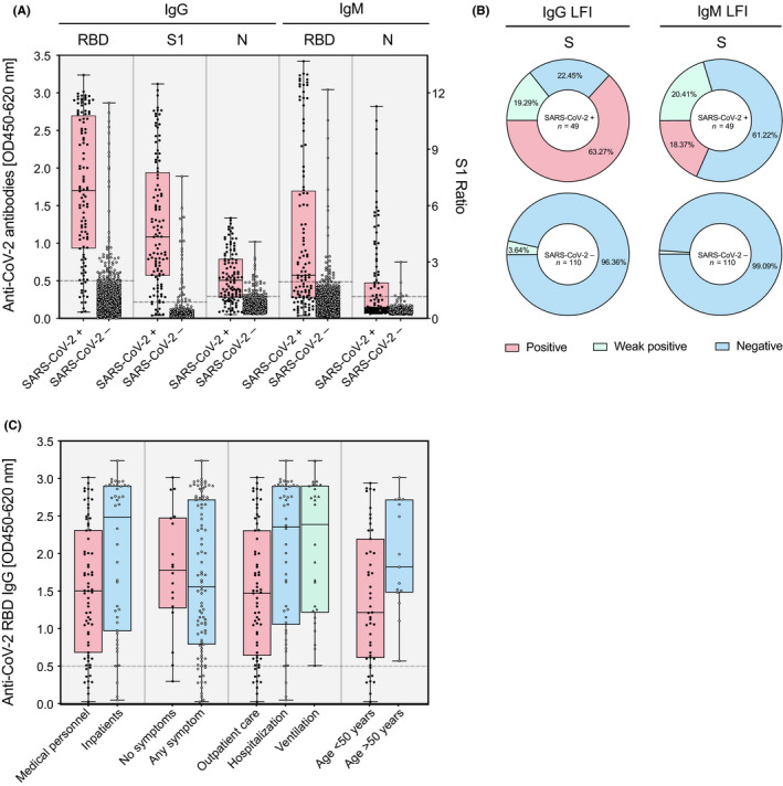
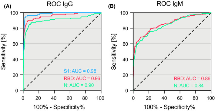
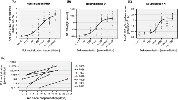
Results: One thousand four hundred and seventy-seven individuals were included comprising 112 SARS-CoV-2 positives (defined as a positive real-time PCR result; prevalence 7.6%). IgG seroconversion occurred between day 0 and day 21. While the ELISAs showed sensitivities of 88.4% for RBD, 89.3% for S1, and 72.9% for N protein, the specificity was above 94% for all tests. Out of 54 SARS-CoV-2 positive individuals, 96.3% showed full neutralization of live SARS-CoV-2 at serum dilutions ≥ 1:16, while none of the 6 SARS-CoV-2-negative sera revealed neutralizing activity.

Conclusions: ELISAs targeting RBD and S1 protein of SARS-CoV-2 are promising immunoassays which shall be further evaluated in studies verifying diagnostic accuracy and protective immunity against SARS-CoV-2.

Keywords: Antibodies; COVID-19 [Supplementary Concept]; COVID-19 diagnostic testing [Supplementary Concept]; Enzyme-Linked Immunosorbent Assay [Mesh]; Neutralizing [Mesh]; Severe Acute Respiratory Syndrome Coronavirus 2 [Supplementary Concept].


Conflict of interest statement

All authors declare that there is no conflict of interests.

Figures

Figure 1
Figure 1
Flowchart of study cohort and study design. Only RT‐PCR‐positive inpatients were considered in the current phase of the study (*). Consecutive patients admitted with confirmed SARS‐CoV‐2 infection were prospectively followed alongside medical staff and biobank samples from winter 2018/2019 (pooled data were used for calculation of diagnostic accuracy). RT‐PCR, real‐time PCR; ELISA, enzyme‐linked immunosorbent assay; LFI, lateral flow immunoassay
Figure 2
Figure 2
Seroconversion rate since symptoms and positive RT‐PCR result. The percentage of consecutive patients (n = 25) positively tested for anti‐SARS‐CoV‐2 protein antibodies is shown as a function of time (since symptom onset: red; since positive RT‐PCR result: blue line) for IgG in the RBD (A), IgG in the S1 (B), IgG in the N (C), IgM in the RBD (D), and IgM in the N ELISA (E). Curves were calculated using nonlinear fitting
Figure 3
Figure 3
Temporal pattern of antibody responses against SARS‐CoV‐2 since seroconversion. IgG and IgM antibody responses of consecutive patients (n = 25) as measured by three ELISAs targeting different proteins of SARS‐CoV‐2: (A) IgG against the receptor‐binding domain (RBD), the S1 domain of the spike protein (S1), and the nucleoprotein (N); (B) IgM against the receptor‐binding domain (RBD) and the nucleoprotein (N). Data are shown as mean ± SEM. Curves were calculated using nonlinear fitting
Figure 4
Figure 4
Distribution of immunoassay results among SARS‐CoV‐2‐positive and negative individuals. Consecutive patients admitted with confirmed SARS‐CoV‐2 infection were prospectively followed alongside medical staff and biobank samples from winter 2018/2019 (pooled data). (A) IgG and IgM responses against the receptor‐binding domain (RBD), the S1 domain of the spike protein (S1), and the nucleoprotein (N) of SARS‐CoV‐2 in SARS‐CoV‐2‐positive (n = 112) and negative (n = 1365) individuals as measured by ELISA. Data are shown as individual data points with a box and whiskers plot indicating minimum to maximum response. (B) IgG and IgM responses against the S1 domain of the spike protein of SARS‐CoV‐2 as measured by LFI in RT‐PCR‐positive and negative patients. Positive (red), weak positive (green), and negative (blue) responses are shown as percentage of the whole in a pie chart. (C) Antibody response in salient subgroups of RT‐PCR‐positive individuals (inpatients vs. medical personnel, patients with symptoms vs. patients without, hospitalized patients vs. outpatients, patients with ventilation vs. patients without, patients above 50 years vs. patients below 50 years of age)
Figure 5
Figure 5
Accuracy of three different SARS‐CoV‐2 ELISAs. Receiver operating characteristics curves of IgG (A) and IgM (B) measurements against the receptor‐binding domain (RBD), the S1 domain of the spike protein (S1), and the nucleoprotein (N) of SARS‐CoV‐2 in SARS‐CoV‐2‐positive (n = 112) and negative (n = 1365) individuals
Figure 6
Figure 6
Live SARS‐CoV‐2 neutralization. Individual antibody responses (gray dots) against RBD (A), S1 (B) and N protein (C) in sera of 54 SARS‐CoV‐2‐positive and 6 SARS‐CoV‐2‐negative individuals as measured by ELISA are shown together with the corresponding serum dilution at which full neutralization of SARS‐CoV‐2 is observed. Nonlinear curve fitting was calculated based on the means of each serum dilution group (red circles). (D) Changes in the serum dilution for full neutralization of SARS‐CoV‐2 over time are depicted for seven individual SARS‐CoV‐2 patients. no NT: no neutralization detectable

References

    1. Krammer F, Simon V. Serology assays to manage COVID‐19. Science. 2020;368(6495):1060‐1061. - PubMed
    1. Tang YW, Schmitz JE, Persing DH, Stratton CW. The Laboratory Diagnosis of COVID‐19 Infection: Current Issues and Challenges. J Clin Microbiol. 2020.58(6):e00512–20. - PMC - PubMed
    1. Winter AK, Hegde ST. The important role of serology for COVID‐19 control. Lancet Infect Dis. 2020;20(7):758‐759. - PMC - PubMed
    1. Yong SEF, Anderson DE, Wei WE, et al. Connecting clusters of COVID‐19: an epidemiological and serological investigation. Lancet Infect Dis. 2020;20(7):809‐815. - PMC - PubMed
    1. Pung R, Chiew CJ, Young BE, et al. Investigation of three clusters of COVID‐19 in Singapore: implications for surveillance and response measures. Lancet. 2020;395(10229):1039‐1046. - PMC - PubMed

Publication types

Substances