Review
. 2021 Jul;40(7):1427-1443.
doi: 10.1002/jum.15517. Epub 2020 Sep 30.

Ultrasound-Guided Breast Biopsies: Basic and New Techniques

Affiliations
Review

Ultrasound-Guided Breast Biopsies: Basic and New Techniques

Asha A Bhatt et al. J Ultrasound Med. 2021 Jul.

Abstract

Ultrasound-guided breast biopsies can be challenging to perform, especially when the target is adjacent to the nipple, skin, or implant or when the target is small and in very posterior, dense fibroglandular tissue. Oftentimes, a slightly modified approach can result in a diagnostic biopsy specimen with minimal complications. After a brief review of basic techniques for ultrasound-guided breast biopsies that includes a review of conventional breast biopsy devices, a presentation of procedural modifications and techniques to consider for more challenging cases is described. In particular, novel open-trough and tandem-needle techniques are detailed. Several cases using these techniques are then presented.

Keywords: breast biopsy; breast intervention; hydrodissection; tandem needle; ultrasound-guided.


Figures

Figure 1
Figure 1
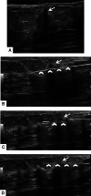
Spring‐loaded core needle biopsy device using a 2‐staged approach to confirm positioning of the sampling trough. A 77‐year‐old woman presented for US‐guided CB of a palpable mass, which was performed with a 14‐gauge SLD (Achieve; Cardinal Health, Dublin, OH). A, Prebiopsy image shows a hypoechoic mass (arrow) with angular margins and posterior acoustic shadowing. B, Prefire image shows the biopsy needle (arrowheads) directed toward the targeted mass and parallel to the chest wall. C, In this 2‐staged approach, the postfire image of the first stage shows the mass (arrow) within the needle trough (arrowheads). D, During the second stage, the outer cutting cannula covers the trough (arrowheads), securing a tissue specimen in the trough.
Figure 2
Figure 2
Spring‐loaded core needle biopsy device using a 2‐staged approach to confirm optimal needle positioning in the orthogonal view. A, The needle is parallel and aligned with the footprint of the transducer. B, Corresponding transverse US image from A shows that the needle and open trough (arrowheads) are parallel to the chest wall, and the trough traverses the target (arrow). C, Rotation of the transducer 90° makes the transducer orthogonal to the needle and longitudinal with the lesion. D, Corresponding longitudinal or orthogonal US image shows the needle (arrowhead) nearly centered within the slightly echogenic target (arrow).
Figure 3
Figure 3
Spring‐loaded core needle biopsy device using a 2‐staged approach and maximizing target tissue in the trough. A, Orientation of the needle (arrows) is such that the angle (red angle) between the transducer and the biopsy device is relatively acute. B, Ultrasound image shows the open trough (arrowheads) located below the lesion (arrow). C, To increase target tissue in the trough, a downward force creating torque along with slight lifting force is applied to the device, increasing the angle (red angle) between the transducer and the biopsy needle (arrows). D, Ultrasound image shows how the maneuver results in the trough (arrowheads) being closer to the lesion (arrow), allowing one to better approximate the target into the trough.
Figure 4
Figure 4
Vacuum‐assisted breast biopsy with the correct prebiopsy positioning. An 80‐year‐old woman with a screen‐detected left breast mass presented for US‐guided CB. A 9‐gauge petite Suros VABB device was selected for the procedure. The device (arrows) is positioned below the target. When the device is correctly positioned, 2 echogenic lines corresponding to the 2 edges of the trough/aperture can be seen.
Figure 5
Figure 5
Spring‐loaded core needle biopsy device open‐trough technique (profile view on a phantom). A, With this technique, the introducer is placed at the posterior margin (deep to the transducer) and slightly beyond the lesion to account for the 8‐mm dead space of the SLD. B and C, The obturator is removed (B), and the biopsy needle (with the sampling notch open) is advanced to the end of the coaxial needle (C, arrow). D, The coaxial needle is then pulled back, exposing the biopsy notch. Upward force or torque on the biopsy needle will help fill the notch with tissue when the sample is obtained. Firing the second stage or outer cutting cannula will collect the core without advancing the needle tip. The coaxial device is then advanced to the far side of the lesion, covering the entire length of the biopsy needle which is then removed. This process can be repeated multiple times as needed.
Figure 6
Figure 6
Open‐trough technique. A, To perform an open‐trough technique, an introducer is used and is placed deep to the target. B, Ultrasound image of the phantom and target shows the introducer (arrowheads) deep to and just past the target (arrow). C, The inner stylet is removed, and the biopsy device with the trough open (arrowheads) is inserted into the introducer. D, The biopsy device should be advanced until approximately 2 cm remain (arrow), at which point the “dead space” of the device will be visible on the US image. E, At this point, the introducer can be retracted (arrow) with one's index finger. F, Corresponding US image will reveal a portion of the trough of the biopsy device (arrowhead). G–J, The introducer is fully retracted to reveal the trough of the SLD, which will be centered below the target. K, From this position, the target can be sampled.
Figure 7
Figure 7
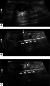
Superficial lesion sampling with an SLD. A 75‐year‐old woman presented for biopsy of a palpable mass. A, Preprocedural image shows an irregularly shaped hypoechoic mass (arrow) with posterior shadowing just deep to the dermis. The procedure was performed with a 16‐gauge Achieve needle. B, Subsequent image shows an introducer (arrowheads) placed below the target (arrow). C, The SLD with an open trough (arrowheads) is exposed by retracting the introducer cannula. D, Final image shows sampling of the lesion (arrow) with the cutting cannula (arrowheads) released.
Figure 8
Figure 8
Superficial lesion sampling after hydrodissection. A and B, Artist renderings of a breast biopsy shows lidocaine being instilled between the dermis and the target lesion (A) to create a safe zone between the dermis and the biopsy target (B) (Used with permission from the Mayo Foundation for Medical Education and Research; all rights reserved.)
Figure 9
Figure 9
Superficial lesion sampling with an SLD. A 75‐year‐old woman with a history of breast cancer presented for biopsy of a palpable lesion, which was performed with a 16‐gauge Achieve needle. A, Prebiopsy image shows a hypoechoic oval mass (arrow) with irregular margins superficially located with extension to the dermis. B, Initial procedural image shows the tip of the introducer (arrowheads) at the edge of the target (arrow). C, Subsequent image with lesion with in the open trough (arrowheads). D, Final image shows sampling of the lesion (arrow) with the cutting cannula released (arrowheads).
Figure 10
Figure 10
Sampling with the tandem‐needle technique. Artist rendering shows a cross‐sectional image of a VABB device with a mass being pulled (arrow) into the aperture. In this illustration, the hydrodissection needle is placed orthogonal to the biopsy needle and is located above the mass. (Used with permission from the Mayo Foundation for Medical Education and Research; all rights reserved.)
Figure 11
Figure 11
Superficial lesion sampling with a tandem‐needle technique. An 88‐year‐old woman with a new screen‐detected mass presented for US‐guided CB. A, Preprocedural image shows a complex solid and cystic mass (arrow) in the subareolar breast. A 9‐gauge petite Suros VABB device was selected for the procedure with a tandem‐needle approach. B, The superficial needle (arrow) is located above the targeted lesion, and the VABB device (arrowheads) is located below the target. The superficial tandem‐needle acts as a barrier and allows for the infusion of saline or lidocaine, which acts as a second shield for the dermis. C, During the procedure, saline or lidocaine is actively instilled above the target (arrow) as the VABB device (arrowheads) continues to take samples.
Figure 12
Figure 12
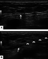
Deep lesion sampling with hydrodissection. A, Preprocedural image shows a hypoechoic oval mass (arrow) with slightly irregular margins immediately above the pectoralis muscle. B, A 25‐gauge spinal needle (arrowheads) is used to instill lidocaine (arrow) posterior to the lesion to lift the target off the pectoral muscle. Once the lesion has been elevated, either an SLD or a VABB device can be used to sample the mass. The histologic diagnosis was focal atypical ductal hyperplasia in a background of a fibroadenomatoid nodule.
Figure 13
Figure 13
A 43‐year‐old woman after mastectomy with implant reconstruction presented for biopsy of a right breast mass. A, Preprocedural image shows a hypoechoic circumscribed mass immediately adjacent to the implant (arrow) and within a few millimeters of the skin. B, A CB could not be performed, but fine‐needle aspiration was performed with 25‐gauge needles (arrowheads). Histologic findings were most consistent with benign scar tissue. The fine‐needle aspiration cytologic examination showed no evidence of malignancy.
Figure 14
Figure 14
Open‐trough technique for a lesion on an implant. A 52‐year‐old woman with a history of right nipple‐sparing mastectomy and implant reconstruction presented for biopsy of a painful mass right breast mass. A, Preprocedural image shows an oval mass with irregular margins (arrowhead) located between the dermis and the implant. An open‐trough technique with a coaxial needle was performed. B, Subsequent image shows placement of the introducer (arrowheads) below the target (arrow). C, The outer introducer is retracted, revealing the SLD with the trough (arrowheads) positioned below the target (arrow). D, Final image shows the outer cutting cannula deployed (arrowheads) sampling through the lesion (arrow). The histologic diagnosis was invasive ductal carcinoma.
Figure 15
Figure 15
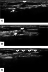
Mass adherent to an implant capsule in a 44‐year‐old woman with an area of palpable concern in the left breast. A, The mass (arrowhead) is immediately adjacent to the anterior surface of the patient's implant in the region of the valve (arrow) of the saline implant. The procedure was performed with a 16‐gauge Temno needle (Merit Medical, Jordan, UT). B, The needle is advanced through the superficial (arrowheads) aspect of the target (arrow). C, The outer cutting cannula (arrowheads) is deployed, sampling the superficial aspect of the mass. The histologic diagnosis was grade I invasive ductal carcinoma.
Figure 16
Figure 16
Implant and hydrodissection. A, Artist rendering shows how hydrodissection can be used to lift a lesion off an implant. B, Then lidocaine can be instilled superficially to the mass to create a safe zone between the lesion and the skin surface. C, These steps created room for the biopsy device. (Used with permission from the Mayo Foundation for Medical Education and Research; all rights reserved.)
Figure 17
Figure 17
Mastectomy and hydrodissection. A, Artist rendering shows how hydrodissection can be used to lift a lesion off the pectoral muscle. B, Then lidocaine can be instilled superficially to the mass to create a safe zone between the lesion and the skin surface. C, Having lidocaine above and below the lesion creates enough space for the use of a biopsy device. (Used with permission from the Mayo Foundation for Medical Education and Research; all rights reserved.)
Figure 18
Figure 18
Lesion seen in a single plane in a 61‐year‐old woman with asymmetry in the left breast and a vague hypoechoic suspicious left breast lesion identified on an outside US examination. A and B, Preprocedural grayscale images show a vague area of shadowing (arrows) at the 12‐o'clock position, with an adjacent cyst (arrowhead) seen in the longitudinal plane (A). Biopsy was performed with a 14‐gauge Achieve device; samples were taken in 2 stages. C, The first stage shows the open trough (arrowheads) in the region of the target (arrow). D, Orthogonal image shows the needle (arrow) at the edge of the vague lesion; the cyst (arrowhead) was used as a landmark. E, In this plane, the needle can be moved to the desired location (asterisk). The histologic diagnosis was ductal carcinoma in situ, cribriform and solid types, intermediate nuclear grade.
Figure 19
Figure 19
Biopsy entering the same tract. A 54‐year‐old woman with a new screen‐detected mass presented for US‐guided biopsy performed with a 14‐gauge Achieve needle. A, Preprocedural image shows an indistinct hypoechoic mass with posterior acoustic shadowing (arrow). B, Initial sampling shows the needle (arrowheads) traversing the mass (arrow). C, Orthogonal image shows the needle (arrowhead) centered within the mass. Subsequent sampling results in small cores because the biopsy device continues to enter the same tract. D, After the first stage of sampling, rotate the device 90° (open pie shape) in the tract to sample a different part of the lesion. E, Continue to rotate the device (open pie shape) for subsequent samples. The histologic diagnosis was pseudoangiomatous stromal hyperplasia.
Figure 20
Figure 20
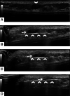
Mobile lesion. A 95‐year‐old woman unable to raise her arm above her head presented with a palpable right breast mass. A, Preprocedural image shows a hypoechoic mass (arrow) with internal cystic components. The mass and breast tissue were highly mobile, and the patient was unable to raise her arm above her head for proper positioning. B, An introducer (arrowheads) was used to immobilize the lesion, passing through the lesion, which was fairly soft. C, The biopsy was then performed with an open trough (arrow), and the introducer (arrowheads) was slowly retracted. D, With the introducer retracted, the trough of the SLD (arrowhead) can be easily visualized. The histologic diagnosis was invasive ductal carcinoma with mucinous features, Nottingham grade I (of III).

References

    1. Parker SH, Jobe WE, Dennis MA, et al. US‐guided automated large‐core breast biopsy. Radiology 1993; 187:507–511. - PubMed
    1. Parker SH, Burbank F, Jackman RJ, et al. Percutaneous large‐core breast biopsy: a multi‐institutional study. Radiology 1994; 193:359–364. - PubMed
    1. Caruncho MV, Branas F. Mirror pain as a complication of large core biopsy of the breast. Breast J 2006; 12:393–395. - PubMed
    1. Harvey JA, Moran RE. US‐guided core needle biopsy of the breast: technique and pitfalls. Radiographics 1998; 18:867–877. - PubMed
    1. Brenner RJ, Fajardo L, Fisher PR, et al. Percutaneous core biopsy of the breast: effect of operator experience and number of samples on diagnostic accuracy. AJR Am J Roentgenol 1996; 166:341–346. - PubMed