. 2020 Sep 28;18(10):495.
doi: 10.3390/md18100495.

Astaxanthin Inhibits p70 S6 Kinase 1 Activity to Sensitize Insulin Signaling

Affiliations

Astaxanthin Inhibits p70 S6 Kinase 1 Activity to Sensitize Insulin Signaling

Chunmei Li et al. Mar Drugs. .

Abstract

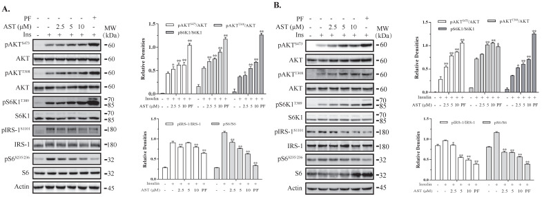
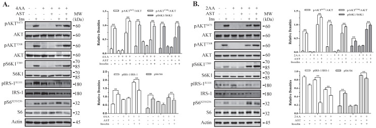
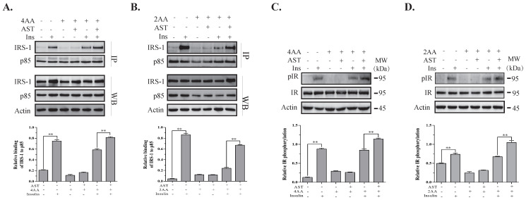
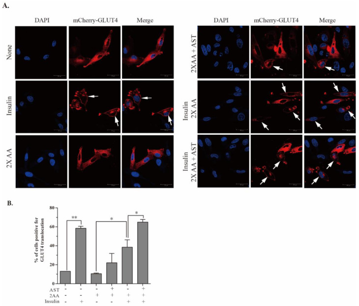
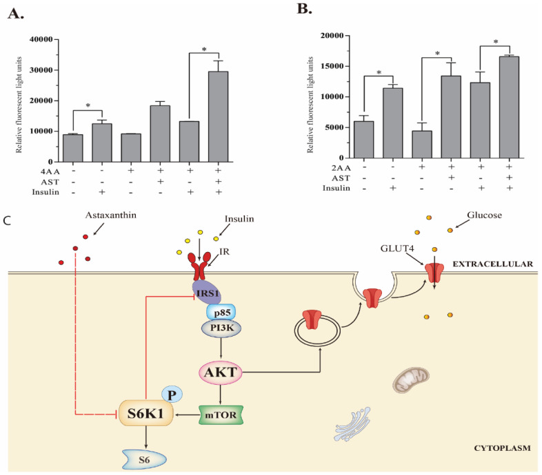
Astaxanthin (AST) is a carotenoid with therapeutic values on hyperglycemia and diabetic complications. The mechanisms of action of AST remain incompletely understood. p70 S6 kinase 1 (S6K1) is a serine/threonine kinase that phosphorylates insulin receptor substrate 1 (IRS-1)S1101 and desensitizes the insulin receptor (IR). Our present study aims to determine if AST improves glucose metabolisms by targeting S6K1. Western blot analysis revealed that AST inhibited the phosphorylation of two S6K1 substrates, S6S235/236 and IRS-1S1101, but enhanced the phosphorylation of AKTT308, AKTS473, and S6K1T389 by feedback activation of the phosphatidylinositol-3 (PI-3) kinase in 3T3-L1 adipocytes and L6 myotubes. In vitro kinase assays revealed that AST inhibited S6K1 activity with an IC50 value of approximately 13.8 μM. AST increased insulin-induced IR tyrosine phosphorylation and IRS-1 binding to the p85 subunit of PI-3 kinase. Confocal microscopy revealed that AST increased the translocation of the glucose transporter 4 (GLUT4) to the plasma membrane in L6 cells. Glucose uptake assays using a fluorescent dye, 2-NBDG (2-N-(Nitrobenz-2-oxa-1,3-diazol-4-yl)amino)-2-deoxyglucose), revealed that AST increased glucose uptake in 3T3-L1 adipocytes and L6 myotubes under insulin resistance conditions. Our study identifies S6K1 as a previously unrecognized molecular target of AST and provides novel insights into the mechanisms of action of AST on IR sensitization.

Keywords: AKT; GLUT4; PI-3 kinase; S6K1; astaxanthin; glucose uptake; insulin receptor.


Conflict of interest statement

The authors declare no conflict of interest with the contents of this article.

Figures

Figure 1
Figure 1
Astaxanthin (AST) induces feedback activation of the PI-3 kinase pathway. 3T3-L1 adipocytes (A) and L6 myotubes (B) were starved of serum overnight and for 2 h, respectively. After preincubation in the absence or presence of the indicated concentrations of AST or PF-4708671 (10 μM) for 4 h, the cells were left unstimulated or stimulated with (100 nM) for 10 min. Cell lysates were prepared and analyzed for the phosphorylation of IRSS1101, AKTT308, AKTS473, S6K1T389, and S6S235/236 with their specific antibodies and re-probed with antibodies against total proteins. The density of the phosphorylated protein bands was analyzed by using NIH Image-J software and normalized by the arbitrary units of total proteins. Data are expressed as the mean ± standard derivation (SD) of three experiments. * p < 0.05, ** p < 0.01, compared to the insulin-stimulated controls.
Figure 2
Figure 2
AST inhibits S6K1 activity. (A) The ability of AST to inhibit S6K1 activity was assayed by an ADP-GloTM kinase kit. (B) Chemical structure of AST; in vitro S6K1 kinase assay. (C) The predictive model of S6K1 (red: β-folds; green: α-helices). The model was built using a Swiss-Model Server. (D) Ramachandran plot of the S6K1 3D model. The Ramachandran plot was performed with a PROCHECK program. (E) AST binds to S6K1 in the ATP binding pocket (orange). (F) Interactions between AST and S6K1. AST binds to S6K1 by two hydrogen bonds, and the amino acid residues involved in the formation of hydrogen bonds were Glu173 and Arg298. The molecular docking diagram was performed by using MGLTools 1.5.6., PyMOL 1.2, and AutoDock Vina 1.1.2. ** p < 0.01, compared to the untreated control.
Figure 3
Figure 3
AST induces feedback activation of the PI-3 kinase pathway under insulin resistance conditions. 3T3-L1 adipocytes (A) and L6 myotubes (B) were starved of serum overnight and for 2 h, respectively. These cells were incubated for 4 h in an essential balanced salt solution (EBSS) in the absence or presence of AST (10 μM), minus or plus high concentrations of amino acids (AA). The cells were then left unstimulated or stimulated with insulin (100 nM) for 10 min. Cell lysates were prepared and analyzed for the phosphorylation of IRSS1101, AKTT308, AKTS473, S6K1T389, and S6S235/236 with their specific antibodies and then re-probed with antibodies against their total proteins. The density of the phosphorylated protein bands was analyzed by using NIH Image-J software and normalized by the arbitrary units of total proteins. Data represent the mean ± SD of three experiments. ** p < 0.01, compared to insulin-stimulated controls.
Figure 4
Figure 4
AST sensitizes the insulin receptor (IR). 3T3-L1 adipocytes (A,C) and L6 myotubes (B,D) were starved of serum overnight and 2 h, respectively. 3T3-L1 adipocytes and L6 myotubes were then incubated for 4 h in EBSS or EBSS containing 4× and 2× AA, respectively, and in the absence or presence of 10 μM AST. The cells were then left unstimulated or stimulated with insulin (100 nM) for 10 min. Cell lysates were immunoprecipitated (A,B) with an anti-p85 antibody and probed with anti-p85 and anti-insulin receptor substrate 1 (IRS-1) antibodies or analyzed for IRY1146 phosphorylation (C,D). The density of the phosphorylated protein bands was analyzed by using NIH Image-J software and normalized by the arbitrary units of total proteins, ** p < 0.01. Data are expressed as the mean ± SD of three independent experiments.
Figure 5
Figure 5
AST promotes GLUT4 translocation to the plasma membrane. (A) L6 cells transiently transfected with mCherry-GLUT4-myc were left untreated or treated with AST (10 μM) for 2 h in EBSS (containing 5% FBS) minus or plus 2× AA. The cells were then left unstimulated or stimulated with insulin (100 nM) for 10 min. The cells were fixed and stained with 4,6-diamidino-2-phenylindole (DAPI). mCherry-tagged GLUT4 red fluorescence was visualized under a Leica confocal microscope. GLUT4 translocation to the cytoplasm membrane was marked with arrows. (B) Quantification of GLUT4 translocation to the plasma membrane. The data represent the mean ± SD from one of three experiments with similar results. * p < 0.05; ** p < 0.01.
Figure 6
Figure 6
AST improves glucose uptake. 3T3-L1 adipocytes (A) and L6 myotubes (B) were starved of serum for 12 and 2 h, respectively. The cells were then incubated in the absence or presence of 4× or 2× AA or plus AST (10 μM) in EBSS for 1 h. The cells were pulsed with 2-NBDG (50 μM) in darkness for another 1 h and then left unstimulated or stimulated with insulin (100 nM) for 10 min. After aspirating the media, fluorescent signals were read in a microplate reader with excitation and emission wavelengths of 485 and 535 nm, respectively. Data represent the mean ± SD of one experiment in triplicate. * p < 0.05. (C) Schematic model depicting the mechanism of action of AST. S6K1 phosphorylates IRS-1S1101 and attenuates the activation of the PI-3 kinase pathway. AST inhibits S6K1, leading to decreased IRS-1S1101 phosphorylation and the sensitization of the PI-3 kinase pathway. AKT activation enhances GLUT4 translocation into the plasma membrane and improves glucose uptake.

Similar articles

Cited by

References

    1. Zimmet P., Alberti K.G., Magliano D.J., Bennett P.H. Diabetes mellitus statistics on prevalence and mortality: Facts and fallacies. Nat. Rev. Endocrinol. 2016;12:616–622. doi: 10.1038/nrendo.2016.105. - DOI - PubMed
    1. Zimmet P.Z., Magliano D.J., Herman W.H., Shaw J.E. Diabetes: A 21st century challenge. Lancet Diabetes Endocrinol. 2014;2:56–64. doi: 10.1016/S2213-8587(13)70112-8. - DOI - PubMed
    1. Saeedi P., Salpea P., Karuranga S., Petersohn I., Malanda B., Gregg E.W., Unwin N., Wild S.H., Williams R. Mortality attributable to diabetes in 20–79 years old adults, 2019 estimates: Results from the International Diabetes Federation Diabetes Atlas, 9th edition. Diabetes Res. Clin. Pract. 2020;162:108086. doi: 10.1016/j.diabres.2020.108086. - DOI - PubMed
    1. Roden M., Shulman G.I. The integrative biology of type 2 diabetes. Nature. 2019;576:51–60. doi: 10.1038/s41586-019-1797-8. - DOI - PubMed
    1. Saeedi P., Petersohn I., Salpea P., Malanda B., Karuranga S., Unwin N., Colagiuri S., Guariguata L., Motala A.A., Ogurtsova K., et al. Global and regional diabetes prevalence estimates for 2019 and projections for 2030 and 2045: Results from the International Diabetes Federation Diabetes Atlas, 9 (th) edition. Diabetes Res. Clin. Pract. 2019;157:107843. doi: 10.1016/j.diabres.2019.107843. - DOI - PubMed