. 2020 Sep 28;11(10):1141.
doi: 10.3390/genes11101141.

Limited Availability of General Co-Repressors Uncovered in an Overexpression Context during Wing Venation in Drosophila melanogaster

Affiliations

Limited Availability of General Co-Repressors Uncovered in an Overexpression Context during Wing Venation in Drosophila melanogaster

Anja C Nagel et al. Genes (Basel). .

Abstract

Cell fate is determined by the coordinated activity of different pathways, including the conserved Notch pathway. Activation of Notch results in the transcription of Notch targets that are otherwise silenced by repressor complexes. In Drosophila, the repressor complex comprises the transcription factor Suppressor of Hairless (Su(H)) bound to the Notch antagonist Hairless (H) and the general co-repressors Groucho (Gro) and C-terminal binding protein (CtBP). The latter two are shared by different repressors from numerous pathways, raising the possibility that they are rate-limiting. We noted that the overexpression during wing development of H mutants HdNT and HLD compromised in Su(H)-binding induced ectopic veins. On the basis of the role of H as Notch antagonist, overexpression of Su(H)-binding defective H isoforms should be without consequence, implying different mechanisms but repression of Notch signaling activity. Perhaps excess H protein curbs general co-repressor availability. Supporting this model, nearly normal wings developed upon overexpression of H mutant isoforms that bound neither Su(H) nor co-repressor Gro and CtBP. Excessive H protein appeared to sequester general co-repressors, resulting in specific vein defects, indicating their limited availability during wing vein development. In conclusion, interpretation of overexpression phenotypes requires careful consideration of possible dominant negative effects from interception of limiting factors.

Keywords: C-terminal binding protein; Drosophila; Groucho; Hairless; Notch signaling; Suppressor of Hairless; co-repressor; repressor complex; sequestration; wing venation.


Conflict of interest statement

The authors declare no conflict of interest. The funders had no role in the design of the study; in the collection, analyses, or interpretation of data; in the writing of the manuscript; or in the decision to publish the results.

Figures
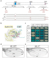
Figure 1
Figure 1
Hairless mutants affecting Su(H) and co-repressor binding. (a) Scheme of the H protein with the Su(H)-binding domain (SBD, cyan), the Gro-binding domain (GBD, blue), and the CtBP-binding domain (CBD, purple) indicated. Numbering is according to [55]. The C2 deletion is shown in grey. The NT-box is part of the SBD and contains amino acids in direct contact with Su(H) (bold) [53]. Residues involved in the binding of Gro or CtBP are likewise shown in bold [9]. Amino acid replacements are indicated in red: HLD impairs the interaction with Su(H), H*G prevents binding to Gro, and H*C to CtBP [9,40]. H* mutations used in this study are depicted underneath. (b) Structure of the H-Su(H) repressor complex (PDB ID: 5E24) [53]. The picture focuses on the C-terminal domain (CTD) of Su(H) in yellow with amino acids important for H binding shown in pink (L343, L445, L514). The domain in H interacting with Su(H) (H NT, aa 232–269) is shown in cyan; L235 is labelled. (c) Pairwise protein–protein interactions assayed by yeast two-hybrid: H wild type or H* mutant constructs in pEG vector served as bait, whereas Su(H) in pJG vector, and either Gro or CtBP in VP16 vector served as prey. Blue colonies reveal pairwise protein interactions, whereas white colonies show respective lack of interaction. Empty vectors served as controls. (d) Control wing of a female fly: longitudinal veins L1-L5 and anterior and posterior cross veins (acv, pcv) are labelled (QE-Gal4::UAS-GFP). (e) Wing of a female fly overexpressing HC2 in the wing blade (QE-Gal4::UAS-H-C2). Note plexus of ectopic veins along the L2 and L5 veins (arrowheads).
Figure 2
Figure 2
Overexpression of H protein variants in the central wing anlagen using omb-Gal4. (ad’) The transgenes, as indicated, were overexpressed in the central part of the wing anlagen using omb-Gal4 at 20 °C. Female wings are shown. (a) UAS-GFP served as control. (b) Wing incisions (asterisk), proliferation defects, and vein thickening (arrowhead) were typical consequences of an inhibition of Notch activity resulting from overexpression of wild type H protein. (c,d) Overexpression of either HdNT or HLD mutants did not affect the wing margin. Note, however, ectopic wing vein formation (arrowheads mark examples) and slightly reduced wing size. (a’d’) Combined overexpression of Su(H) and H variants. (a’) Whereas the overexpression Su(H) within the wing anlagen resulted in crumbled wings, a combination with wild type H was lethal as a consequence of excessive repressor complex formation (b’) [9,40,52,53]. In combination with either (c’) HdNT or (d’) HLD, however, the wings resembled those of the sole Su(H) overexpression.
Figure 3
Figure 3
Overexpression of H protein variants in the presumptive wing tissue using QE-Gal4. (ad’) UAS-H* transgenes, as indicated, were overexpressed in the presumptive wing tissue using QE-Gal4 at 18 °C. (a) UAS-GFP served as control; longitudinal veins are numbered. (b) Overexpression of wild type H protein affected wing size and tissue adhesion (asterisk). The most prominent phenotype, however, was a thickening of veins and formation of ectopic veinlets (arrowheads point to examples). (c,d) The latter was also observed upon the overexpression of HdNT (c) or HLD (d) variants compromised in Su(H) binding (arrowheads point to examples). (a’d’) UAS-Su(H) plus the respective UAS-H* transgenes were overexpressed in combination in the developing wing field with QE-Gal4. (a’) Su(H) overexpression alone caused a loss of distal parts of longitudinal veins (arrow). (b’) A combined overexpression of UAS-Su(H) with UAS-H resulted in a transformation of intervein to vein tissue, as well as the emergence of ectopic bristle organs (open arrow, see enlargement). Combination of Su(H) with either HdNT (c’) or HLD (d’), however, gave a mixed phenotype with erased distal wing veins (arrow) plus ectopic veinlets (arrowheads), indicating lack of repressor complex formation. Female wings are shown.
Figure 4
Figure 4
Loss of co-repressor binding inhibited ectopic vein formation by H protein overexpressed in the central wing anlagen. (ad’) UAS-H* transgenes, as indicated, were overexpressed in the central part of the wing anlagen using omb-Gal4 at 25 °C. Note enhancement of phenotypes at the higher temperature (compare with Figure 2). (a) UAS-GFP served as control. (b) Wing incisions (asterisk) are very deep when H is induced. Overexpression of either HdNT (c) or HLD (d) isoforms, however, induced a lattice of veinlets (arrowheads point to examples). The margin was unaffected, however. (b’d’) Overexpression of the respective H* protein isoforms lacking Gro and CtBP co-repressor binding in addition to compromised Su(H) binding (*GC; see Figure 1). (b’) Loss of co-repressors impeded H repressor activity. Accordingly, mild wing incisions were seen when UAS-H*GC was overexpressed. In contrast, overexpression of either HdNT*GC (c) or HLD*GC (d) variants affecting both Su(H) and co-repressor binding neither affected wing margin nor vein development. Female wings are shown.
Figure 5
Figure 5
Overexpression of H* protein lacking Su(H) and co-repressor binding ability in the developing wing blade. (ad’) UAS-H* transgenes, as indicated, were induced at 25 °C with QE-Gal4 in presumptive wing tissue. (a) UAS-GFP served as control. (b) Whereas wild type H induced wing blisters (asterisk) and thickened veins (arrowheads), HdNT*GC (c) and HLD*GC (d) isoforms induced ectopic veinlets (arrowheads point to examples). (b’d’) Overexpression of the respective H* protein isoforms that in addition lack Gro and CtBP co-repressor binding (*GC; see Figure 1a). Overall, the wings resembled the control. However, minute veinlets may appear in the developing wing blade. Examples are shown for (c’) HdNT*GC and (d’) HLD*GC (arrows). Female wings are shown.
Figure 6
Figure 6
Model explaining the ectopic veinlet phenotype. (a) General co-repressors Gro and CtBP are shared by numerous regulators acting in different signaling pathways, including Brinker (Brk), Capicua (Cic), and E(spl) and Hairy (HES)-type transcription factors. Moreover, Hairless recruits both co-repressors for Su(H)-H repressor complex formation. (b) Overexpression of H* variants defective for Su(H)-binding does not influence Notch target gene expression any longer. H* variants, however, may compete with other transcription factors for the binding to co-repressors, thereby limiting their availability. As a consequence, H* overexpression may intercept with signaling pathways involved in vein formation that are specifically sensitive towards a mitigation of co-repressor availability.

Similar articles

Cited by

References

    1. Bray S.J. Notch signalling: A simple pathway becomes complex. Nat. Rev. Mol. Biol. 2006;7:678–689. doi: 10.1038/nrm2009. - DOI - PubMed
    1. Louvi A., Artavanis-Tsakonas S. Notch and disease: A growing field. Sem. Cell Dev. Biol. 2012;23:473–480. doi: 10.1016/j.semcdb.2012.02.005. - DOI - PMC - PubMed
    1. Kovall R.A., Blacklow S.C. Mechanistic insights into Notch receptor signaling from structural and biochemical studies. Curr. Top. Dev. Biol. 2010;92:31–71. doi: 10.1016/S0070-2153(10)92002-4. - DOI - PubMed
    1. Wang H., Zang C., Liu X.S., Aster J.C. The role of Notch receptors in transcriptional regulation. J. Cell Physiol. 2015;230:982–988. doi: 10.1002/jcp.24872. - DOI - PMC - PubMed
    1. Bray S.J. Notch signalling in context. Nat. Rev. Mol. Biol. 2016;17:722–735. doi: 10.1038/nrm.2016.94. - DOI - PubMed

Publication types

MeSH terms

LinkOut - more resources