. 2020 Sep 28;21(19):7160.
doi: 10.3390/ijms21197160.

Caffeoyl-Prolyl-Histidine Amide Inhibits Fyn and Alleviates Atopic Dermatitis-Like Phenotypes via Suppression of NF-κB Activation

Affiliations

Caffeoyl-Prolyl-Histidine Amide Inhibits Fyn and Alleviates Atopic Dermatitis-Like Phenotypes via Suppression of NF-κB Activation

Hayan Jeong et al. Int J Mol Sci. .

Abstract

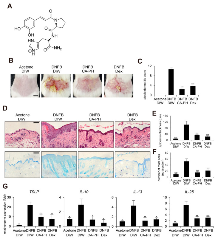
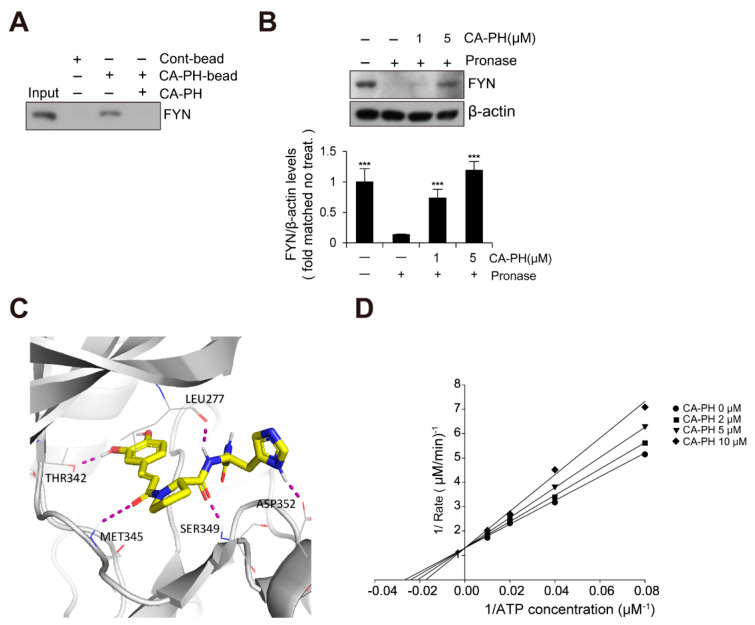
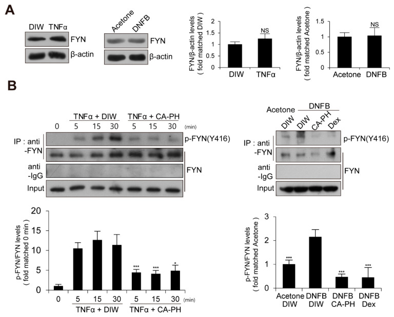
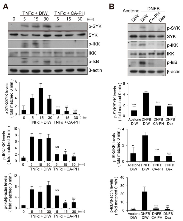
Caffeic acid (CA) is produced from a variety of plants and has diverse biological functions, including anti-inflammation activity. It has been recently demonstrated that caffeoyl-prolyl-histidine amide (CA-PH), which is CA conjugated with proline-histidine dipeptide, relieves atopic dermatitis (AD)-like phenotypes in mouse. In this study, we investigated the molecular mechanism underlying CA-PH-mediated alleviation of AD-like phenotypes using cell line and AD mouse models. We confirmed that CA-PH suppresses AD-like phenotypes, such as increased epidermal thickening, infiltration of mast cells, and dysregulated gene expression of cytokines. CA-PH suppressed up-regulation of cytokine expression through inhibition of nuclear translocation of NF-κB. Using a CA-PH affinity pull-down assay, we found that CA-PH binds to Fyn. In silico molecular docking and enzyme kinetic studies revealed that CA-PH binds to the ATP binding site and inhibits Fyn competitively with ATP. CA-PH further suppressed spleen tyrosine kinase (SYK)/inhibitor of nuclear factor kappa B kinase (IKK)/inhibitor of nuclear factor kappa B (IκB) signaling, which is required for nuclear factor kappa-light-chain-enhancer of activated B cells (NF-κB) activation. In addition, chronic application of CA-PH, in contrast with that of glucocorticoids, did not induce up-regulation of regulated in development and DNA damage response 1 (REDD1), reduction of mammalian target of rapamycin (mTOR) signaling, or skin atrophy. Thus, our study suggests that CA-PH treatment may help to reduce skin inflammation via down-regulation of NF-κB activation, and Fyn may be a new therapeutic target of inflammatory skin diseases, such as AD.

Keywords: CA-PH; Fyn; NF-κB; SYK; atopic dermatitis; skin atrophy.


Conflict of interest statement

The authors declare no conflict of interest. The funders had no role in the design of the study; in the collection, analyses, or interpretation of data; in the writing of the manuscript, or in the decision to publish the results.

Figures

Figure 1
Figure 1
CA-PH alleviates AD-like phenotypes induced by DNFB treatment. Topical application of CA-PH alleviates AD-like phenotypes induced by DNFB treatment in mouse skin. Dex was used as the positive control. Acetone and DIW were used as solvents for DNFB and CA-PH, respectively. (A) Structure of CA-PH. (B) After mice were sensitized with DNFB for 7 days, DNFB was further topically applied to the shaved dorsal skin with or without CA-PH for 12 days (n = 6/group). Mice were photographed and skin tissues were harvested. Representative images are shown. Scale bar, 0.5 cm (C) CA-PH alleviates the severity of AD-like phenotypes induced by DNFB treatment in mouse skin. The severity of dermatitis was determined by a scoring index of AD (see Material and Methods). (D) Tissue sections from the back skin were stained with hematoxylin and eosin (H&E) or toluidine blue (TB). Representative images are shown. Scale bar, 50 μm. (E) CA-PH suppresses the increased epidermis thickness induced by DNFB treatment in mouse skin. Epidermis thicknesses were measured (n = 6/group). (F) CA-PH suppresses infiltration of mast cells induced by DNFB treatment in mouse skin. The number of mast cells was counted from five randomly selected low-power fields (n = 6/group). (G) CA-PH suppresses DNFB-induced up-regulation of cytokine expression in mouse skin. Transcripts of TSLP, IL-10, IL-13, IL-25, and RPLP0 from mouse skin were quantified using real-time PCR (n = 6/group). All data represent means ± S.E.M. Significance values were * p ≤ 0.05, ** p ≤ 0.01, and *** p ≤ 0.005. Dex, dexamethasone; DIW, deionized water.
Figure 2
Figure 2
CA-PH inhibits NF-κB signaling in TNFα-treated keratinocytes. (A) CA-PH suppresses DNFB-induced up-regulation of cytokine expression in TNFα-treated HaCaT keratinocytes. Dex was used as the positive control. DIW was used as a solvent for TNFα and CA-PH. Transcripts of TSLP, IL-10, IL-13, IL-25, and RPLP0 were quantified using real-time PCR (n = 5). (B) CA-PH suppresses the activity of the luciferase reporter containing NF-κB binding sites in TNFα-treated HaCaT keratinocytes. Dex was used as the positive control. DIW was used as the solvent for TNFα and CA-PH. Following transfection of the luciferase reporter vector containing NF-κB binding sites and the control Renilla luciferase expression vector into HaCaT keratinocytes, luciferase activity was measured in cell extracts (n = 5). Reporter activity is represented as the fold activation relative to Renilla luciferase activity. (C) CA-PH inhibits nuclear translocation of NF-κB p65 in TNFα-treated HaCaT keratinocytes. Dex was used as the positive control. DIW was used as the solvent for TNFα and CA-PH. Cells were immunostained with anti-NF-κB p65 antibody (n = 5). Nuclei were identified using DAPI staining. Representative images are shown. Scale bar, 20 μm. (D) TNFα induces the occupancy of NF-κB p65 at the NF-κB binding sites of TSLP, IL-10, IL-13, and IL-25 gene promoters in HaCaT keratinocytes. However, CA-PH decreases the occupancy of NF-κB p65 in the gene promoters in HaCaT keratinocytes treated with TNFα. Dex was used as positive control. DIW was used as solvent for TNFα and CA-PH. ChIP assay was performed using anti-NF-κB p65 antibody (n = 5). The occupancy of each protein was quantified using real-time PCR in the gene promoters encompassing the NF-κB binding sites. All data represent means ± S.E.M. Significance values were * p ≤ 0.05, ** p ≤ 0.01, and *** p ≤ 0.005. Dex, dexamethasone; DIW, deionized water; TNFα, tumor necrosis factor alpha.
Figure 3
Figure 3
CA-PH binds to and inhibits Fyn. (A) CA-PH interacts with Fyn. CA-PH beads were prepared by binding of CA-PH to polystyrene beads (See Materials and Methods). CA-PH-bound beads were mixed with protein extract from HaCaT cells. After extensive washing, Western blotting was performed using anti-Fyn antibody. Free excess CA-PH was added as a competitor. (B) The drug affinity responsive target stability (DARTS) assay reveals that CA-PH-bound Fyn is resistant to protease digestion. Protein extract from HaCaT cells was mixed with CA-PH and treated with Pronase at room temperature. Western blot analysis was performed using anti-Fyn antibody. As a loading control, the reaction mixture without incubation was immunoblotted with anti-β actin antibody. Representative images are shown. Data were quantitatively assessed and are additionally depicted in graphs (n = 3). (C) Molecular docking shows that CA-PH binds to the ATP binding site of Fyn (∆G = −8.74 kcal/mol). CA-PH can form 5 hydrogen bonds with Leu277, Thr342, Met345, Ser349, and Asp352 residues within the ATP binding site of the catalytic domain. (D) CA-PH inhibits Fyn competitively with ATP. Double-reciprocal plots of the inhibitory activity of CA-PH. Fyn kinase activity was measured at the indicated concentrations of CA-PH and ATP (n = 3). The reciprocal velocity was plotted versus 1/(ATP). Km = 40.4 μM, Ki = 16 μM. Fyn kinase assay was performed using a Kinase Enzyme System. For enzyme kinetics, the Lineweaver–Burk method was applied. All data represent means ± S.E.M. Significance value was *** p ≤ 0.005.
Figure 4
Figure 4
Activation of Fyn in TNFα-treated keratinocytes and DNFB-induced AD-like mouse skin. (A) Expression levels of Fyn are not changed in TNFα-treated HaCaT keratinocytes or DNFB-treated mouse skin. DIW and acetone were used as solvents for TNFα and DNFB, respectively. Western blot analysis was performed using anti-Fyn antibody. As a loading control, anti-β actin antibody was used. Representative images are shown. Data were quantitatively assessed and additionally depicted in graphs (n = 3). (B) Increased kinase activity of Fyn in TNFα-treated HaCaT keratinocytes or DNFB-treated mouse skin. Fyn was immunoprecipitated with anti-Fyn antibody and immunoblotted with anti-phospho SRC (Y416) antibody. Data were quantitatively assessed and are additionally depicted in graphs (n = 3). All data represent mean ± S.E.M. Significance values were * p ≤ 0.05, and *** p ≤ 0.005. Dex, dexamethasone; DIW, deionized water; TNFα, tumor necrosis factor alpha; NS, no significance.
Figure 5
Figure 5
CA-PH suppresses NF-κB signaling in TNFα-treated keratinocytes and DNFB-induced AD-like mouse skin. (A,B) CA-PH inhibits phosphorylation of SYK, IKK, and IκB in TNFα-treated HaCaT keratinocytes or DNFB-treated mouse skin. Western blot analysis was performed using anti-SYK, anti-phospho SYK (Y525/526), anti-IKK, anti-phospho IKK (S176/177), and anti-phospho IκBα (S32) antibodies. As a loading control, Western blot analysis was performed using anti-β actin antibody. DIW was used as the solvent for TNFα and CA-PH. Acetone was used as the solvent for DNFB. Data were quantitatively assessed and are additionally depicted in graphs (n = 5). Representative images are shown. All data represent mean ± S.E.M. Significance values were * p ≤ 0.05, ** p ≤ 0.01, and *** p ≤ 0.005. Dex, dexamethasone; DIW, deionized water; TNFα, tumor necrosis factor alpha; NS, no significance.
Figure 6
Figure 6
Chronic treatment of CA-PH does not induce skin atrophy. (A,B) Dex up-regulates regulated in development and DNA damage response 1 (REDD1) gene expression and inhibits mTOR signaling. However, CA-PH does not induce increased expression of REDD1 and inhibit mTOR signaling in HaCaT keratinocytes or mouse skin. Transcripts of REDD1 and RPLP0 were quantified using real-time PCR (n = 5). Western blot analysis was performed using anti-REDD1, anti-phospho mTOR (S2448), anti-phospho p70S6K (T389), anti-phospho 4EBP1 (S65), and anti-β actin antibodies. DIW was used as the solvent for CA-PH. Data were quantitatively assessed and are additionally depicted in graphs (n = 5). Representative images are shown. (C,D) Chronic application of dexamethasone (Dex) induces skin atrophy. However, CA-PH does not induce skin atrophy. Dexamethasone or CA-PH was treated for 1 week in mouse skin. DIW was used as the solvent for CA-PH. Tissue sections from the skin were stained with hematoxylin and eosin (H&E). Representative images are shown. Scale bar, 100 μm. Epidermis thicknesses were measured (n = 6/group). All data represent means ± S.E.M. Significance values were * p ≤ 0.05, ** p ≤ 0.01, and *** p ≤ 0.005. Dex, dexamethasone; DIW, deionized water.

Similar articles

Cited by

References

    1. Janbaz K., Saeed S., Gilani A. Studies on the protective effects of caffeic acid and quercetin on chemical-induced hepatotoxicity in rodents. Phytomedicine. 2004;11:424–430. doi: 10.1016/j.phymed.2003.05.002. - DOI - PubMed
    1. Yamada Y., Yasui H., Sakurai H. Suppressive Effect of Caffeic Acid and its Derivatives on the Generation of UVA-induced Reactive Oxygen Species in the Skin of Hairless Mice and Pharmacokinetic Analysis on Organ Distribution of Caffeic Acid in ddY Mice. Photochem. Photobiol. 2006;82:1668. doi: 10.1111/j.1751-1097.2006.tb09829.x. - DOI - PubMed
    1. Genaro-Mattos T.C., Maurício Â.Q., Rettori D., Alonso A., Hermes-Lima M. Antioxidant Activity of Caffeic Acid against Iron-Induced Free Radical Generation—A Chemical Approach. PLoS ONE. 2015;10:e0129963. doi: 10.1371/journal.pone.0129963. - DOI - PMC - PubMed
    1. Lima V.N., Oliveira-Tintino C.D., Santos E.S., Morais L.P., Tintino S.R., Freitas T.S., Geraldo Y.S., Pereira R.L., Cruz R.P., Menezes I.R., et al. Antimicrobial and enhancement of the antibiotic activity by phenolic compounds: Gallic acid, caffeic acid and pyrogallol. Microb. Pathog. 2016;99:56–61. doi: 10.1016/j.micpath.2016.08.004. - DOI - PubMed
    1. Hirose M., Takesada Y., Tanaka H., Tamano S., Kato T., Shirai T. Carcinogenicity of antioxidants BHA, caffeic acid, sesamol, 4-methoxyphenol and catechol at low doses, either alone or in combination, and modulation of their effects in a rat medium-term multi-organ carcinogenesis model. Carcinogenesis. 1998;19:207–212. doi: 10.1093/carcin/19.1.207. - DOI - PubMed

MeSH terms

LinkOut - more resources