. 2020 Sep 28;8(10):386.
doi: 10.3390/biomedicines8100386.

Thymoquinone-Loaded Polymeric Films and Hydrogels for Bacterial Disinfection and Wound Healing

Affiliations

Thymoquinone-Loaded Polymeric Films and Hydrogels for Bacterial Disinfection and Wound Healing

Anika Haq et al. Biomedicines. .

Abstract

The purpose of this study was to synthesize and characterize novel biocompatible topical polymeric film and hydrogel systems that have the potential to deliver the antibacterial agent thymoquinone (TQ) directly to the skin target site to manage the local wound infection and thereby wound healing. The polyvinyl pyrrolidone (PVP) matrix-type films containing TQ were prepared by the solvent casting method. In vitro skin permeation studies on human cadaver skin produced a mean flux of 2.3 µg TQ/cm2/h. Human keratinocyte monolayers subjected to a scratch wound (an in vitro wound healing assay) showed 85% wound closure at day 6 in the TQ group (100 ng/mL TQ) as compared to 50% in the vehicle control group (p = 0.0001). In a zone-of-inhibition (ZOI) assay, TQ-containing films and hydrogels completely wiped out Staphylococcus aureus in 10 cm diameter Tryptic Soy Agar plates while 500 µg/mL gentamicin containing filters gave 10 mm of ZOI. In an ex vivo model, TQ-containing films eradicated bacterial colonization on human cadaver skin. Furthermore, in a full-thickness wound infection model in mice, TQ-containing films showed significant activity in controlling Staphylococcus aureus infection, thereby disinfecting the skin wound. In summary, TQ-containing PVP films and hydrogels developed in this study have the potential to treat and manage wound infections.

Keywords: Staphylococcus aureus; bacterial skin infections; polymeric film and hydrogel; thymoquinone; topical/transdermal drug delivery; wound disinfection.


Conflict of interest statement

The authors declare no conflict of interest.

Figures

Figure 1
Figure 1
Flow chart depicting the experimental design for in-vivo evaluation of TQ film in a mouse model of bacterial infection. The number of mice used in each experimental group and the timeline for treatment. The time of treatment and sample collection of bacteria (red letters) is shown for each group (green letter indicates on TQ film treatment on day 10).
Figure 2
Figure 2
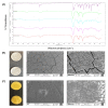
Physicochemical characterization of TQ films. (A) FTIR spectrum of TQ pure drug, PVP, physical mixture of drug and polymer, freshly prepared films containing drug and polymer, stored films containing drug and polymer; (B) Control films (i), Field emission scanning electron microscopic (FESEM) images showing the surface morphology of control film (ii–iii) at different magnifications (scale bar = 100 µm), and (C) TQ films (i), FESEM images showing the surface morphology of TQ films (ii–iii) at different magnifications (scale bar = 100 µm).
Figure 3
Figure 3
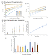
Rheological characterization of TQ hydrogel formulations (F1–F10). (A) Oscillation frequency sweep data. The elastic modulus (i); The viscous modulus (ii) were plotted against angular frequency. TQ permeation and skin deposition from film and gel formulations (B). TQ permeation profile for different hydrogel formulations (i). Time points were measured at 1, 2, 3, 4, 5, 6, and 8 h. Each point represents the five experiments; TQ permeation from film formulation across human cadaver skin (n = 5) (ii); Amount of TQ detected after 8 h in human cadaver skin (n = 5) using different TQ hydrogel formulations (iii).
Figure 4
Figure 4
The cytocompatibility study of TQ film. Cell viability of TQ film with HDF and HaCaT cells using alamarBlue® assay.
Figure 5
Figure 5
Bacterial inhibition study. (A) Inhibition of bacterial growth on agar plate by Control negative (i); Gentamicin positive control 50 µg/mL (ii) right upper and 500 µg/mL (ii) right lower; Control film (iii); TQ hydrogel (iv) and TQ film (v) against S. aureus; (B) Ex vivo antibacterial activity by Control (i); Control film (ii); Gentamicin sulfate USP, 0.1% marketed cream (iii); TQ hydrogel (iv); TQ film (v) and Log of bacterial reduction with different treatment groups (vi). Data represent of four replicates. *** p ≤ 0.001 and ^^^ p ≤ 0.05.
Figure 6
Figure 6
Effect of TQ treatment on the wound healing of human fibroblasts. (A) Representative micrographs (10×) from fibroblast cell migration including different treatment groups (Control, 1 ng/mL, and 100 ng/mL of TQ) showing the original wound and the wound after 12 h and 24 h; (B) Quantitative analysis of wound closure as a function of time. The wound area was determined as the wound area at a given time relative to the original wound area (n = 6). ** p < 0.01 and *** p < 0.001 (Control vs. 100 ng/mL) and ^ p < 0.05 (Control vs. 1 ng/mL); (C) Quantitative measurement of cell number migrating in the corresponding scratched wound areas at 24 h in the different treatment groups (n = 6). *** p < 0.001 (100 ng/mL vs. control and 1 ng/mL).
Figure 7
Figure 7
Effect of TQ treatment on the wound healing of keratinocytes using scratch assay. (A) Representative micrographs (10×) from Control (+Ve), Control, 1 ng/mL, and 100 ng/mL of TQ showing the original wound at day 0 and the wounds after 3 and 6 days of keratinocyte cell migration; (B) Quantitative analysis of wound closure as a function of time. The wound area was determined as the wound area at a given time relative to the original wound area at day 0 (n = 5–6). *** p < 0.001 (Control vs. Control + Ve), ^^ p < 0.01 (Control vs. 1 ng/mL) and ### p < 0.001 (Control vs. 100 ng/mL).
Figure 8
Figure 8
Antibacterial effects of TQ films on full-thickness skin wounds infected with S. aureus. (A) Photographs of skin wounds with different treatments over 21 days. The wounds were infected with bacteria as shown above as a pale biofilm in the bacteria wound group and other related groups. Scale bar = 1 cm. (B) Log of bacterial reduction at each timepoint up to 7 days assessed in different experimental groups. *** p < 0.001 (Bacterial wound vs. TQ Film) and ^^^ p < 0.001 (Bacterial wound vs. Gentamicin). (C) Percentage of wound closure as a function of time in all experimental groups at different time points post-wounding and treatments. ** p < 0.01 (Control wound vs. Control Film) and # p < 0.05 (Control Film vs. Gentamicin).

References

    1. Hadgraft J. Skin, the final frontier. Int. J Pharm. 2001;224:1–18. doi: 10.1016/S0378-5173(01)00731-1. - DOI - PubMed
    1. Brown M.B., Martin G.P., Jones S.A., Akomeah F.K. Dermal and transdermal drug delivery systems: Current and future prospects. Drug Deliv. 2006;13:175–187. doi: 10.1080/10717540500455975. - DOI - PubMed
    1. Kumar S., Kang H.J., Berthiaume F. 40 - Scaffolds for epidermal tissue engineering. In: Mozafari M., Sefat F., Atala A., editors. Handbook of Tissue Engineering Scaffolds: Volume Two. Woodhead Publishing; Cambridge, UK: 2019. [(accessed on 27 September 2020)]. pp. 173–191. Available online: - DOI
    1. Hahn B.L., Onunkwo C.C., Watts C.J., Sohnle P.G. Systemic dissemination and cutaneous damage in a mouse model of staphylococcal skin infections. Microb. Pathog. 2009;47:16–23. doi: 10.1016/j.micpath.2009.04.007. - DOI - PMC - PubMed
    1. Naimi T.S., LeDell K.H., Como-Sabetti K., Borchardt S.M., Boxrud D.J., Etienne J., Johnson S.K., Vandenesch F., Fridkin S., O’Boyle C., et al. Comparison of community- and health care-associated methicillin-resistant Staphylococcus aureus infection. JAMA. 2003;290:2976–2984. doi: 10.1001/jama.290.22.2976. - DOI - PubMed

LinkOut - more resources