Comparative Study
. 2020 Sep 30;21(1):256.
doi: 10.1186/s13059-020-02167-0.

Mustache: multi-scale detection of chromatin loops from Hi-C and Micro-C maps using scale-space representation

Affiliations
Comparative Study

Mustache: multi-scale detection of chromatin loops from Hi-C and Micro-C maps using scale-space representation

Abbas Roayaei Ardakany et al. Genome Biol. .

Abstract

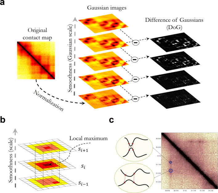
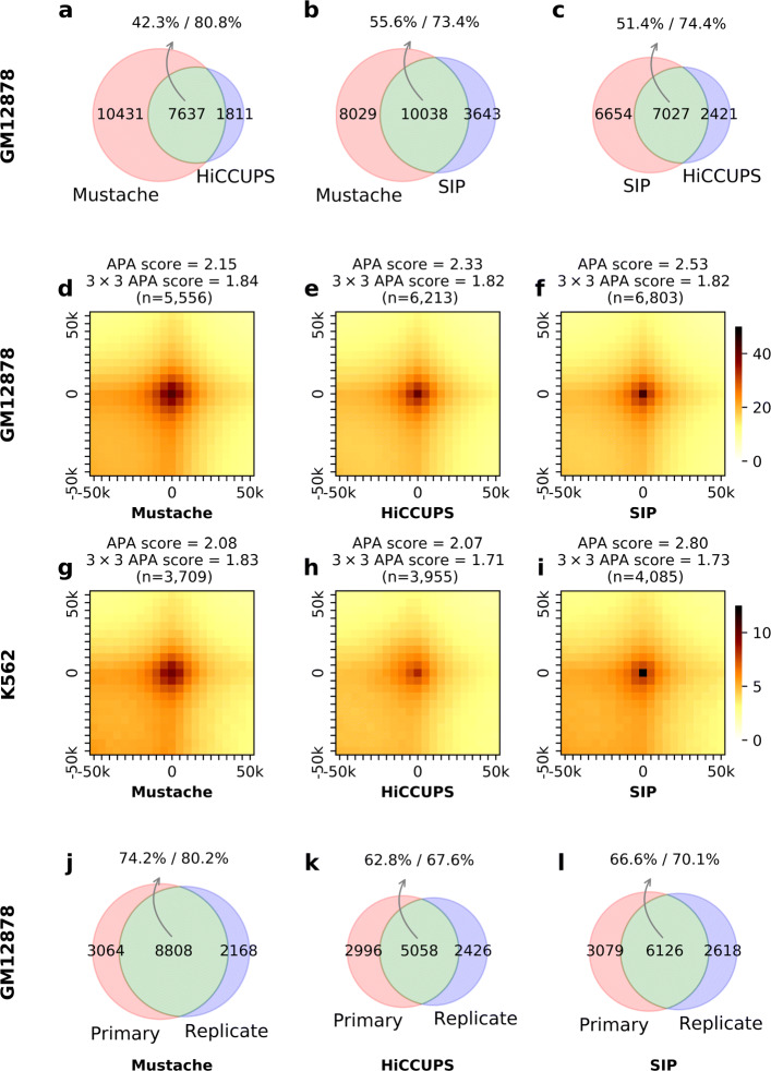
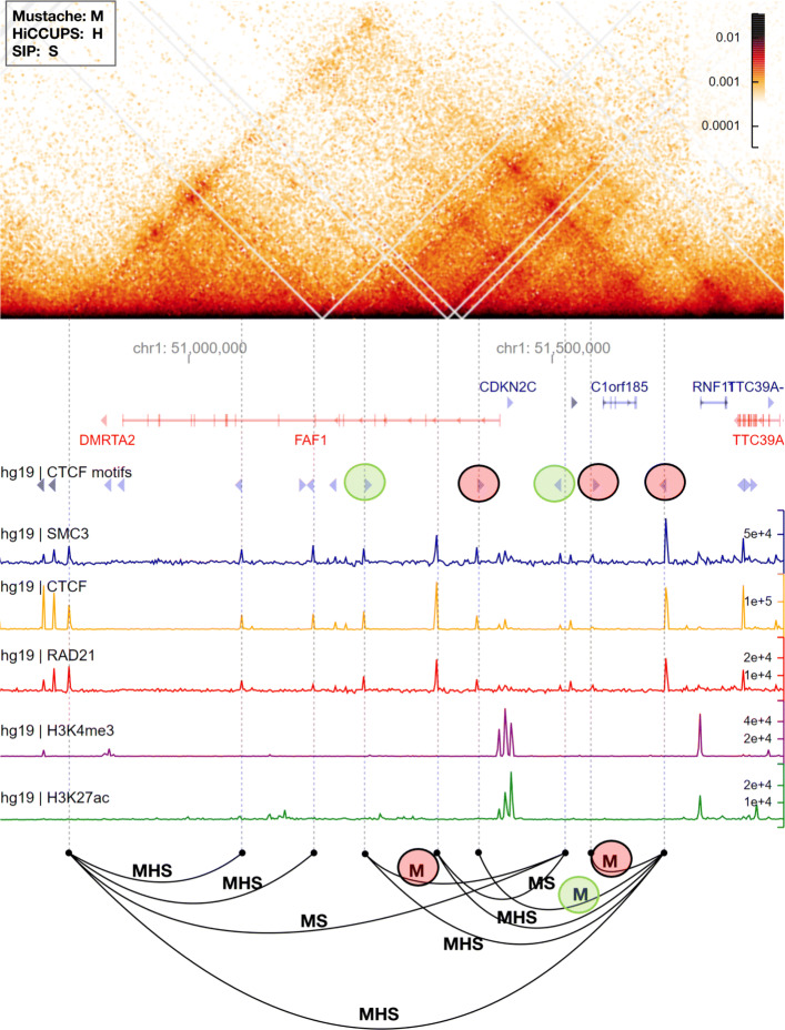
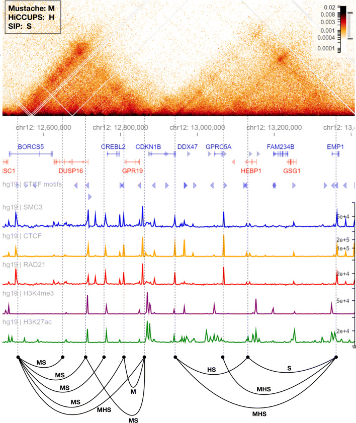
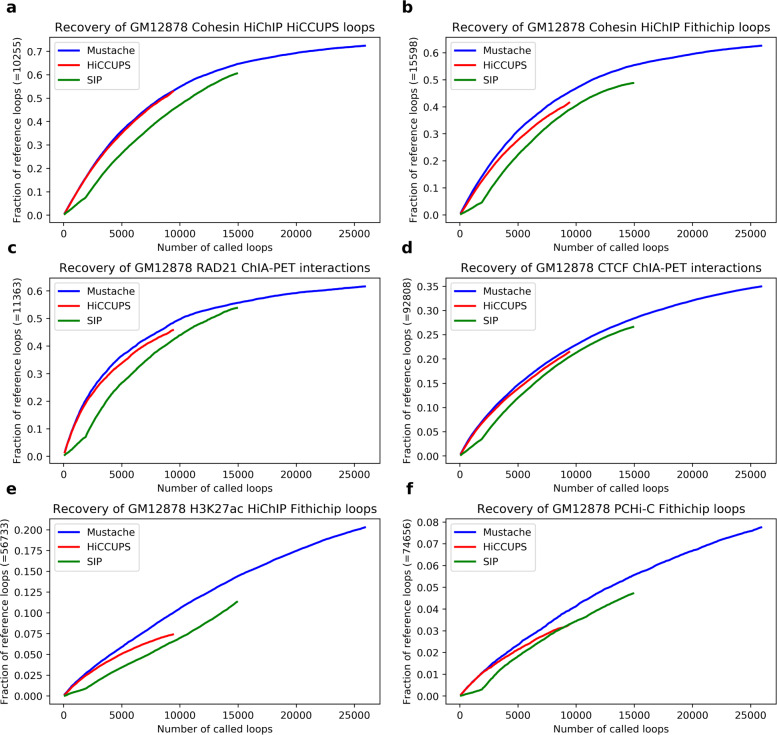
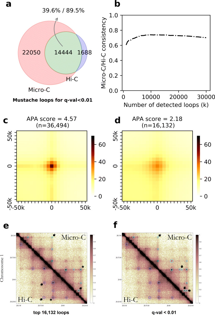
We present MUSTACHE, a new method for multi-scale detection of chromatin loops from Hi-C and Micro-C contact maps. MUSTACHE employs scale-space theory, a technical advance in computer vision, to detect blob-shaped objects in contact maps. MUSTACHE is scalable to kilobase-resolution maps and reports loops that are highly consistent between replicates and between Hi-C and Micro-C datasets. Compared to other loop callers, such as HiCCUPS and SIP, MUSTACHE recovers a higher number of published ChIA-PET and HiChIP loops as well as loops linking promoters to regulatory elements. Overall, MUSTACHE enables an efficient and comprehensive analysis of chromatin loops. Available at: https://github.com/ay-lab/mustache .

Keywords: CTCF; ChIA-PET; Chromatin loops; Cohesin; Contact maps; Genome architecture; Hi-C; HiChIP; Micro-C; Promoter-enhancer interactions.


Conflict of interest statement

The authors declare that they have no competing interests.

Figures

Fig. 1
Fig. 1
a The initial contact map is repeatedly convolved with increasing 2D Gaussians to produce a scale-space representation of the image (shown on the left). Pairwise adjacent Gaussian images are subtracted to produce the difference-of-Gaussian (DoG) images (on the right). b Maxima of the difference-of-Gaussian images are detected by comparing each pixel to its 3 ×3×3 neighborhood in (x,y,σ) space. Note that DoG is a local maximum at (x,y) location at scales si and si+1 but not at scale si−1, therefore passing the first filtering step criteria. c Chromatin loops can be caused by the contact between pairs of DNA segments at different scales
Fig. 2
Fig. 2
Comparison of loops detected by Mustache, HiCCUPS, and SIP from the GM12878 cell line Hi-C data. The agreement between a Mustache and HiCCUPS, b Mustache and SIP, and c SIP and HiCCUPS loops is shown as Venn diagrams for replicate-combined Hi-C contact maps. The overlap between the two loop sets is shown in green, and the percentages of overlap with respect to each set are reported separately. di APA plots for Mustache, HiCCUPS, and SIP loops in GM12878 and K562 cell lines. The APA score calculated with respect to the enrichment of (i) the center pixel and (ii) a 3×3 neighborhood in the center is reported above each plot. The overlap between reported loops on two replicates of GM12878 cell line is shown for j Mustache, k HiCCUPS, and l SIP. For Mustache and SIP, a q value threshold of 0.05 was used. For jl, when replicates are analyzed separately, we use 10 kb resolution Hi-C contact maps
Fig. 3
Fig. 3
A comparison between Mustache, SIP, and HiCCUPS reported loop calls in a region of chromosome 1 for the GM12878 cell line (50.75–51.75 Mb). The Hi-C contact map is rotated 45 such that the main diagonal is horizontal (top). Below the contact map, we report genomic coordinates, gene annotations (genes on the negative strand are shown in red color), CTCF motifs and their orientation, and ChIP-seq signals for SMC3, CTCF, RAD21, H3K4me3, and H3K27ac (coverage tracks plotted by HiGlass). The bottom row demonstrates loop calls as arcs connecting two loci labeled by the initial letter of the methods by which they are detected (“M” for Mustache, “S” for SIP, and “H” for HiCCUPS)
Fig. 4
Fig. 4
A comparison between Mustache, SIP, and HiCCUPS reported loop calls in a region of chromosome 1 for the GM12878 cell line (12.5–13.3 Mb). The display order is similar to that of Fig. 3
Fig. 5
Fig. 5
Comparison of the recovery of several reference loop sets by MUSTACHE, HiCCUPS, and SIP applied on the 5-kb resolution GM12878 Hi-C data. Recovery of a GM12878 cohesin HiChIP HiCCUPS loops, b GM12878 cohesin HiChIP FitHiChIP loops, c GM12878 RAD21 ChIA-PET interactions, d GM12878 CTCF ChIA-PET interactions, e GM12878 H3K27ac HiChIP FitHiChIP loops, and f GM12878 PCHi-C FitHiChIP loops
Fig. 6
Fig. 6
Four example regions showing Mustache, HiCCUPS, and SIP reported loops. Mustache, HiCCUPS, and SIP loops are represented by blue circles, red filled circles, and green squares on the lower diagonal matrices, respectively. The upper diagonal matrices were left untouched in order to allow visualization of contact patterns in the underlying Hi-C data. The loop calls of the three methods using GM12878 Hi-C data are shown for a 50.75–51.75 Mb region of chromosome 1, b 12.5–13.4 Mb region of chromosome 12, c 88.7–88.5 Mb region of chromosome 4, and d 67.2–68Mb region of chromosome 1
Fig. 7
Fig. 7
a The consistency between MUSTACHE loops detected using Micro-C and Hi-C data in HFFc6 cell line using a fixed q value threshold of 0.01, shown as a Venn diagram. b The consistency plot for MUSTACHE results between Hi-C and Micro-C for the top-k reported interactions for each contact map. The APA plots for MUSTACHE loops in HFFc6 cell line for c Hi-C and d Micro-C data. The APA score for the enrichment of center is reported above each plot. MUSTACHE reported loops in Hi-C (lower triangular) and Micro-C (upper triangular) of HFFc6 for e the top 16,132 significant interactions, and f a fixed q value threshold of 0.01. The loop call marked by a “ ⋆” was in common between Hi-C and Micro-C, but was detected at a smaller scale and has a stronger enrichment in Micro-C compared to Hi-C. The loops that are uniquely detected in either by Hi-C or by Micro-C are denoted by black arrows

References

    1. Pederson T. Chromatin structure and the cell cycle. Proc Natl Acad Sci U S A. 1972;69(8):2224–8. doi: 10.1073/pnas.69.8.2224. - DOI - PMC - PubMed
    1. Dixon JR, Xu J, Dileep V, Zhan Y, Song F, Le VT, Yardımcı GG, Chakraborty A, Bann DV, Wang Y, et al. Integrative detection and analysis of structural variation in cancer genomes. Nat Genet. 2018;50(10):1388–98. doi: 10.1038/s41588-018-0195-8. - DOI - PMC - PubMed
    1. Dileep V, Ay F, Sima J, Vera DL, Noble WS, Gilbert DM. Topologically associating domains and their long-range contacts are established during early G1 coincident with the establishment of the replication-timing program. Genome Res. 2015;25(8):1104–13. doi: 10.1101/gr.183699.114. - DOI - PMC - PubMed
    1. Beagrie RA, Pombo A. Cell cycle: continuous chromatin changes. Nature. 2017;547(7661):34–35. doi: 10.1038/547034a. - DOI - PubMed
    1. Bonev B, Cavalli G. Organization and function of the 3D genome. Nat Rev Genet. 2016;17(11):661–78. doi: 10.1038/nrg.2016.112. - DOI - PubMed

Publication types