

Cells expressing PAX8 are the main source of homeostatic regeneration of adult mouse endometrial epithelium and give rise to serous endometrial carcinoma
- PMID: 32998907
- PMCID: PMC7648606
- DOI: 10.1242/dmm.047035
Cells expressing PAX8 are the main source of homeostatic regeneration of adult mouse endometrial epithelium and give rise to serous endometrial carcinoma
Abstract
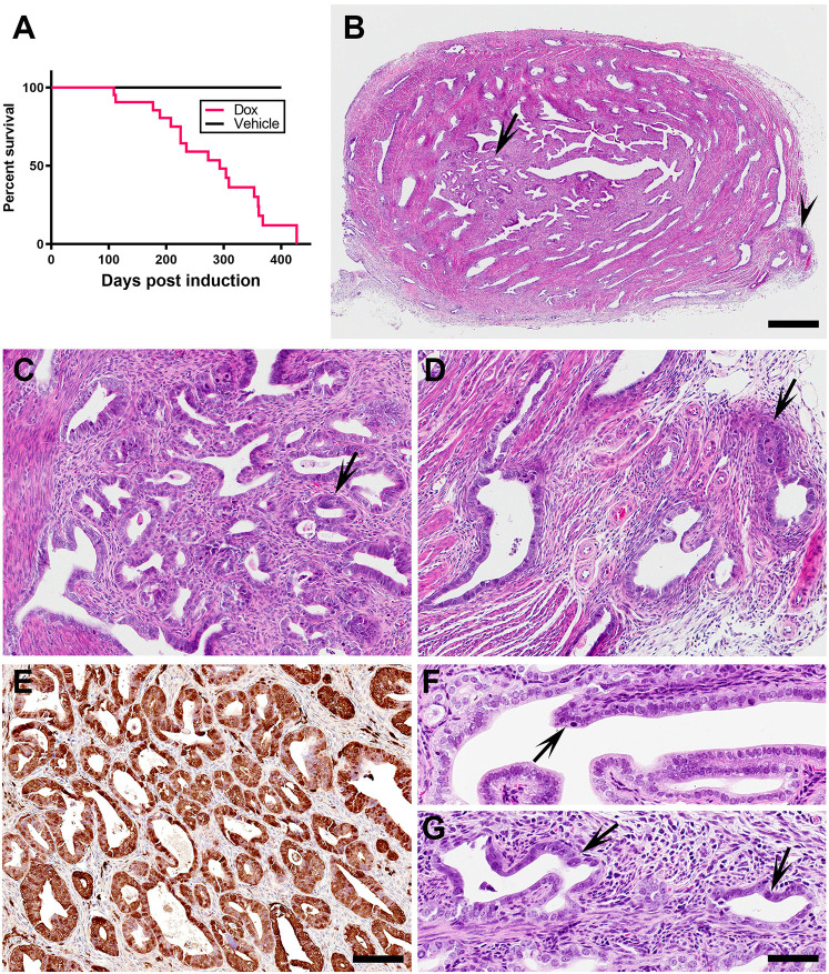
Humans and mice have cyclical regeneration of the endometrial epithelium. It is expected that such regeneration is ensured by tissue stem cells, but their location and hierarchy remain debatable. A number of recent studies have suggested the presence of stem cells in the mouse endometrial epithelium. At the same time, it has been reported that this tissue can be regenerated by stem cells of stromal/mesenchymal or bone marrow cell origin. Here, we describe a single-cell transcriptomic atlas of the main cell types of the mouse uterus and epithelial subset transcriptome and evaluate the contribution of epithelial cells expressing the transcription factor PAX8 to the homeostatic regeneration and malignant transformation of adult endometrial epithelium. According to lineage tracing, PAX8+ epithelial cells are responsible for long-term maintenance of both luminal and glandular epithelium. Furthermore, multicolor tracing shows that individual glands and contiguous areas of luminal epithelium are formed by clonal cell expansion. Inactivation of the tumor suppressor genes Trp53 and Rb1 in PAX8+ cells, but not in FOXJ1+ cells, leads to the formation of neoplasms with features of serous endometrial carcinoma, one of the most aggressive types of human endometrial malignancies. Taken together, our results show that the progeny of single PAX8+ cells represents the main source of regeneration of the adult endometrial epithelium. They also provide direct experimental genetic evidence for the key roles of the P53 and RB pathways in the pathogenesis of serous endometrial carcinoma and suggest that PAX8+ cells represent the cell of origin of this neoplasm.
Keywords: Endometrial cancer; Mouse models; Single-cell transcriptome; Stem cells.
© 2020. Published by The Company of Biologists Ltd.
Conflict of interest statement
Competing interestsThe authors declare no competing or financial interests.
Figures


Similar articles
-
Loss of PAX8 in high-grade serous ovarian cancer reduces cell survival despite unique modes of action in the fallopian tube and ovarian surface epithelium.Oncotarget. 2016 May 31;7(22):32785-95. doi: 10.18632/oncotarget.9051. Oncotarget. 2016. PMID: 27129161 Free PMC article.
-
[Significance and expression of PAX8, PAX2, p53 and RAS in ovary and fallopian tubes to origin of ovarian high grade serous carcinoma].Zhonghua Fu Chan Ke Za Zhi. 2017 Oct 25;52(10):687-696. doi: 10.3760/cma.j.issn.0529-567X.2017.10.008. Zhonghua Fu Chan Ke Za Zhi. 2017. PMID: 29060967 Chinese.
-
Lineage tracing suggests that ovarian endosalpingiosis does not result from escape of oviductal epithelium.J Pathol. 2019 Oct;249(2):206-214. doi: 10.1002/path.5308. Epub 2019 Jul 8. J Pathol. 2019. PMID: 31131879 Free PMC article.
-
A proposed model for endometrial serous carcinogenesis.Am J Surg Pathol. 2011 Jan;35(1):e1-e14. doi: 10.1097/PAS.0b013e318202772e. Am J Surg Pathol. 2011. PMID: 21164282 Review.
-
The role of mesenchymal-epithelial transition in endometrial function.Hum Reprod Update. 2019 Jan 1;25(1):114-133. doi: 10.1093/humupd/dmy035. Hum Reprod Update. 2019. PMID: 30407544 Review.
Cited by
-
Single-cell analysis of mouse uterus at the invasion phase of embryo implantation.Cell Biosci. 2022 Feb 5;12(1):13. doi: 10.1186/s13578-022-00749-y. Cell Biosci. 2022. PMID: 35123575 Free PMC article.
-
Pre-ciliated tubal epithelial cells are prone to initiation of high-grade serous ovarian carcinoma.Nat Commun. 2024 Oct 5;15(1):8641. doi: 10.1038/s41467-024-52984-1. Nat Commun. 2024. PMID: 39366996 Free PMC article.
-
Paired box proteins as diagnostic biomarkers for endocervical adenocarcinoma.World J Clin Cases. 2024 Sep 16;12(26):5854-5858. doi: 10.12998/wjcc.v12.i26.5854. World J Clin Cases. 2024. PMID: 39286388 Free PMC article.
-
Deciphering mouse uterine receptivity for embryo implantation at single-cell resolution.Cell Prolif. 2021 Nov;54(11):e13128. doi: 10.1111/cpr.13128. Epub 2021 Sep 23. Cell Prolif. 2021. PMID: 34558134 Free PMC article.
-
Cell-Cell Communication at the Embryo Implantation Site of Mouse Uterus Revealed by Single-Cell Analysis.Int J Mol Sci. 2021 May 13;22(10):5177. doi: 10.3390/ijms22105177. Int J Mol Sci. 2021. PMID: 34068395 Free PMC article.
References
Publication types
MeSH terms
Substances
Grants and funding
LinkOut - more resources
Full Text Sources
Molecular Biology Databases
Research Materials
Miscellaneous
