. 2020 Sep 30;12(563):eaba0565.
doi: 10.1126/scitranslmed.aba0565.

Paenibacillus infection with frequent viral coinfection contributes to postinfectious hydrocephalus in Ugandan infants

Affiliations

Paenibacillus infection with frequent viral coinfection contributes to postinfectious hydrocephalus in Ugandan infants

Joseph N Paulson et al. Sci Transl Med. .

Abstract

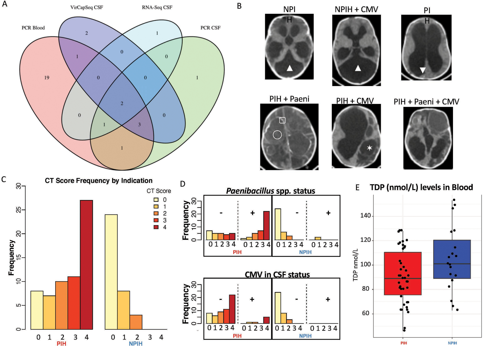
Postinfectious hydrocephalus (PIH), which often follows neonatal sepsis, is the most common cause of pediatric hydrocephalus worldwide, yet the microbial pathogens underlying this disease remain to be elucidated. Characterization of the microbial agents causing PIH would enable a shift from surgical palliation of cerebrospinal fluid (CSF) accumulation to prevention of the disease. Here, we examined blood and CSF samples collected from 100 consecutive infant cases of PIH and control cases comprising infants with non-postinfectious hydrocephalus in Uganda. Genomic sequencing of samples was undertaken to test for bacterial, fungal, and parasitic DNA; DNA and RNA sequencing was used to identify viruses; and bacterial culture recovery was used to identify potential causative organisms. We found that infection with the bacterium Paenibacillus, together with frequent cytomegalovirus (CMV) coinfection, was associated with PIH in our infant cohort. Assembly of the genome of a facultative anaerobic bacterial isolate recovered from cultures of CSF samples from PIH cases identified a strain of Paenibacillus thiaminolyticus This strain, designated Mbale, was lethal when injected into mice in contrast to the benign reference Paenibacillus strain. These findings show that an unbiased pan-microbial approach enabled characterization of Paenibacillus in CSF samples from PIH cases, and point toward a pathway of more optimal treatment and prevention for PIH and other proximate neonatal infections.


Conflict of interest statement

Competing interests: None.

Figures

Figure 1.
Figure 1.. Comparative location of cases within map of Uganda.
The village centroid GPS locations are mapped to the 0.1 × 0.1 degree grid frequently used in satellite rainfall estimation (54), and where village name was uncertain, the centroid of the administrative parish or sub-county was used. The groups shown are mapped by clinical status (NPIH and PIH), and by organism type (Paenibacillus, PAENI; cytomegalovirus, CMV). Both the PAENI and CMV mappings included all NPIH and PIH cases with such diagnoses. Using Fisher’s linear discrimination analysis, group comparisons by latitude and longitude mapping could significantly discriminate NPIH from PIH, or NPIH from PAENI, at the p < 0.01 level parametrically (Wilk’s lambda), and at the p= 0.03 and p=0.01 level respectively using a bootstrap method (see Figure S2). The number of cases mapped to a 0.1 × 0.1 degree grid (11 km per edge at the equator) are indicated with colored circles as 1, 2, or 3.
Figure 2.
Figure 2.. Detection and typing of bacteria using 16S rDNA.
A) Agarose gels showing 16S rDNA amplification products for PIH (red) and NPIH (blue), along with PCR positive (+) and negative (−) controls (green) and 10 separate extraction reagent controls (yellow). Brightness and contrast were adjusted for gels to maximize visibility of faint bands and smears. Asterisk (*) denotes lanes where faint non-specific amplification (smears) or bands of unexpected size were observed. All amplification products including non-specific amplifications were subjected to subcloning and sequencing. B) Paenibacillus qPCR quantification. C-D) Stacked bar relative abundance plots of (C) 16S V4 and (D) V1-V2 regions in microbial communities for the most dominant bacteria observed within (C) 16S positive samples from (B) and in (D) all 100 samples. Star underneath PIH sample (C, D) highlights individual PIH sample with group B streptococcal infection. E) Scatterplot of cumulative sum scaling normalized Paenibacillus abundance for (abscissa) V4 16S sequencing of fresh frozen CSF samples and (ordinate) V1-V2 16S sequencing of biological replicates from preserved CSF samples. F) Receiver-operating-characteristic (ROC) curve using the number of Paenibacillus reads as the predictor for PIH or NPIH status. Area under the curve was 70.88% (95% DeLong CI = 60.61%−81.15%). Sensitivity and specificity are maximized at 47.5 reads in a given sample, consistent with the threshold employed of 50 reads.
Figure 3.
Figure 3.. Viral detection, CT findings, thiamine levels, and infection status.
A) Venn diagram of CMV detection using PCR on blood, and VirCap-Seq, RNA-Sequencing and PCR on CSF samples. B) Representative images of patients with NPIH or PIH, and CMV or Paenibacillus (Paeni) status. Both NPIH demonstrate Dandy-Walker cyst malformations (▲) without history or surgical findings reflective of prior infection. Note the presence in the Paenibacillus PIH cases of multiple loculations of CSF (✶), higher density fluid collections reflective of debris or blood within the CSF (▼), ectopic calcification within the brain (▢), and abscess formation (◯). C) CT scores stratified by clinical indication, PIH or NPIH, and D) as a function of CMV and Paenibacillus infection status, positive or negative respectively (see also Fig S6). E) Levels, in nmol/L, of thiamine diphosphate (TDP) in PIH (n=42) vs NPIH (n=19) cases. We observed that TDP levels are lower in PIH patients, (t-test, p<0.05). Boxplots display the median and upper and lower quartiles with whiskers forming the 1.5x the interquartile range.
Figure 4.
Figure 4.. Clinical signs associate with Paenibacillus detection.
A) Boxplots of (ordinate) log2 normalized Paenibacillus 16S rDNA abundance by age (categorized into 0–6 weeks and >6 weeks) and by indication (see also Fig S6). B) Boxplots of (ordinate) CSF WBC count (cell/μL) by Paenibacillus status (+/−) and by indication (see also Fig S6). Cell count values less than 5 were mapped to 5. Cell count values greater than 250 were mapped to 250. C) Boxplots of (ordinate) corrected weight-for-age z-scores (WAZ), after calculating and subtracting excess CSF volume-for-age, by Paenibacillus status (+/−) and by indication. Boxplots display the median and upper and lower quartiles with whiskers forming the 1.5x the interquartile range.
Figure 5.
Figure 5.. Whole genome sequencing and assembly of P. thiaminolyticus CSF isolate strain Mbale.
From the cultured CSF samples, three isolates were identified as Paenibacillus (marked with asterisks in panel C). One isolate showed high 16S rDNA sequence identity to our V1-V2 and V4 sequencing results thus making it the isolate of high interest. A) P. thiaminolyticus Mbale Gram stain from the chocolate agar subculture of the lytic anaerobic bottle at 1,000x magnification. Weak or negative Gram staining, despite a Gram positive cell structure, is characteristic of Paenibacillus species (55). B) To classify the P. thiaminolyticus clinical isolate, an extensive genome analysis was performed using both long-read and next generation sequencing along with optical mapping (GenBank Accession CP041404). The resulting draft circular genome shown was created using CGView which features coding sequences (CDS), tRNA, rRNA, phage insertions and GC content (53%). C) Phylogenetic tree of Paenibacillus spp. based on 40 marker genes. Cultured CSF isolates are indicated by yellow marks; the isolate 2033 was renamed P. thiaminolyticus strain Mbale.
Figure 6.
Figure 6.. Comparative murine histology of reference and Mbale strains of Paenibacillus thiaminolyticus.
Murine model contrasting the virulence of the Mbale strain of P. thiaminolyticus vs the reference P. thiaminolyticus type strain NRRL B-4156T following intraperitoneal injection into C56BL/6J littermates, in comparison with saline control injections. No significant lesions are seen (top row) in the spleen of mice inoculated with normal saline (n=6) or reference strain (n=10). However, tingible body macrophage necrosis (black arrows) with intracytoplasmic apoptotic bodies were present in spleen of mice inoculated with the Mbale strain (9/10), suggestive of toxin induced apoptosis and a marked immune response with a high lymphocyte turnover. Likewise, the kidneys (middle row) of mice inoculated with normal saline or the reference strain had no significant lesions. In contrast, pyknotic nuclei (arrow head) of proximal tubule epithelial cells with cell sloughing and loss were observed within mice inoculated with the Mbale strain (9/10), indicative of acute tubular necrosis. In the brain (bottom row), there was no substantial evidence of infection-associated pathology (normal saline n=6/6, reference strain n=10/10, Mbale strain n=10/10).

References

    1. Dewan MC, Rattani A, Mekary R, Glancz LJ, Yunusa I, Baticulon RE, Fieggen G, Wellons JC 3rd, Park KB, Warf BC, Global hydrocephalus epidemiology and incidence: systematic review and meta-analysis. J Neurosurg, 1–15 (2018). - PubMed
    1. Ranjeva SL, Warf BC, Schiff SJ, Economic burden of neonatal sepsis in sub-Saharan Africa. BMJ Glob Health 3, e000347 (2018). - PMC - PubMed
    1. Warf BC, Hydrocephalus in Uganda: the predominance of infectious origin and primary management with endoscopic third ventriculostomy. Journal of neurosurgery 102, 1–15 (2005). - PubMed
    1. Warf BC, Alkire BC, Bhai S, Hughes C, Schiff SJ, Vincent JR, Meara JG, Costs and benefits of neurosurgical intervention for infant hydrocephalus in sub-Saharan Africa. Journal of neurosurgery Pediatrics 8, 509–521 (2011). - PubMed
    1. Aziz IA, Hydrocephalus in the Sudan. Journal of the Royal College of Surgeons of Edinburgh 21, 222–224 (1976). - PubMed

Publication types

Supplementary concepts