. 2020 Nov 23;94(24):e01350-20.
doi: 10.1128/JVI.01350-20. Print 2020 Nov 23.

Interferon-Induced Transmembrane Protein 3 Is a Virus-Associated Protein Which Suppresses Porcine Reproductive and Respiratory Syndrome Virus Replication by Blocking Viral Membrane Fusion

Affiliations

Interferon-Induced Transmembrane Protein 3 Is a Virus-Associated Protein Which Suppresses Porcine Reproductive and Respiratory Syndrome Virus Replication by Blocking Viral Membrane Fusion

Angke Zhang et al. J Virol. .

Abstract

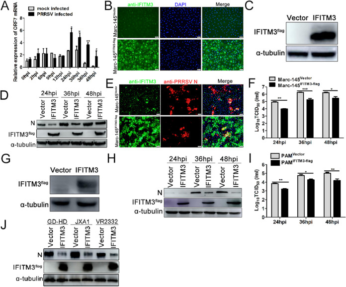
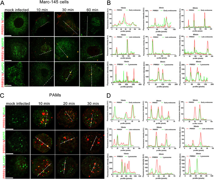
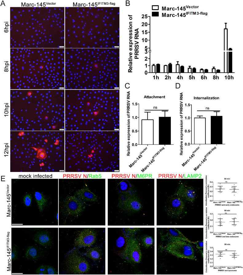
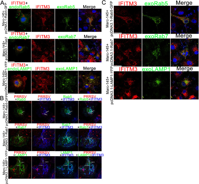
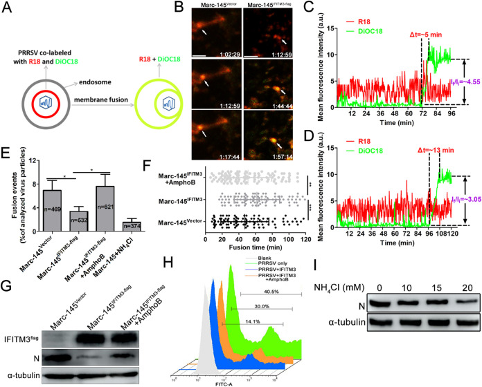
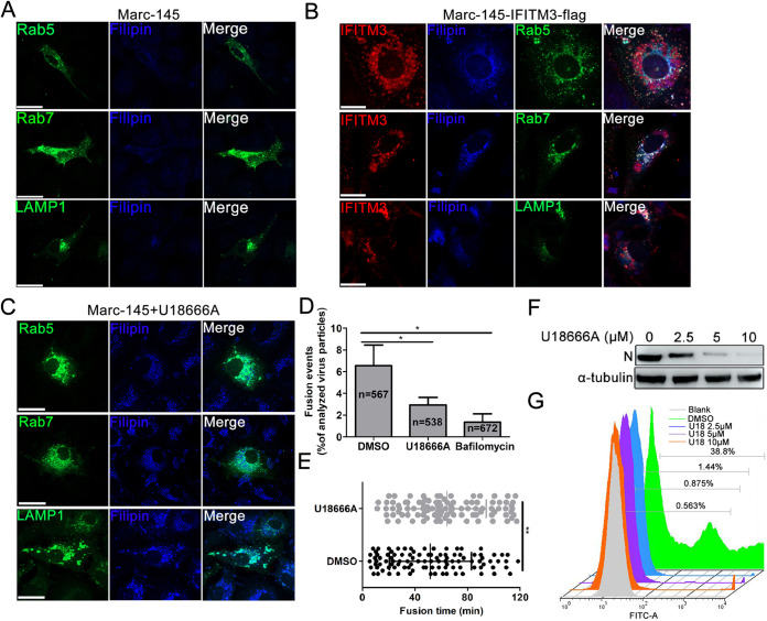
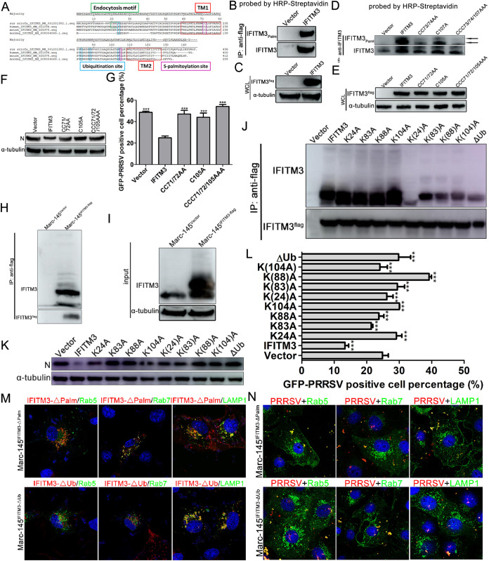
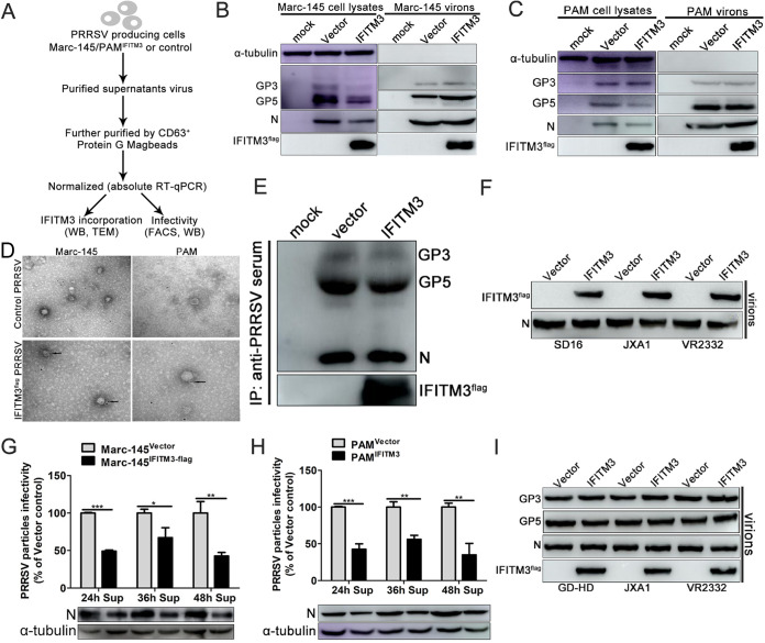
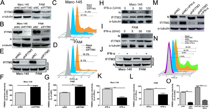
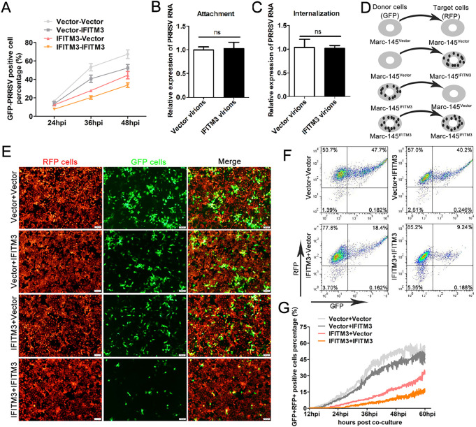
Porcine reproductive and respiratory syndrome virus (PRRSV) infection eliminates production of type I interferons (IFNs) in host cells, which triggers an antiviral immune response through the induction of downstream IFN-stimulated genes (ISGs), thus escaping the fate of host-mediated clearance. The IFN-induced transmembrane 3 (IFITM3) has recently been identified as an ISG and plays a pivotal role against enveloped RNA viruses by restricting cell entry. However, the role of IFITM3 in PRRSV replication is unknown. The present study demonstrated that overexpression of IFITM3 suppresses PRRSV replication, while silencing of endogenous IFITM3 prominently promoted PRRSV replication. Additionally, it was shown that IFITM3 undergoes S-palmitoylation and ubiquitination modification, and both posttranslational modifications contribute to the anti-PRRSV activity of IFITM3. Further study showed that PRRSV particles are transported into endosomes and then into lysosomes during the early stages of infection, and confocal microscopy results revealed that PRRSV particles are transported to IFITM3-positive cellular vesicles. By using a single virus particle fluorescent labeling technique, we confirmed that IFITM3 can restrict PRRSV membrane fusion by inducing accumulation of cholesterol in cellular vesicles. Additionally, we found that both endogenous and exogenous IFITM3 are incorporated into newly producing PRRS virions and diminish viral intrinsic infectivity. By using cell coculture systems, we found that IFITM3 effectively restricted PRRSV intercellular transmission, which may have been caused by disrupted membrane fusion and reduced viral infectivity. In conclusion, our results demonstrate, for the first time, that swine IFITM3 interferes with the life cycle of PRRSV, and possibly other enveloped arteritis viruses, at multiple steps.IMPORTANCE Porcine reproductive and respiratory syndrome (PRRS), which is caused by PRRS virus (PRRSV), is of great economic significance to the swine industry. Due to the complicated immune escape mechanisms of PRRSV, there are no effective vaccines or therapeutic drugs currently available against PRRS. Identification of cellular factors and underlying mechanisms that establish an effective antiviral state against PRRSV can provide unique strategies for developing antiviral vaccines or drugs. As an interferon (IFN)-stimulated gene, the role of IFN-induced transmembrane 3 (IFITM3) in PRRSV infection has not been reported as of yet. In the present study, it was shown that IFITM3 can exert a potent anti-PRRSV effect, and PRRS virions are trafficked to IFITM3-containing cell vesicles, where viral membrane fusion is impaired by cholesterol accumulation that is induced by IFITM3. Additionally, both endogenous and exogenous IFITM3 are incorporated into newly assembled progeny virions, and this decreased their intrinsic infectivity.

Keywords: IFITM3; PRRSV; cellular vesicles; cholesterol; membrane fusion.


Figures

FIG 1
FIG 1
Overexpression of IFITM3 suppresses PRRSV replication in vitro. (A) Expression of IFITM3 mRNA in PRRSV-infected PAMs. (B) IFA or (C) Western blotting verification of stable expression of IFITM3 in Marc-145 cells. Marc-145-Vector or Marc-145-IFITM3-flag cells were infected with PRRSV (0.1 MOI) at 37°C for 1 h, and cells were subsequently maintained in 3% FBS+DMEM. (D and F) Cells were collected after 24, 36, and 48 hpi for detection of IFITM3 or N protein expression using (D) Western blotting, and supernatant virus titers using (F) TCID50. (E) A portion of cells were fixed using 70% 20°C prechilled alcohol 24 hpi for IFA. (G) PAMs were transduced with recombinant lentivirus expressing IFITM3-flag or control lentivirus for 36 h, and cells were harvested for IFITM3 analysis. (H and I) PAMs transduced with recombinant lentivirus expressing IFITM3-flag or control lentivirus were infected with 0.1 MOI of PRRSV, and cells and supernatants were collected at 24, 36, and 48 hpi to determine (H) IFITM3 and N protein expression and (I) supernatant virus titers. (J) Marc-145 cells were infected with GD-HD-, JXA1-, and VR2332-PRRSV (0.1 MOI). At 48 hpi, cells were harvested and analyzed by Western blotting. IFA, indirect immunofluorescence assay; MOI, multiplicity of infection; hpi, hours postinfection; PAM, porcine alveolar macrophage; IFITM, interferon-induced transmembrane; PRRSV, porcine reproductive and respiratory syndrome virus.
FIG 2
FIG 2
PRRSV particles are transported along the endosome-lysosome pathway during an early stage of infection. Marc-145 cells or PAMs were prechilled on ice for 30 min before incubation with 1.0 MOI of PRRSV. Cells were further incubated on ice for 1 h to ensure abundant adsorption of virus. Subsequently, cells were washed extensively with ice-cold PBS to remove unabsorbed virus and incubated at 37°C for the indicated time points. Cells were fixed with 70% −20°C prechilled alcohol, and cellular compartments and viral proteins were stained with specific antibodies against early endosomes (Rab5), late endosomes (M6PR), lysosomes (LAMP2), and capsid protein (N protein), followed by staining with Alexa Fluor 488-conjugated goat anti-rabbit IgG (H&L) antibody to visualize cellular compartments (green) and Alexa Fluor-594 conjugated goat anti-mouse IgG (H&L) antibody to visualize virions (red). Fluorescent images were acquired with a confocal laser scanning microscope. Scale bar, 10 μm. (A and C) Confocal immunofluorescence images showing PRRSV particles colocalized with endosomes and lysosomes at different time points. (B and D) Colocalization analysis corresponding to the PRRSV particles and cellular compartments. PAM, porcine alveolar macrophage; PRRSV, porcine reproductive and respiratory syndrome virus; MOI, multiplicity of infection.
FIG 3
FIG 3
IFITM3 does not affect PRRSV particle attachment, entry, or access to endosomes or lysosomes. (A) Marc-145-Vector or Marc-145-IFITM3-flag cells were incubated with PRRSV at an MOI of 1.0 for 6, 8, 10, and 12 h. The infected cells were fixed and stained with anti-PRRSV N antibody and counterstained with DAPI to visualize the nuclei. (B) Marc-145-Vector or Marc-145-IFITM3-flag cells were infected with 1.0 MOI of PRRSV for 1 h at 37°C, and then cells were washed with PBS and cultured with 3% FBS+DMEM. Cells were harvested 1, 2, 4, 5, 6, 8, or 10 hpi, and PRRSV genome levels were determined. Prechilled Marc-145-Vector or Marc-145-IFITM3-flag cells were infected with PRRSV (1.0 MOI) and further chilled on ice for 1 h. (C) For the attachment assay, after washing 3 times using ice-cold PBS, cells were harvested for PRRSV genome abundance analysis. (D) For the entry assay, cells were washed with PBS three times and treated with 37°C prewarmed DMEM and incubated at 37°C for 1 h, followed by trypsin treatment for 30 sec and 3 washes with PBS. Cells were collected for PRRSV genome content analysis. (E) Marc-145-Vector or Marc-145-IFITM3-flag cells were inoculated with 1.0 MOI PRRSV for 1 h at 37°C, and then cells were fixed and stained with anti-PRRSV N, -Rab5, -M6PR, and -LAMP2 antibodies, followed by an Alexa Fluor 488-conjugated goat anti-rabbit IgG (H&L) antibody and an Alexa Fluor 594-conjugated goat anti-mouse IgG (H&L) antibody. Colocalization of PRRSV particles with cellular vesicles was quantified, and representative images are presented. Scale bar, 10 μm. Horizontal bars represent the mean of the Pearson’s correlation coefficient (Rr) calculated based on 10 fields of view, with error bars marking the 95% confidence intervals. PRRSV, porcine reproductive and respiratory syndrome virus; MOI, multiplicity of infection; hpi, hours postinfection.
FIG 4
FIG 4
PRRSV particles are transported to IFITM3-containing endosomes or lysosomes. (A) Marc-145-IFITM3-flag cells transfected with pcDNA3.1-Rab5-GFP, -Rab7, and -LAMP1-YFP plasmids for 48 h were fixed with 4% paraformaldehyde and permeabilized with 0.3% Triton X-100. Cells were labeled with anti-IFITM3 and -Rab7 antibodies followed by Alexa Fluor 594-conjugated goat anti-mouse IgG (H&L) antibody for IFITM3 (red) and Alexa Fluor 488-conjugated goat anti-rabbit IgG (H&L) antibody for Rab7 (green), and cell nuclei were counterstained using DAPI. Rab5 and LAMP1 (green) were observed directly using a confocal microscope. (B) PRRSV were allowed to bind to prechilled Marc-145-IFITM3-flag cells for 1 h on ice, unbound virions were washed, and cells were warmed to 37°C for 45 min. IFITM3 was stained using an anti-IFITM3 antibody and the corresponding fluorescent second antibody (blue), and PRRSV was stained using an anti-N antibody and a fluorescent second antibody (red). (C) Marc-145-IFITM3-flag cells were infected with 0.1 MOI of PRRSV for 12 h, and then cells were transfected with pcDNA3.1-Rab5-GFP, -Rab7, and -LAMP1-YFP plasmids for 36 h and fixed with 4% paraformaldehyde and permeabilized with 0.3% Triton X-100. Cells were labeled as described in Fig. 4A. Scale bar, 10 μM. PRRSV, porcine reproductive and respiratory syndrome virus; IFITM, interferon-induced transmembrane.
FIG 5
FIG 5
IFITM3 restricts PRRSV membrane fusion. (A) Schematic illustration of the single-virus fusion assay. PRRS virions were colabeled with DiOC18 (green) and R18 (red). In this model, viral membrane fusion led to a decrease of DiOC18 concentration, which appeared as a sudden increase of the green signal (dequenching), while the red fluorescence intensity remained almost unchanged. PRRSV particles colabeled with DiOC18 and R18 were prebound to Marc-145-Vector or Marc-145-IFITM3-flag cells for 1 h on ice and then incubated at 37°C for 2 h. Particle fusion with endosomes or lysosomes (white arrows) exhibited a marked increase in green signal and thus appeared yellow. (B) Time-lapse images from the single PRRSV fusion event showed the increase of green signal at around 72 min in Marc-145-Vector cells and 104 min in Marc-145-IFITM3-flag cells, indicating the fusion events. Scale bar, 4 μm. (C and D) Particle fluorescence intensities obtained by tracking virions in Marc-145-Vector or Marc-145-IFITM3-flag cells. Δt represents the time for complete dequenching. (E) Single PRRSV fusion efficiency in Marc-145-Vector or Marc-145-IFITM3-flag cells. (F) PRRSV fusion events following synchronized infection in Marc-145-Vector or Marc-145-IFITM3-flag cells (100 dequenching particles tracked in 5 independent experiments). (G and H) Marc-145-IFITM3-flag cells were pretreated with AmphoB (1 μM) for 1 h and then infected with 0.1 MOI of GFP-PRRSV. N protein- and GFP-PRRSV-positive cells were analyzed using Western blotting and FACS, respectively. (I) Marc-145 cells were infected with 0.1 MOI of PRRSV for 1 h on ice, and then cells were treated with 10, 15, and 20 mM NH4Cl for 36 h. N protein was analyzed using Western blotting. PRRSV, porcine reproductive and respiratory syndrome virus; IFITM, interferon-induced transmembrane; MOI, multiplicity of infection; DiOC18, 3,3′-dioctadecyloxacarbocyanine; R18, octadecyl rhodamine B.
FIG 6
FIG 6
Accumulation of cholesterol in cell vesicles induced by IFITM3 effectively impairs viral membrane fusion. (A) Cholesterol distribution in Marc-145 cells transfected with pcDNA3.1-Rab5-GFP, -Rab7, and -LAMP1-YFP plasmids. (B) Marc-145-IFITM3-flag cells were transfected with pcDNA3.1-Rab5-GFP, -Rab7, and -LAMP1-YFP at 37°C for 48 h followed by fixing, permeabilizing, and labeling with anti-IFITM3 and -Rab7 antibodies and the corresponding fluorescent secondary antibodies. Subsequently, cells were stained using 50 μg/ml filipin for 2 h at room temperature in the dark for detection of cholesterol. Images showing colocalization between IFITM3, endosomes, lysosomes, and cholesterol were obtained using a confocal microscope. (C) Marc-145 cells transfected with pcDNA3.1-Rab5-GFP, -Rab7, and -LAMP1-YFP for 36 h were treated with 10 μM U18666A for 12 h, and then cells were fixed and endogenous cholesterol was stained using 50 μg/ml filipin in the dark for 2 h at room temperature. Scale bar, 10 μm. (D) Disturbance of intracellular cholesterol efflux reduced PRRSV membrane fusion. R18 and DiOC18 double labeled PRRS virions were prebound in the cold to DMSO- and U18666A-treated Marc-145 cells, and virus entry was initiated by transfer to 37°C for 2 h. Control experiments were performed by pretreating Marc-145 cells with 10 μM BafA1 for 1 h. Viral fusion events were normalized to the total number of cell-bound particles from 10 independent experiments. (E) PRRSV fusion events following synchronized infection of DMSO-treated or 10 μM U18666A-treated Marc-145 cells (100 dequenching particles tracked in 10 independent experiments). (F and G) Marc-145 cells were pretreated with 0, 2.5, 5, and 10 μM U18666A followed by inoculating with 0.1 MOI of GFP-PRRSV for 1 h at 37°C. Cells were collected 36 hpi for GFP-PRRSV-positive cell analysis using FACS, and for N protein expression detection using Western blotting. PRRSV, porcine reproductive and respiratory syndrome virus; IFITM, interferon-induced transmembrane; MOI, multiplicity of infection; hpi, hours postinfection; DiOC18, 3,3′-dioctadecyloxacarbocyanine; R18, octadecyl rhodamine B.
FIG 7
FIG 7
Both S-palmitoylation and ubiquitination of IFITM3 are critical for inhibition of PRRSV infection. (A) Alignment of human, mouse, porcine, and monkey IFITM3 using the ClustalV method. (B) IFITM3 palmitoylation was detected in Marc-145-Vector or Marc-145-IFITM3-flag cells using streptavidin-HRP following an ABE assay and immunoprecipitation with anti-flag MagBeads. A portion of the immunoprecipitation products was probed with an anti-IFITM3 antibody. (C) Another portion of cell lysates served as input. (D) Detection of palmitoylation of IFITM3 and its CC71/72AA, C105A, and CCC71/72/105AAA mutants in Marc-145-IFITM3-flag cells using streptavidin-HRP following an ABE assay and immunoprecipitation with anti-IFITM3 MagBeads. A portion of the immunoprecipitation products was probed with anti-IFITM3 antibody. (E) Another portion of cell lysates served as input. (F and G) Marc-145-Vector, Marc-145-IFITM3-flag cells, and their corresponding mutants were infected with 0.1 MOI GFP-PRRSV, and cells were harvested at 36 hpi for Western blotting and FACS analysis. (H) IFITM3 ubiquitination was determined using anti-flag MagBeads followed by Western blotting detection using anti-IFITM3 antibody. (I) A portion of cell lysates served as input. (J) Immunoprecipitation of IFITM3 and its K24A, K83A, K88A, K104A, K(24)A, K(83)A, K(88)A, K(104)A, and ΔUb mutants in recombinant cells was performed using anti-flag MagBeads, and then Western blotting was performed using anti-IFITM3 antibody. (K and L) Marc-145-Vector, Marc-145-IFITM3-flag, and their corresponding mutants infected with 0.1 MOI GFP-PRRSV were collected at 36 hpi and subjected to Western blotting or FACS detection. (M) Marc-145-IFITM3-ΔPal-flag or Marc-145-IFITM3-ΔUb-flag cells were transfected with pcDNA3.1-Rab5-GFP, -Rab7, or -LAMP1-YFP plasmids followed by staining with anti-Rab7 antibody 48 h posttransfection. Fluorescent images were acquired with a confocal laser scanning microscope. Scale bar, 10 μm. (N) Marc-145-IFITM3-ΔPal-flag or Marc-145-IFITM3-ΔUb-flag cells were transfected with pcDNA3.1-Rab5-GFP, -Rab7, or -LAMP1-YFP plasmids followed by inoculation with 1.0 MOI PRRSV for 1 h at 37°C, and then cells were fixed and stained with anti-PRRSV N antibody and Alexa Fluor 594-conjugated goat anti-mouse IgG (H&L) antibody. Scale bar, 10 μm. PRRSV, porcine reproductive and respiratory syndrome virus; IFITM, interferon-induced transmembrane; MOI, multiplicity of infection; HRP, horseradish peroxidase; ABE, acyl-biotin exchange; hpi, hours postinfection; for K(24)A, K(83)A, K(88)A, K(104)A, the numbers is parentheses indicate the one lysine of four that is not mutated to alanine.
FIG 8
FIG 8
IFITM3 is a PRRS virion-associated protein. (A) Representation of the experimental scheme used. (B) Marc-145-Vector or Marc-145-IFITM3-flag cells were infected with 0.1 MOI PRRSV. At 48 hpi, both cell lysates and supernatants purified by ultracentrifugation through sucrose density gradient were collected and analyzed by Western blotting. (C) As described above, PAM lysates and purified virions were analyzed. (D) Virions produced as described above were analyzed using immunogold electron microscopy. Unfixed viral preparations produced from IFITM3-overexpressing or control cells were purified and incubated with anti-flag antibodies, followed by incubation with 6 nm gold-conjugated secondary antibody. Representative pictures are presented. Scale bar, 100 nm. (E) Viral supernatants produced from IFITM3-overexpressing or control cells were concentrated and purified, followed by immunoprecipitation using swine antiserum. Western blotting was performed using an anti-flag antibody to detect IFITM3 and using swine antiserum to detect PRRSV GP3 and GP5. (F) Virions of different PRRSV strains obtained as described above were subjected to Western blotting using anti-IFITM3 antibody, and N protein was used as the loading control. (G and H) Incorporation of IFITM3 into PRRS virions decreased viral infectivity. Newly produced virions from IFITM3-overexpressing or control cells were collected 24, 36, or 48 hpi, purified, and normalized prior to infectivity analysis. (I) IFITM3 did not affect PRRSV major envelope protein processing and incorporation into viral particles. Equal amounts of N protein were loaded into each lane, and Western blotting was performed using anti-flag, swine antiserum, and anti-N antibodies. PRRSV, porcine reproductive and respiratory syndrome virus; IFITM, interferon-induced transmembrane; MOI, multiplicity of infection; PAM, porcine alveolar macrophage; hpi, hours postinfection.
FIG 9
FIG 9
Endogenous IFITM3 is incorporated into PRRSV particles and reduces the virions’ intrinsic infectivity. (A) Western blotting of endogenous IFITM3 expression in Marc-145-shIFITM3 cells or PAMs transfected with 100 nM IFITM3-specific siRNA using an anti-IFITM3 antibody. (B to D) Marc-145-shIFITM3 cells or (B and D) PAMs transfected with IFITM3 siRNA were infected with 0.1 MOI GFP-PRRSV. At 36 hpi, cells were harvested for analysis of N protein expression using Western blotting or for analysis of GFP-PRRSV-positive cells using FACS. (E to G) Marc-145-shIFITM3 cells or PAMs transfected with IFITM3-specific siRNA were challenged with GFP-PRRSV at an MOI of 0.1 at 37°C for 1 h and subsequently incubated in 3% FBS+DMEM after extensive cell washing to remove input virus. At 36 hpi, newly produced virions were collected, purified, and normalized prior to Western blotting and infectivity analysis (expressed as GFP-PRRSV positive cells). (H and I) Western blotting of PRRSV infection in Marc-145 cells or PAMs pretreated with 0, 5, 50, or 100 U/ml IFN-α for 24 h. (J to L) Marc-145 cells or PAMs were treated with 100 U/ml IFN-α and then challenged with 0.1 MOI GFP-PRRSV. Virions retrieved from infected cell supernatants were purified and normalized for IFITM3 protein or infectivity assay. (M and N) Marc-145-shNC or Marc-145-shIFITM3 cells pretreated with 100 U/ml IFN-α for 24 h were infected with GFP-PRRSV at an MOI of 0.1. Cells were collected at 36 hpi for N protein or GFP-PRRSV-positive cell analysis. (O) Virions retrieved from the supernatants shown in panel M were purified and normalized for intrinsic infectivity detection. PRRSV, porcine reproductive and respiratory syndrome virus; IFN-α, interferon-α; IFITM, IFN-induced transmembrane; MOI, multiplicity of infection; PAM, porcine alveolar macrophage; siRNA, small interfering RNA; hpi, hours postinfection; NC, negative control.
FIG 10
FIG 10
IFITM3 in virus-producing cells restricts PRRSV cell-to-cell transmission. (A) GFP-PRRSV particles retrieved from Marc-145-Vector or Marc-145-IFITM3-flag cells were purified, normalized, and then infected with Marc-145-Vector or Marc-145-IFITM3-flag cells. At 24, 36, or 48 hpi, cells were collected for GFP-PRRSV-positive cell analysis using FACS. (B) PRRSV particles from Marc-145-Vector or Marc-145-IFITM3-flag cells were purified and normalized, and Marc-145 cells were challenged with the viral particles for 1 h on ice. Unabsorbed virus was washed away using ice-cold PBS, and cells were harvested for PRRSV genome analysis using RT-qPCR. In another parallel group, cells were incubated with 37°C prewarmed DMEM and shifted to 37°C for 1 h after washing using PBS. After trypsin treatment and washes with PBS, cells were used for PRRSV genome detection via RT-qPCR (C). Marc-145-Vector cells were infected with GFP-PRRSV (0.1 MOI). When ∼60% of the cells were GFP positive, cells were sorted and used as donor cells to coculture with Marc-145-IFITM3-flag-RFP cells (red, target cells) for 2 h. Target cells were sorted and cultured, and transmission was monitored by the fraction of GFP+RFP+ target cells at the indicated time points. (D) Schematic illustration of the four combinations of coculture systems. (E) Representative images of coculture cell viral transmission experiments were extracted 48 hpi. (F) Flow cytometry analysis of GFP+RFP+ double positive cells in the various coculture systems, 48 hpi. (G) Real-time time-lapse microscopy analysis of GFP-PRRSV intercellular spread. Marc-145-Vector-RFP or Marc-145-IFITM3-flag-RFP cells were infected with GFP-PRRSV at an MOI of 0.1. When ∼60% of the cells were GFP-PRRSV positive, the cells were digested, sorted, and counted as donor cells to coculture with target cells to produce four coculture conditions. Images were taken every 5 min for 60 hpi. The area of GFP+RFP+ pixels was quantified and used as a marker of target cell infection. A total of three fields of view in two independent experiments were analyzed and plotted as the mean ± the standard error of the mean; one representative experiment is shown. PRRSV, porcine reproductive and respiratory syndrome virus; IFITM, interferon-induced transmembrane; MOI, multiplicity of infection; RT-qPCR, reverse transcription-quantitative PCR; hpi, hours postinfection.

References

    1. Schneider WM, Chevillotte MD, Rice CM. 2014. Interferon-stimulated genes: a complex web of host defenses. Annu Rev Immunol 32:513–545. doi:10.1146/annurev-immunol-032713-120231. - DOI - PMC - PubMed
    1. Schoggins JW. 2014. Interferon-stimulated genes: roles in viral pathogenesis. Curr Opin Virol 6:40–46. doi:10.1016/j.coviro.2014.03.006. - DOI - PMC - PubMed
    1. Smith SE, Weston S, Kellam P, Marsh M. 2014. IFITM proteins: cellular inhibitors of viral entry. Curr Opin Virol 4:71–77. doi:10.1016/j.coviro.2013.11.004. - DOI - PMC - PubMed
    1. Diamond MS, Farzan M. 2013. The broad-spectrum antiviral functions of IFIT and IFITM proteins. Nat Rev Immunol 13:46–57. doi:10.1038/nri3344. - DOI - PMC - PubMed
    1. Perreira JM, Chin CR, Feeley EM, Brass AL. 2013. IFITMs restrict the replication of multiple pathogenic viruses. J Mol Biol 425:4937–4955. doi:10.1016/j.jmb.2013.09.024. - DOI - PMC - PubMed

Publication types

MeSH terms