. 2020 Oct 13;117(41):25543-25552.
doi: 10.1073/pnas.2007622117. Epub 2020 Sep 30.

Asbestos induces mesothelial cell transformation via HMGB1-driven autophagy

Affiliations

Asbestos induces mesothelial cell transformation via HMGB1-driven autophagy

Jiaming Xue et al. Proc Natl Acad Sci U S A. .

Abstract

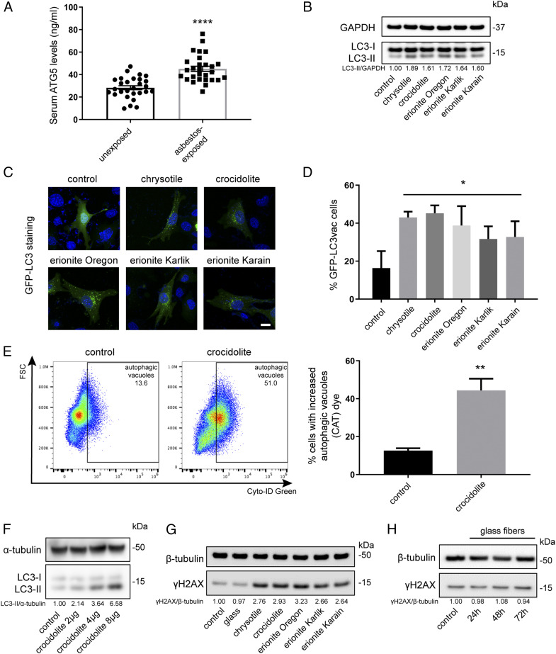
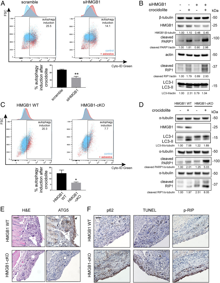
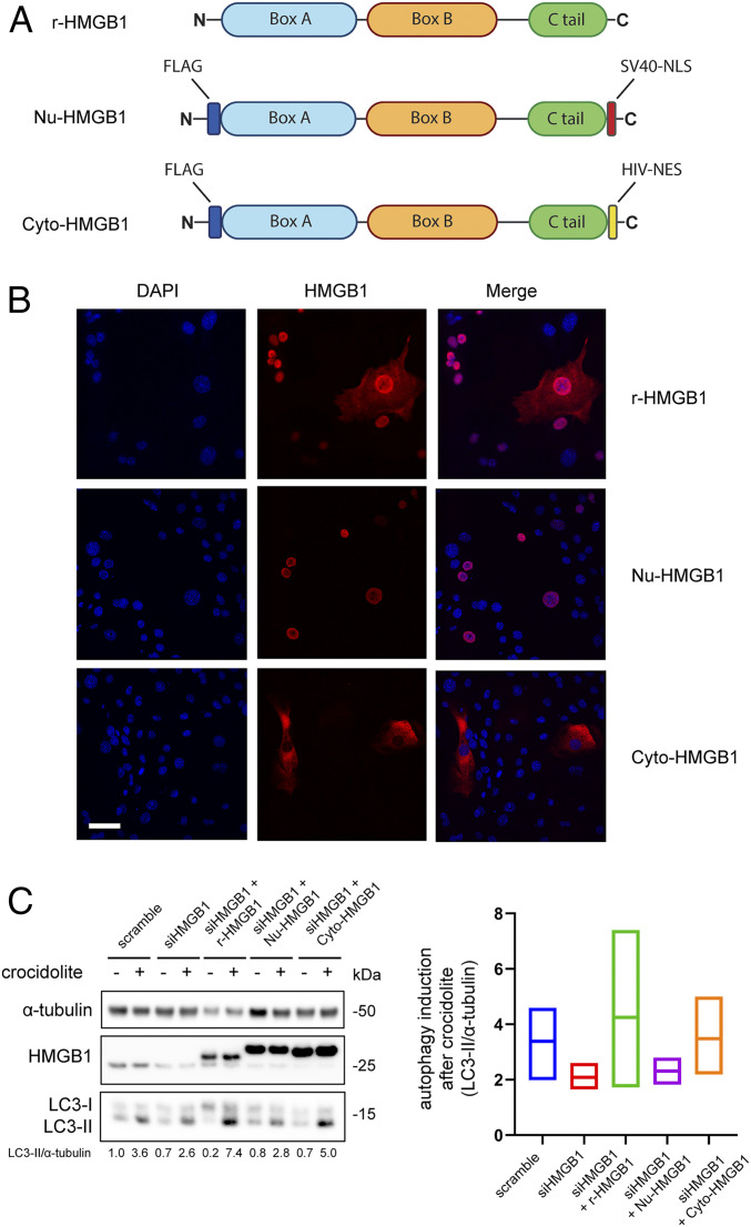
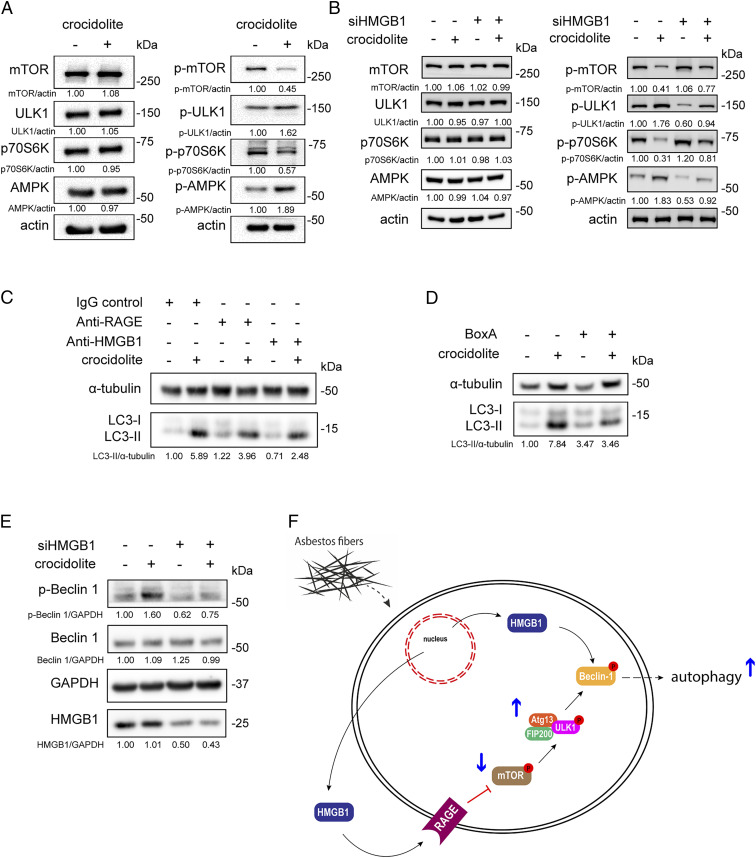
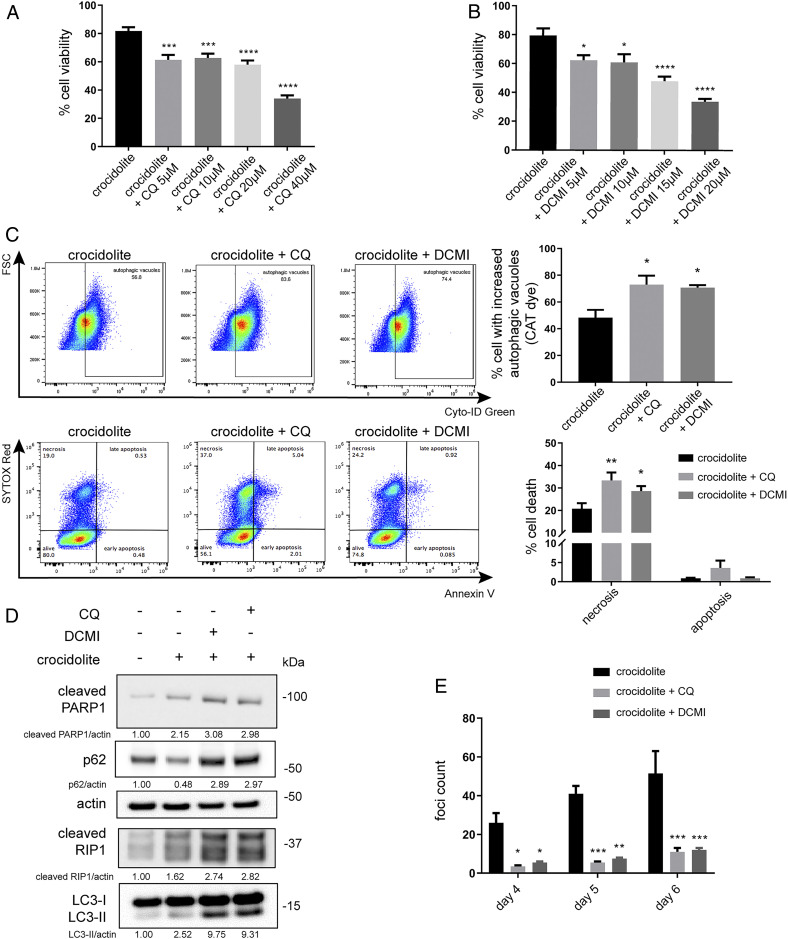
Asbestos causes malignant transformation of primary human mesothelial cells (HM), leading to mesothelioma. The mechanisms of asbestos carcinogenesis remain enigmatic, as exposure to asbestos induces HM death. However, some asbestos-exposed HM escape cell death, accumulate DNA damage, and may become transformed. We previously demonstrated that, upon asbestos exposure, HM and reactive macrophages releases the high mobility group box 1 (HMGB1) protein that becomes detectable in the tissues near asbestos deposits where HMGB1 triggers chronic inflammation. HMGB1 is also detectable in the sera of asbestos-exposed individuals and mice. Searching for additional biomarkers, we found higher levels of the autophagy marker ATG5 in sera from asbestos-exposed individuals compared to unexposed controls. As we investigated the mechanisms underlying this finding, we discovered that the release of HMGB1 upon asbestos exposure promoted autophagy, allowing a higher fraction of HM to survive asbestos exposure. HMGB1 silencing inhibited autophagy and increased asbestos-induced HM death, thereby decreasing asbestos-induced HM transformation. We demonstrate that autophagy was induced by the cytoplasmic and extracellular fractions of HMGB1 via the engagement of the RAGE receptor and Beclin 1 pathway, while nuclear HMGB1 did not participate in this process. We validated our findings in a novel unique mesothelial conditional HMGB1-knockout (HMGB1-cKO) mouse model. Compared to HMGB1 wild-type mice, mesothelial cells from HMGB1-cKO mice showed significantly reduced autophagy and increased cell death. Autophagy inhibitors chloroquine and desmethylclomipramine increased cell death and reduced asbestos-driven foci formation. In summary, HMGB1 released upon asbestos exposure induces autophagy, promoting HM survival and malignant transformation.

Keywords: HMGB1; asbestos; autophagy; cell death; mesothelioma.


Conflict of interest statement

Competing interest statement: M.C. has a patent issued for BAP1. M.C. and H.Y. have two patents issued for HMGB1. M.C. is a board-certified pathologist who provides consultation for pleural pathology, including medical–legal consultation.

Figures

Fig. 1.
Fig. 1.
Asbestos promotes autophagy in primary HM. (A) Serum levels of the autophagic marker ATG5 from 30 unexposed individuals and 29 asbestos-exposed individuals. ATG5 levels were significantly elevated in asbestos-exposed individuals (45.11 ± 12.14 ng/mL) compared to the unexposed group (28.29 ± 9.4 ng/mL) (ANOVA, ****P < 0.0001). (B) Representative Western blot showed increased LC3 lipidation (conversion of LC3-I, upper band, to LC3-II, lower band) 48 h after exposure to 4 μg/cm2 of the indicated carcinogenic fibers, compared to unexposed control cells. The densitometry ratios of LC3-II (an autophagy marker) over GAPDH were reported relative to the control. (C and D) HM were transduced with GFP-LC3 adenovirus, followed by exposure to the indicated carcinogenic fibers. Autophagy puncta were visualized by fluorescence microscopy (C), and the percentages of LC3 vacuolated cells per field were quantified (ANOVA, *P < 0.05) (D). (Scale bar in C, 10 μm.)(E) Asbestos-induced autophagy was measured with Cyto-ID Green staining (fluorescent cationic amphiphilic tracer [CAT]) by flow cytometry. HM undergoing autophagy (indicated by autophagic vacuoles) were quantified in the graph on the right (two-tailed unpaired t test, **P < 0.01). (F) Crocidolite-induced autophagy in HM correlated with fiber density: autophagy (LC3-II) became detectable after exposure to concentrations of 2 μg/cm2 of crocidolite and increased up to 8 μg/cm2. (G) Representative Western blot measuring levels of γH2AX (a marker of DNA damage/repair). HM exposed to 4 μg/cm2 of the indicated carcinogenic fibers for 48 h showing increased γH2AX levels compared to unexposed HM or to HM exposed to noncarcinogenic glass fibers. (H) HM exposed to noncarcinogenic glass fibers at 24, 48, and 72 h: no changes in γH2AX were observed at any time point.
Fig. 2.
Fig. 2.
Loss of HMGB1 reduces asbestos-induced autophagy and promotes cell death upon crocidolite asbestos exposure. (A) Flow cytometry with Cyto-ID Green labeling of HM 48 h post exposure to 4 μg/cm2 of crocidolite. In the overlay dot plots with the corresponding histograms (upper top graph), blue represents autophagy in unexposed control and red represents autophagy after crocidolite exposure. The difference between blue and red was reported as “autophagy induction.” Silencing of HMGB1 led to a significant decrease of asbestos-induced autophagy (bottom graph) (two-tailed unpaired t test, **P < 0.01). (B) Western blot showing that HMGB1 silencing (siHMGB1) led to the reduction of LC3-II (autophagy) in HM exposed to crocidolite while markers for apoptotic and necrotic pathways increased, as shown by the higher levels of cleaved PARP1 (apoptosis) and cleaved RIP1 (necrosis). (C) Flow cytometry of Cyto-ID–labeled primary murine mesothelial cells exposed to crocidolite. Asbestos-induced autophagy was significantly reduced in mesothelial cells isolated from HMGB1-cKO mice compared to cells from HMGB1 WT mice (two-tailed unpaired t test, *P < 0.05). In the overlay dot plots with the corresponding histograms as in A, blue represents autophagy in unexposed control cells and red represents autophagy in HM exposed to crocidolite. The difference between blue and red was reported as “autophagy induction.” (D) Representative Western blot of primary mesothelial cells from HMGB1-cKO mice showing decreased LC3-II and elevated cleaved PARP1 and cleaved RIP1 compared to cells from HMGB1 WT upon crocidolite exposure. (E) Representative H&E (hematoxylin and eosin) and ATG5 staining of peritoneal/diaphragm biopsies from mice exposed to crocidolite. Note the prominent chronic inflammation and foreign body giant-cell formation near fiber deposits in all of the microphotographs. HMGB1 WT mice displayed prominent ATG5 staining in hyperplastic mesothelial cells (arrow), while HMGB1-cKO mice did not. See also SI Appendix, Fig. S5D. (Scale bar, 50 µm.) (F) Compared to biopsies from HMGB1 WT, HMGB1-cKO mice mesothelial cells showed increased apoptosis (TUNEL, brown staining) and necrosis (p-RIP, brown staining) and reduced autophagy (increased p62, an autophagy marker, brown staining). (Scale bar, 50 µm.)
Fig. 3.
Fig. 3.
Cytoplasmic HMGB1 mediates asbestos-induced autophagy. (A) Schematic representation of the three HMGB1 constructs. R-HMGB1: recombinant human HMGB1. Nu-HMGB1: HMGB1 modified by insertion of the nuclear localization signal (NLS) from the SV40 virus. Cyto-HMGB1: HMGB1 bearing the nuclear export signal (NES) of HIV. (B) Immunofluorescence staining of the HMGB1-KO cell line transduced with one of the three designated HMGB1 constructs via adenovirus. R-HMGB1 was present in both nucleus and cytoplasm, whereas Nu-HMGB1 could be seen only inside the nucleus and Cyto-HMGB1 only inside in the cytoplasm. (Scale bar, 50 µm.) (C) HM were silenced with siHMGB1 for 48 h followed by transduction of the indicated HMGB1 constructs for an additional 24 h before crocidolite exposure. Silencing of HMGB1 led to a reduction in crocidolite-induced autophagy (LC3-II), which was rescued by transducing with either r-HMGB1 or Cyt-HMGB1, but not Nu-HMGB1 (Left). Rescue experiments were repeated three times on different HM, and the averages of autophagy induction after crocidolite exposure were quantified (Right). The middle lines of each bar represent the mean values.
Fig. 4.
Fig. 4.
Both cytoplasmic and extracellular HMGB1 modulate asbestos-induced autophagy. (A) Representative Western blot showing the activation of the mTOR-ULK1 autophagic pathway in HM after 48 h exposure to 4 μg/cm2 of crocidolite. The reduction of mTOR phosphorylation was accompanied by ULK1 and AMPK hyperphosphorylation. The level of p-p70S6K, a target of mTOR, was reduced, consistent with decreased mTOR activity. (B) HMGB1 silencing in HM blunted the activation of mTOR-ULK1 following crocidolite exposure. (C) Asbestos-induced autophagy (LC3-II) in HM treated with mouse IgG control (1.7 μg/mL), anti-RAGE (1.7 μg/mL), or anti-HMGB1 (1 μg/mL). LC3-II levels were partially reduced when using the neutralizing antibodies against either RAGE or HMGB1. (D) Representative Western blot of HM exposed to crocidolite in the presence or absence of 100 ng/mL BoxA. BoxA blocked the crocidolite-induced increase of LC3-II in HM. (E) Representative Western blot of p-Beclin 1 in HM silenced for HMGB1 and then exposed to crocidolite. Crocidolite induced the activation of Beclin 1 (p-Beclin 1), which was inhibited when HMGB1 was silenced. (F) Schematic representation of the asbestos-HMGB1-autophagy pathway. Asbestos induced HMGB1 translocation from the nucleus to the cytoplasm where HMGB1 is also released extracellularly. Extracellular HMGB1 exerted its effect through the RAGE-mTOR-ULK pathway, which converged with cytoplasmic HMGB1 to activate Beclin 1 and induce autophagy.
Fig. 5.
Fig. 5.
The autophagy inhibitor CQ and the antidepressant DCMI sensitize HM to asbestos toxicity and reduce asbestos-induced HM transformation. (A and B) MTS assays revealed that pretreatment with CQ (A) or DCMI (B) for 24 h, followed by crocidolite exposure for 48 h, reduced HM viability (ANOVA, *P < 0.05, ***P < 0.001, ****P < 0.0001). (C) Flow cytometry of HM pretreated with 10 μM of CQ or DCMI before crocidolite exposure. The cotreatment with CQ or DCMI and crocidolite increased the accumulation of autophagic vacuoles due to the inhibition of autophagic flux (Top). In parallel, we observed increased cell death following CQ or DCMI cotreatment with crocidolite (Bottom). Quantifications of autophagy and cell death are shown (Right Top and Bottom) (ANOVA, *P < 0.05; **P < 0.01). (D) Western blot for markers of autophagy (LC3-II and p62), apoptosis (cleaved PARP1), and necrosis (cleaved RIP1) in HM pretreated with CQ or DCMI before crocidolite exposure. Cotreatment with CQ or DCMI and crocidolite led to increased expressions of both LC3-II and p62, evidence that the autophagy flux was blocked. Elevations of cleaved PARP1 and cleaved RIP1 demonstrated increased cell death, consistent with the flow cytometry results shown in C. (E) Foci formation assay. HM were pretreated for 24 h with CQ or DCMI and then exposed to 5 μg/cm2 of crocidolite. Fresh medium containing CQ or DCMI was replaced every 48 h thereafter. The number of tridimensional foci formation was significantly lower in CQ or DCMI treated and crocidolite-exposed HM, compared to HM exposed only to crocidolite (ANOVA, *P < 0.05, **P < 0.01, ***P < 0.001).

References

    1. Carbone M. et al. ., Mesothelioma: Scientific clues for prevention, diagnosis, and therapy. CA Cancer J. Clin. 69, 402–429 (2019). - PMC - PubMed
    1. Baumann F., Ambrosi J. P., Carbone M., Asbestos is not just asbestos: An unrecognised health hazard. Lancet Oncol. 14, 576–578 (2013). - PubMed
    1. Brims F. J., Asbestos: A legacy and a persistent problem. J. R. Nav. Med. Serv. 95, 4–11 (2009). - PubMed
    1. Baumann F. et al. ., The presence of asbestos in the natural environment is likely related to mesothelioma in young individuals and women from Southern Nevada. J. Thorac. Oncol. 10, 731–737 (2015). - PMC - PubMed
    1. Carbone M. et al. ., Erionite exposure in North Dakota and Turkish villages with mesothelioma. Proc. Natl. Acad. Sci. U.S.A. 108, 13618–13623 (2011). - PMC - PubMed

Publication types