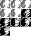
Pulmonary Sparganosis: Tunnel Sign and Migrating Sign on Computed Tomography
- PMID: 32999230
- PMCID: PMC7946513
- DOI: 10.2169/internalmedicine.5304-20
Pulmonary Sparganosis: Tunnel Sign and Migrating Sign on Computed Tomography
Abstract
A 77-year-old woman presented at our hospital to undergo a close examination of an abnormal shadow which was observed on a chest radiograph. Contrast-enhanced computed tomography (CT) images in the lung window revealed a tortuous tunnel structure (tunnel sign), which was suspected to be the migration path of a parasite. Furthermore, CT images in the mediastinal window showed a linear filling defect from the right inferior pulmonary vein to the venous ostium in the left atrium (migrating sign), which was suspected to be a migrating parasite in the pulmonary vein. Tunnel and migrating signs on chest CT images were helpful in diagnosing pulmonary sparganosis.
Keywords: computed tomography; migrating sign; pulmonary sparganosis; tunnel sign.
Conflict of interest statement
Figures


Similar articles
-
'Natural Conduit' between two atria associated with atrial septal defect.Int J Cardiovasc Imaging. 2005 Aug;21(4):379-82. doi: 10.1007/s10554-004-6135-y. Int J Cardiovasc Imaging. 2005. PMID: 16047117
-
The Tunnel Sign Revisited: A Novel Observation of Cerebral Melioidosis Mimicking Sparganosis.J Radiol Case Rep. 2018 Aug 31;12(8):1-11. doi: 10.3941/jrcr.v12i8.3441. eCollection 2018 Aug. J Radiol Case Rep. 2018. PMID: 30651915 Free PMC article.
-
A case with pseudo-scimitar syndrome: "scimitar sign" with normal pulmonary venous drainage.Jpn Circ J. 1987 Jun;51(6):642-6. doi: 10.1253/jcj.51.642. Jpn Circ J. 1987. PMID: 3669271
-
Computed Tomography Angiography and Magnetic Resonance Angiography of Congenital Anomalies of Pulmonary Veins.J Comput Assist Tomogr. 2019 May/Jun;43(3):399-405. doi: 10.1097/RCT.0000000000000857. J Comput Assist Tomogr. 2019. PMID: 31082945 Review.
-
Scimitar syndrome versus meandering pulmonary vein: evaluation with three-dimensional computed tomography.Acta Radiol. 2006 Nov;47(9):927-32. doi: 10.1080/02841850600885401. Acta Radiol. 2006. PMID: 17077042 Review.
Cited by
-
Case report: A suspected case of chronic pulmonary sparganosis characterized by migrating cavities and tunnel sign.Front Med (Lausanne). 2024 Sep 19;11:1453043. doi: 10.3389/fmed.2024.1453043. eCollection 2024. Front Med (Lausanne). 2024. PMID: 39364014 Free PMC article.
-
A Human Case of Lumbosacral Canal Sparganosis in China.Korean J Parasitol. 2021 Dec;59(6):635-638. doi: 10.3347/kjp.2021.59.6.635. Epub 2021 Dec 22. Korean J Parasitol. 2021. PMID: 34974670 Free PMC article.
References
-
- Baily G, Garcia HH. Other cestode infections: intestinal cestodes, cysticercosis, other larval cestode infections. In: Manson's Tropical Diseases. 23rd ed. Elsevier, London, 2014: 820-832.
-
- Chung SW, Kim YH, Lee EJ, Kim DH, Kim GY. Two cases of pulmonary and pleural sparganosis confirmed by tissue biopsy and immunoserology. Braz J Infect Dis 16: 200-203, 2012. - PubMed
-
- Li N, Xiang Y, Feng Y, Li M, Gao BL, Li QY. Clinical features of pulmonary sparganosis. Am J Med Sci 350: 436-441, 2015. - PubMed
-
- Kołodziej-Sobocińsk M, Miniuk M. Sparganosis-neglected zoonosis and its reservoir in wildlife. Med Weter 74: 224-227, 2018.
-
- Lo Presti A, Aguirre DT, De Andrés P, Daoud L, Fortes J, Muñiz J. Cerebral sparganosis: case report and review of the European cases. Acta Neurochir (Wien) 157: 1339-1343, 2015. - PubMed
Publication types
MeSH terms
LinkOut - more resources
Full Text Sources
Other Literature Sources

