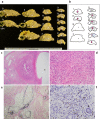
Type 1 Autoimmune Pancreatitis Extending along the Main Pancreatic Duct: IgG4-related Pancreatic Periductitis
- PMID: 32999241
- PMCID: PMC7990648
- DOI: 10.2169/internalmedicine.5754-20
Type 1 Autoimmune Pancreatitis Extending along the Main Pancreatic Duct: IgG4-related Pancreatic Periductitis
Abstract
We herein report a unique form of autoimmune pancreatitis (AIP) spreading along the main pancreatic duct (MPD). A 70-year-old man was referred for a small lesion at the pancreatic neck, accompanying an adjacent cyst and dilated upstream MPD. Four years earlier, health checkup images had shown a pancreatic cyst but no mass lesion. Endoscopic ultrasonography showed a contrast-enhanced, tumorous lesion, mainly occupying the MPD. With a preoperative diagnosis of ductal neoplasms mainly spreading in the MPD, Whipple's resection was performed. The resected specimens showed MPD periductitis with IgG4-related pathology, indicating type 1 AIP. Clinicians should practice caution concerning the various AIP forms.
Keywords: IgG4; autoimmune pancreatitis; diagnosis; intraductal neoplasms; periductitis.
Conflict of interest statement
Figures


References
-
- Okazaki K, Tomiyama T, Mitsuyama T, Sumimoto K, Uchida K. Diagnosis and classification of autoimmune pancreatitis. Autoimmun Rev 13: 451-458, 2014. - PubMed
-
- Shimosegawa T, Chari ST, Frulloni L, et al. . International consensus diagnostic criteria for autoimmune pancreatitis: guidelines of the International Association of Pancreatology. Pancreas 40: 352-358, 2011. - PubMed
-
- Umemura S, Naitoh I, Nakazawa T, et al. . Autoimmune pancreatitis presenting a short narrowing of main pancreatic duct with subsequent progression to diffuse pancreatic enlargement over 24 months; natural history of autoimmune pancreatitis. JOP 15: 261-265, 2014. - PubMed
-
- Koshita S, Noda Y, Ito K, et al. . Branch duct intraductal papillary mucinous neoplasms of the pancreas involving type 1 localized autoimmune pancreatitis with normal serum IgG4 levels successfully diagnosed by endoscopic ultrasound-guided fine-needle aspiration and treated without pancreatic surgery. Intern Med 56: 1163-1167, 2017. - PMC - PubMed
Publication types
MeSH terms
Substances
LinkOut - more resources
Full Text Sources
Other Literature Sources
Medical
Miscellaneous

