. 2020 Sep 30;3(1):542.
doi: 10.1038/s42003-020-01265-w.

Micromanipulation of prophase I chromosomes from mouse spermatocytes reveals high stiffness and gel-like chromatin organization

Affiliations

Micromanipulation of prophase I chromosomes from mouse spermatocytes reveals high stiffness and gel-like chromatin organization

Ronald J Biggs et al. Commun Biol. .

Abstract

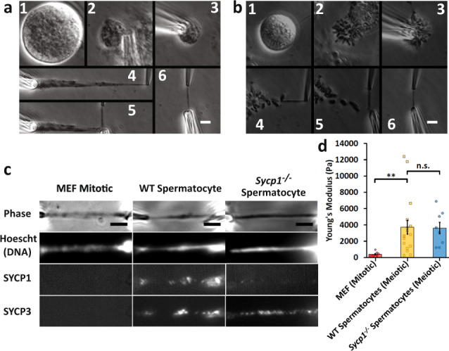
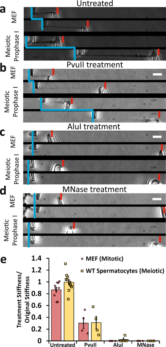
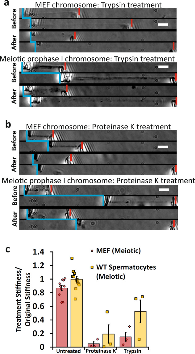
Meiosis produces four haploid cells after two successive divisions in sexually reproducing organisms. A critical event during meiosis is construction of the synaptonemal complex (SC), a large, protein-based bridge that physically links homologous chromosomes. The SC facilitates meiotic recombination, chromosome compaction, and the eventual separation of homologous chromosomes at metaphase I. We present experiments directly measuring physical properties of captured mammalian meiotic prophase I chromosomes. Mouse meiotic chromosomes are about ten-fold stiffer than somatic mitotic chromosomes, even for genetic mutants lacking SYCP1, the central element of the SC. Meiotic chromosomes dissolve when treated with nucleases, but only weaken when treated with proteases, suggesting that the SC is not rigidly connected, and that meiotic prophase I chromosomes are a gel meshwork of chromatin, similar to mitotic chromosomes. These results are consistent with a liquid- or liquid-crystal SC, but with SC-chromatin stiff enough to mechanically drive crossover interference.


Conflict of interest statement

The authors declare no competing interests.

Figures

Fig. 1
Fig. 1. Chromosome isolation and micromanipulation.
Images show steps for isolation of meiotic (a) and mitotic (b) chromosomes. a A large spermatocyte containing chromosomes (subpanel 1) was identified, and lysed with Triton X-100 in a pipette to release the prophase nucleus (2). Another pipette then was used to grab and hold the nucleus (3). A chromosome end at the edge of the nucleus then was grabbed with a soft force-measuring pipette (4); the other chromosome end was captured with a stiff pulling pipette (5), and the rest of the nucleus was removed, leaving only the prophase chromosomes on the pipettes (6). b A rounded mitotic cell with visible dark chromosomes in prometaphase was identified (1), lysed with Triton X-100 sprayed from a pipette to release the prometaphase mitotic bundle (2), and another pipette then grabbed and held the bundle (3). A chromosome end at the edge of the bundle was grabbed with a thin, force pipette and moved away from the bundle (4), where the other end of the chromosome was grabbed by a stiff, pulling pipette (5), and the bundle was removed, leaving one chromosome isolated between two pipettes (6). Scale bars are 5 μm. c Distinguishing mitotic MEF from meiotic prophase spermatocyte chromosomes using fluorescence microscopy. MEF chromosomes appear as rod-like structures in the phase-contrast channel that stain for DNA, but do not immunostain for SYCP1 or SYCP3. Wildtype spermatocyte chromosomes appear as rod-like structures that stain for DNA, SYCP1, and SYCP3. Sycp1−/− spermatocyte chromosomes stain for DNA and SYCP3, but not SYCP1. Scale bar is 5 μm. d Stiffness (Young’s Modulus) of mitotic and meiotic chromosomes. MEF chromosomes have a stiffness of 370 ± 70 Pascals (Pa, N = 10), ten-fold weaker than wildtype spermatocyte chromosomes (3700 ± 800 Pa, N = 17, p = 0.006 relative to MEF). Wildtype and Sycp1−/− spermatocyte chromosomes (Sycp1−/− modulus 3600 ± 700 Pa, N = 8, p = 0.0001 relative to MEF) do not have a statistically significant difference in stiffness (p = 0.93). All averages are reported as mean value ± SEM. All p values calculated via t-test.
Fig. 2
Fig. 2. Mitotic and meiotic chromosomes have a contiguous DNA connection, which is dissolved by 4 bp restriction enzymes, but only weakened by 6 bp restriction enzymes.
Image pairs show pipette positions untreated (native isolated) chromosomes when relaxed and stretched (a), and chromosomes following enzyme treatments (bd). Vertical blue lines mark positions of force pipettes. Force pipette deflection by pulling (horizontal blue lines) indicates mechanical connection; no movement (no horizontal blue line) indicates no mechanical connection. Red notches mark positions of stiff pipettes. Bars are 5 µm. b Both mitotic and meiotic chromosomes were weakened, but not fully digested after treatment with PvuII (cut sequence CAG˅CTG). c Both mitotic and meiotic chromosomes lost connectivity after treatment with AluI (cut sequence AG˅CT; for 1 of 4 trials meiotic chromosomes were not fully digested by AluI). d Both mitotic and meiotic chromosomes lost connectivity when treated with MNase (cleaves all DNA sequences). e Quantification of chromosome stretching elasticity after no treatment or after being treated with PvuII, AluI, and MNase. No treatment caused a 13 ± 4% weakening of mitotic chromosomes (N = 10) and a 1 ± 4% weakening of meiotic chromosomes (N = 10). PvuII treatment caused a 70 ± 8% reduction in stiffness for MEF chromosomes (N = 4) and 70 ± 9% reduction in stiffness for meiotic chromosomes (N = 4). One of four AluI treatments of meiotic chromosomes caused a 90% reduction in stiffness (rather than fully digesting), while AluI treatment digested 4 of 4 mitotic chromosomes. All MNase treatments caused full digestion of mitotic and meiotic chromosomes (N = 4 in both cases). All averages are reported as mean value ± SEM. Bars are 5 µm.
Fig. 3
Fig. 3. Both meiotic and mitotic chromosomes are weakened, but do not dissolve when treated with Trypsin and Proteinase K.
a, b Vertical blue lines indicate force-measuring pipette positions. Force pipette movement means connection (indicated by horizontal blue line connecting relaxed and stretched force pipette positions); no movement means no connection. Red notches mark positions of stiff pipettes. Image pairs show pipettes for relaxed and stretched chromosomes. Before enzyme treatments, the left pipette is deflected indicating force transmitted through the chromosome. Bars are 5 µm. a MEF chromosomes lose all phase contrast after treatment with trypsin; but still are well connected enough to move the force pipette, and are weaker than before treatment (smaller deflection during pulling, after panel). Similarly, meiotic chromosomes lose most definition in the phase-contrast channel, but can move force pipette after digestion when treated with Trypsin, and are weaker than before treatment. b MEF chromosomes lose all definition in the phase-contrast channel, but can move force pipette after digestion when treated with Proteinase K and are weaker than before treatment. Similarly, meiotic chromosomes lose most definition in the phase-contrast channel, but can move force pipette after digestion by Proteinase K and are weaker than before treatment. c Quantification of untreated and protease-treatment weakening of mitotic and meiotic chromosomes. No treatment caused a 13 ± 4% weakening of mitotic chromosomes (N = 10) and a 1 ± 4% weakening of meiotic chromosomes (N = 10). Trypsin treatment caused an 85 ± 6% weakening in mitotic chromosomes (N = 3) and a 48 ± 17% weakening in meiotic chromosomes (N = 3). Proteinase K treatment caused a 95 ± 3% weakening in meiotic chromosomes (N = 3) and an 81 ± 13% weakening in meiotic chromosomes (N = 3). All averages are reported as mean value ± SEM.

References

    1. Bolcun-Filas E, Schimenti JC. Genetics of meiosis and recombination in mice. Int. Rev. Cell Mol. Biol. 2012;298:179–227. doi: 10.1016/B978-0-12-394309-5.00005-5. - DOI - PubMed
    1. Fraune J, Schramm S, Alsheimer M, Benavente R. The mammalian synaptonemal complex: protein components, assembly and role in meiotic recombination. Exp. Cell Res. 2012;318:1340–1346. doi: 10.1016/j.yexcr.2012.02.018. - DOI - PubMed
    1. Hassold T, Hunt P. To err (meiotically) is human: the genesis of human aneuploidy. Nat. Rev. Genet. 2001;2:280–291. doi: 10.1038/35066065. - DOI - PubMed
    1. Morelli MA, Werling U, Edelmann W, Roberson MS, Cohen PE. Analysis of meiotic prophase I in live mouse spermatocytes. Chromosome Res. 2008;16:743–760. doi: 10.1007/s10577-008-1224-8. - DOI - PubMed
    1. Ivanovska I, Orr-Weaver TL. Histone modifications and the chromatin scaffold for meiotic chromosome architecture. Cell Cycle. 2006;5:2064–2071. doi: 10.4161/cc.5.18.3253. - DOI - PubMed

Publication types