. 2021 Feb;15(2):473-488.
doi: 10.1038/s41396-020-00789-7. Epub 2020 Sep 30.

Direct interactions with commensal streptococci modify intercellular communication behaviors of Streptococcus mutans

Affiliations

Direct interactions with commensal streptococci modify intercellular communication behaviors of Streptococcus mutans

Justin R Kaspar et al. ISME J. 2021 Feb.

Abstract

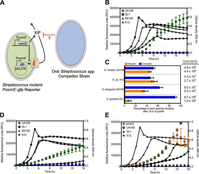
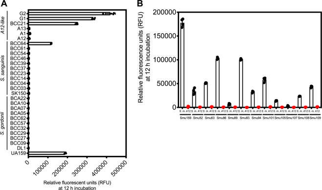
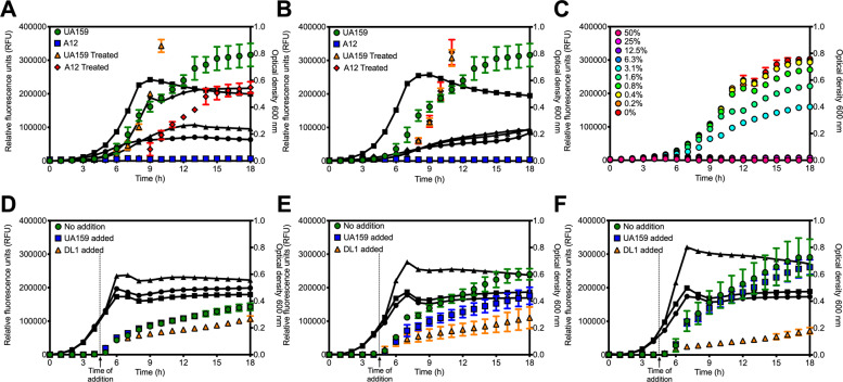
The formation of dental caries is a complex process that ultimately leads to damage of the tooth enamel from acids produced by microbes in attached biofilms. The bacterial interactions occurring within these biofilms between cariogenic bacteria, such as the mutans streptococci, and health-associated commensal streptococci, are thought to be critical determinants of health and disease. To better understand these interactions, a Streptococcus mutans reporter strain that actively monitors cell-cell communication via peptide signaling was cocultured with different commensal streptococci. Signaling by S. mutans, normally highly active in monoculture, was completely inhibited by several species of commensals, but only when the bacteria were in direct contact with S. mutans. We identified a novel gene expression pattern that occurred in S. mutans when cultured directly with these commensals. Finally, mutant derivatives of commensals lacking previously shown antagonistic gene products displayed wild-type levels of signal inhibition in cocultures. Collectively, these results reveal a novel pathway(s) in multiple health-associated commensal streptococci that blocks peptide signaling and induces a common contact-dependent pattern of differential gene expression in S. mutans. Understanding the molecular basis for this inhibition will assist in the rational design of new risk assessments, diagnostics, and treatments for the most pervasive oral infectious diseases.


Conflict of interest statement

The authors declare that they have no conflict of interest.

Figures

Fig. 1
Fig. 1. Loss of S. mutans peptide signaling in presence of competitor.
a An oral Streptococcus spp. competitor strain (blue) was cocultured in chemically defined medium (CDM) with an S. mutans PcomX::gfp reporter strain (green). As cell density of the reporter strain increases during growth, the XIP peptide that originates from the comS gene will be produced and accumulates extracellularly. XIP is then reimported into the cell through the Opp oligopeptide permease, binds to ComR and activates the comX promoter. Additionally, intracellular signaling occurs with ComS binding directly to ComR. The reporter strain harbors a plasmid, pDL278, carrying a copy of gfp that is driven by the comX promoter (PcomX) to monitor ComRS signaling activation. b Cocultures of the S. mutans PcomX::gfp reporter strain grown with either S. mutans UA159 (control, green circles), S. gordonii DL1 (blue squares), S. sanguinis SK150 (orange triangles), or S. sp. A12 (red diamonds). Colored, non-connected symbols represent relative fluorescent units (RFUs) plotted on the left y-axis, while black, connected lines with symbols represent growth of the cocultures over the course of the experiment measured by optical density at 600 nm plotted on the right y-axis. Data are averages from three biological replicates of the experiment. c Percentage of each species remaining within the coculture after 18 h of monitoring, determined by colony forming unit (CFU) plating. The PcomX::gfp reporter strain is represented in the orange bars, while the competitor, listed on the left y-axis, is represented in blue. Average of collected CFUs is shown to the right. Data represent averages from three biological replicates of the experiment that was conducted in panel (b). d Cocultures of the S. mutans PcomX::gfp reporter strain in which 5 µM sXIP was added prior to the start of the experiment. e Cocultures of the S. mutans PcomX::gfp reporter strain that contains a plasmid that overexpresses the XIP peptide precursor, ComS. Control represents the PcomX::gfp reporter that contained an empty vector only.
Fig. 2
Fig. 2. S. mutans peptide signaling in coculture biofilms.
a 3D volume projections of imaged biofilms in the XY-orientation (from the top looking down). Each biofilm contains either S. mutans UA159 with a constitutive gfp reporter plasmid (top row), or the PcomX::gfp reporter plasmid (bottom row) that was cocultured with either S. mutans (control; left), S. gordonii DL1 (middle), or S. sp. A12 (right) who all constitutively produce DsRed2. To the right of each expanded color image is the black and white image capture of each individual channel: blue (top), green (middle), and red (bottom). b Zoomed image frames of PcomX-active cells within cocultured biofilms with S. gordonii DL1. The images captured are a single z plane near or at the biofilm substratum. Two different areas of the biofilm (top and bottom rows) were imaged. Each panel represents one color channel of blue (SYTO 42 stained; total cells), green (PcomX::gfp positive cells), or red (S. gordonii P23::DsRed2) followed by the merged image on the far right. The top panel of (b) is the same area of biofilm shown in Movie S1.
Fig. 3
Fig. 3. Cell contact dependence in signaling inhibition.
a Growth and fluorescence of S. mutans PcomX::gfp reporter strain in spent supernatant fluids of either S. mutans UA159 (control, green circles), S. gordonii DL1 (blue squares), S. sanguinis SK150 (orange triangles), or S. sp. A12 (red diamonds). Depiction on top shows methods used to treat supernatant fluids following harvesting and prior to reporter strain inoculation. Overnight cultures of selected strains where centrifuged, spent supernates removed, filter sterilized, the pH was adjusted to 7.0 and 20 mM additional glucose was added. The PcomX::gfp reporter strain was then inoculated and monitored for 18 h in a Synergy 2 multimode plate reader. b Growth of cocultures in a transwell apparatus. The PcomX::gfp reporter strain was first inoculated in 0.1 mL of CDM medium in a 96-well microtiter plate. The transwell plate was then overlaid on top of the 96-well plate, and 0.1 mL of CDM inoculated with either S. mutans UA159 (control, green circles), S. gordonii DL1 (blue squares), S. sanguinis SK150 (orange triangles), or S. sp. A12 (red diamonds) was added to the top chamber, as shown. Cultures of the reporter strain and competitor were separated by a 0.4 µM pore size polycarbonate filter membrane. Fluorescence (RFUs) of the reporter strain was monitored for 18 h.
Fig. 4
Fig. 4. Conservation of ComRS signaling antagonism across oral isolates.
a Relative fluorescent units (RFUs) of the S. mutans PcomX::gfp reporter strain cocultured with clinical oral isolates of either S. gordonii, S. sanguinis or S. sp. A12-like strains. Relative fluorescent units were recorded after coculture inoculation at 1:1 ratio and 12 h of incubation at 37 °C. Results from four biological replicates of the experiment are shown. b RFUs after 12 h of incubation of the PcomX::gfp reporter harbored in various S. mutans clinical isolates. The PcomX::gfp reporter strain was cocultured with either S. mutans UA159 (control; black dots and bars) or an oral competitor streptococci (S. sp. A12, red dots and bars).
Fig. 5
Fig. 5. Importance of oral competitor cell density in signaling inhibition.
Cocultures of the S. mutans PcomX::gfp reporter strain with untreated or treated cells by either a 0.5 h heat inactivation at 80 °C or b 1 h suspension in 4% paraformaldehyde. Data represent averages from three biological replicates. c Dilution of an oral competitor streptococci (S. sp. A12) in coculture with the S. mutans PcomX::gfp reporter strain. Legend (top left) refers to the amount of S. sp. A12 within the coculture at the time of initial inoculation. Bottom: addition of either control (UA159; blue squares) or an oral competitor streptococci (S. gordonii DL1; orange triangles) at 4.5 h to a growing culture of the S. mutans PcomX::gfp strain when competence activation was d fully detected, e beginning to be detected, or f not yet detected. See Supplementary Fig. 7 for comparisons at 4.5 and 12 h, specifically.
Fig. 6
Fig. 6. Transcriptome Profiling S.mutans culture in supernates or in direct contact.
Volcano plots from transcriptome analysis of a S. mutans UA159 cultured in S. sp. A12 spent supernatant fluid compared to culturing in S. mutans UA159 spent supernatant fluid and b S. mutans UA159 directly cocultured with S. sp. A12 compared to culturing in S. sp. A12 spent supernatant fluid only. Data represent three independent replicates of each condition. Log2 fold changes and false discovery rates (FDR) converted to −log10 p values were calculated from the Degust website using edgeR analysis. Genes of interest that were ≥(−)1.5 log2 fold change and ≥4 −log10 p values were highlighted either in blue (downregulated, upper left quadrant) or red (upregulated, upper right quadrant) and are listed in Supplementary Tables 2 and 3, respectively. qRT-PCR confirmation of selected c upregulated or d downregulated genes from transcriptome analysis. Cocultures of S. mutans UA159 and selected competitors were grown in CDM medium to OD600 = 0.5 before harvesting for RNA extraction. Data represent fold change in gene expression compared to S. mutans UA159 monocultures. Three independent cocultures were analyzed and plotted.
Fig. 7
Fig. 7. Selected competitor mutants still inhibit S. mutans peptide signaling.
Cocultures of the S. mutans PcomX::gfp reporter strain with mutants of selected oral commensal streptococci. Comparison of relative fluorescent units (RFUs) after 12 h of incubation in strains of a S. gordonii DL1, b S. sp. A12, and c in either aerobic (black bars) or anaerobic (red bars) conditions. Data are averages from four biological replicates of the experiment. The changes between parental and selected commensal mutants are all not significant. Statistical analysis shown in (c) was completed using Student’s t test.
Fig. 8
Fig. 8. Model for S. mutans peptide signaling inhibition.
Three different models of direct contact with commensal streptococci (blue cells) leading to S. mutans (direct contact white cells with green outline, noncontact green cells) peptide signaling inhibition and conserved gene expression patterns. a A commensal-derived inhibitory product from the initiation of contact with S. mutans is secreted to modify S. mutans behavior and peptide sensing across the entire S. mutans population. b A subset of S. mutans that is in direct contact with commensals produces an inhibitory signal that modifies behavior for the rest of the population, including those noncontacted S. mutans cells. c Transient contact between an oral commensal (blue) and S. mutans cell (green/green outline), prior to ComRS signaling activation, leads to posttranscriptional modifications in ComRS signaling. In the proposed mechanism shown, contact between S. mutans and commensal leads to sequestering of ComR with an unknown interaction partner, preventing ComR-XIP complex binding to the PcomX promoter and genetic competence activation.

Similar articles

Cited by

References

    1. Ghoul M, Mitri S. The ecology and evolution of microbial competition. Trends Microbiol. 2016;24:833–45. - PubMed
    1. Granato ET, Meiller-Legrand TA, Foster KR. The evolution and ecology of bacterial warfare. Curr Biol. 2019;29:R521–37. - PubMed
    1. Greenberg EP. Bacterial communication and group behavior. J Clin Invest. 2003;112:1288–90. - PMC - PubMed
    1. Kolenbrander PE, Andersen RN, Blehert DS, Egland PG, Foster JS, Palmer RJ. Communication among oral bacteria. Microbiol Mol Biol Rev. 2002;66:486–505. - PMC - PubMed
    1. Waters CM, Bassler BL. Quorum sensing: cell-to-cell communication in bacteria. Annu Rev Cell Dev Biol. 2005;21:319–46. - PubMed

Publication types