

Routine Orthoptic-led Paediatric Fundus Digital Imaging: Benefits to Patients and Healthcare System
- PMID: 32999957
- PMCID: PMC7510548
- DOI: 10.22599/bioj.106
Routine Orthoptic-led Paediatric Fundus Digital Imaging: Benefits to Patients and Healthcare System
Abstract
Aims: Dilated fundus examinations are a vital, yet time-consuming and sometimes distressing part of paediatric ophthalmology examinations. Limited resources, personnel and time can result in prolonged waiting time and increase risk from delayed diagnosis and treatment. Using a Nikon D80 TopCon TRC-NW6S non-mydriatic fundus camera (TopCon (GB) Ltd, Newbury), we aimed to demonstrate the safety and efficacy of orthoptic-led fundus digital imaging and the potential time and cost benefits to the healthcare system.
Methods: We conducted a retrospective review of all digital fundus images taken over a six month period in 2012 (n = 616, age range 2.1-16.5 years, mean age 8.7 years).
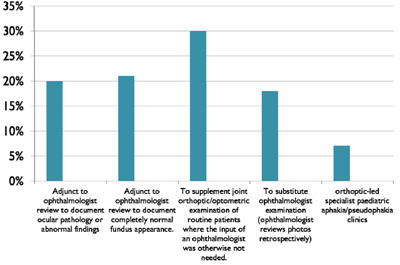
Results: Overall success rate for paediatric fundus digital imaging was 97%. Successful images were achieved in 87% of patients without the need for pupil dilation. Images were taken for a variety of clinical reasons. 45% of patients were discharged immediately, many with copies of photographs to facilitate follow-up with community optometrists.
Conclusions: Orthoptic-led fundus digital imaging is an innovative, speedy, safe and efficient method of documenting fundal appearance, enabling serial documentation of stability/progression of ocular disease. It allows adequate examination of routine patients, freeing up time within busy clinics. Paediatric fundus digital imaging brings a potential positive cost benefit to healthcare systems under pressure, and facilitates skill development for allied health professionals.
Keywords: fundus digital imaging; orthoptic-led; paediatric.
Copyright: © 2018 The Author(s).
Conflict of interest statement
The authors have no competing interests to declare.
Figures


References
-
- Farukhi, F, Dakkouri, C, Wang, H, et al. 2006. Etiology of Vision Loss in Ganglioside GM3 Synthase Deficiency. Ophthalmic Genet., 27 (3): 89–91. September. - PubMed
LinkOut - more resources
Full Text Sources
