

Type A aortic dissection mimicking saddle pulmonary embolism on CT imaging
- PMID: 33000025
- PMCID: PMC7493540
- DOI: 10.1002/emp2.12026
Type A aortic dissection mimicking saddle pulmonary embolism on CT imaging
Abstract
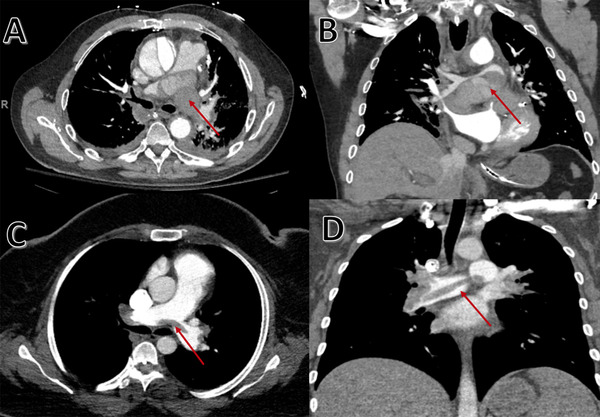
Type A aortic dissection is an uncommon cause of chest pain that carries a high morbidity and mortality rate. A previous history of hypertension and coronary artery bypass grating (CABG) are recognized risk factors for Type A aortic dissection. We present a case of an elderly man who presents with acute onset chest pain and was found to have an acute ruptured Type A aortic dissection that has a "saddle pulmonary embolism"-like appearance on computed tomography (CT) imaging. We also describe the clinical, laboratory, and radiological workup done leading up to the diagnosis of Type A aortic dissection in the emergency setting.
Keywords: Aortic Dissection Detection Risk Score (ADD‐RS); Mediastinal Hematoma; Point of Care Ultrasound (POCUS); Pulmonary Artery Compression; Type A Aortic Dissection.
© 2020 The Authors. JACEP Open published by Wiley Periodicals, Inc. on behalf of the American College of Emergency Physicians.
Conflict of interest statement
The authors have no conflicts of interest to disclose.
Figures


References
-
- Johansson G, Markström U, Swedenborg J. Ruptured thoracic aortic aneurysms: a study of incidence and mortality rates. J Vasc Surg. 1995;21(6):985‐988. - PubMed
-
- Tsai TT, Nienaber CA, Eagle KA. Acute aortic syndromes. Circulation. 2005;112(24):3802‐3813. - PubMed
-
- Nienaber CA, Eagle KA. Aortic dissection: new frontiers in diagnosis and management: Part I: from etiology to diagnostic strategies. Circulation. 2003;108(5):628‐635. - PubMed
-
- Hagan PG, Nienaber CA, Isselbacher EM, et al. The International Registry of Acute Aortic Dissection (IRAD): new insights into an old disease. JAMA. 2000;283(7):897‐903. - PubMed
Publication types
LinkOut - more resources
Full Text Sources
