. 2020 Nov;22(5):4221-4226.
doi: 10.3892/mmr.2020.11510. Epub 2020 Sep 15.

Neuropilin‑1 as a new potential SARS‑CoV‑2 infection mediator implicated in the neurologic features and central nervous system involvement of COVID‑19

Affiliations

Neuropilin‑1 as a new potential SARS‑CoV‑2 infection mediator implicated in the neurologic features and central nervous system involvement of COVID‑19

Julie Davies et al. Mol Med Rep. 2020 Nov.

Abstract

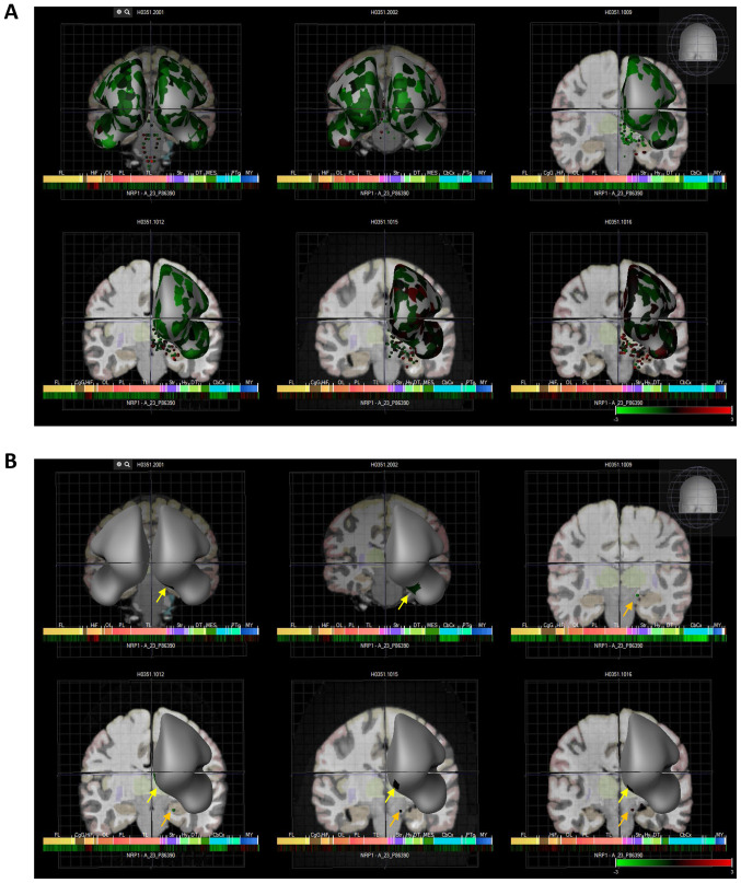
Infection by the severe acute respiratory syndrome (SARS) coronavirus‑2 (SARS‑CoV‑2) is the cause of the new viral infectious disease (coronavirus disease 2019; COVID‑19). Emerging evidence indicates that COVID‑19 may be associated with a wide spectrum of neurological symptoms and complications with central nervous system (CNS) involvement. It is now well‑established that entry of SARS‑CoV‑2 into host cells is facilitated by its spike proteins mainly through binding to the angiotensin‑converting enzyme 2 (ACE‑2). Preclinical studies have suggested that neuropilin‑1 (NRP1), which is a transmembrane receptor that lacks a cytosolic protein kinase domain and exhibits high expression in the respiratory and olfactory epithelium, may also be implicated in COVID‑19 by enhancing the entry of SARS‑CoV‑2 into the brain through the olfactory epithelium. In the present study, we expand on these findings and demonstrate that the NRP1 is also expressed in the CNS, including olfactory‑related regions such as the olfactory tubercles and paraolfactory gyri. This furthers supports the potential role of NRP1 as an additional SARS‑CoV‑2 infection mediator implicated in the neurologic manifestations of COVID‑19. Accordingly, the neurotropism of SARS‑CoV‑2 via NRP1‑expressing cells in the CNS merits further investigation.


Figures

Figure 1.
Figure 1.
NRP1 expression in the human brain. (A) Regarding cell types, based on RNA-Seq data, NRP1 is highly expressed in mature astrocytes (mature astrocytes have multiple functions, including controlling the permeability of the blood brain barrier and maintaining extracellular homeostasis), microglia/macrophages, and endothelial cells. Insert on panel A; FPKM reads. (B) Regarding brain regions, NRP1 exhibits widespread expression in the human brain (including the olfactory regions) based on data from both RNA-Seq expression using CAGE on FANTOM5 and (C) RNA expression (NX) of normalised NRP1 expression levels from 10 brain regions. The insert panel of brain anatomy in panels B and C indicates in colour the different anatomical structures. NRP1, neuropilin-1; RNA-Seq, RNA sequencing; FPKM, fragments per kilobase of transcript per million mapped; CAGE, Cap Analysis of Gene expression; FANTOM5, functional annotation of mammalian genome 5.
Figure 2.
Figure 2.
Single cell analysis of the hypothalamic Arc-ME complex, using the Single Cell Portal, revealed distinct expression of NRP1. (A) Arc-ME single-cell transcriptomics data are displayed as a spectral tSNE plot of 20,921 cells, coloured per density clustering and annotated according to known cell types. (B) Sub-populations of cell types which are enriched for NRP1 expression, with expression intensity demonstrated by a heat map. (C) Single cell analysis of the hypothalamic Arc-ME complex, using the Single Cell Portal, enriched for population subtypes, represented as violin plots. Arc-ME, arcuate and median eminence; tSNE, T-distributed Stochastic Neighbor Embedding.
Figure 3.
Figure 3.
Six human donors and heat map expression of NRP1 as assessed by microarray (Allen brain atlas). (A) Indicates global brain expression of NRP1 as detected by microarray probes, with expression of cerebral areas indicated on the hemispheres and expression in brain nuclei indicated as dots. Heat maps of expression are displayed below individual brains and organised by anterior to posterior regions with the FL, HiF, OL, PL, TL, Str, DT, CbCx and MY marked on the heat map. (B) Shows expression in olfactory accessory tissues with yellow arrows indicating the paraolfactory gyri and orange arrows indicating the olfactory tubercle. NRP1, neuropilin-1; FL, frontal lobe; HiF, hippocampal formation; OL, occipital lobe; PL, parietal lobe; TL, temporal lobe; Str, striatum; DT, dorsal thalamus; CbCx, cerebral cortex, MY, myelencephalon.

References

    1. Domingo P, Mur I, Pomar V, Corominas H, Casademont J, de Benito N. EBioMedicine. Vol. 58. Elsevier B.V.; 2020. (2020). The four horsemen of a viral Apocalypse: The pathogenesis of SARS-CoV-2 infection (COVID-19) - DOI - PMC - PubMed
    1. World Health Organisation (WHO), corp-author Coronavirus disease 2019 (COVID-19): Weekly Epidemiological Update. 21 August 2020. www.who.int/emergencies/diseases/novel-coronavirus-2019/situation-reports. Last accessed August 22, 2020.
    1. Gavriatopoulou M, Korompoki E, Fotiou D, Ntanasis- Stathopoulos I, Psaltopoulou T, Kastritis E, Terpos E, Dimopoulos MA. Organ-specific manifestations of COVID-19 infection. https://doi.org/10.1007/s10238-020-00648-x. Clin Exp Med. - PMC - PubMed
    1. De Felice FG, Tovar-Moll F, Moll J, Munoz DP, Ferreira ST. Severe Acute Respiratory Syndrome Coronavirus 2 (SARS-CoV-2) and the Central Nervous System. Trends Neurosci. 2020;43:355–357. doi: 10.1016/j.tins.2020.04.004. - DOI - PMC - PubMed
    1. Mao L, Jin H, Wang M, Hu Y, Chen S, He Q, Chang J, Hong C, Zhou Y, Wang D, et al. Neurologic manifestations of hospitalized patients with Coronavirus Disease 2019 in Wuhan, China. JAMA Neurol. 2020;77:683. doi: 10.1001/jamaneurol.2020.1127. - DOI - PMC - PubMed