. 2020 Oct 1;9(1):17.
doi: 10.1186/s13619-020-00057-1.

Positive feedback between retinoic acid and 2-phospho-L-ascorbic acid trisodium salt during somatic cell reprogramming

Affiliations

Positive feedback between retinoic acid and 2-phospho-L-ascorbic acid trisodium salt during somatic cell reprogramming

Mengdan Zhang et al. Cell Regen. .

Abstract

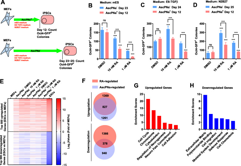
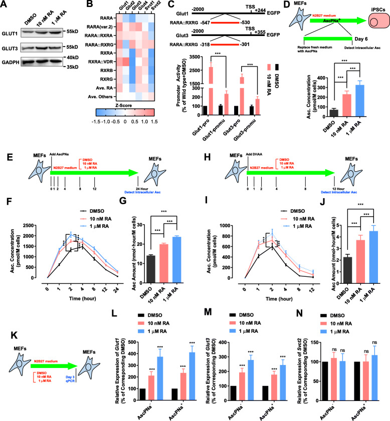
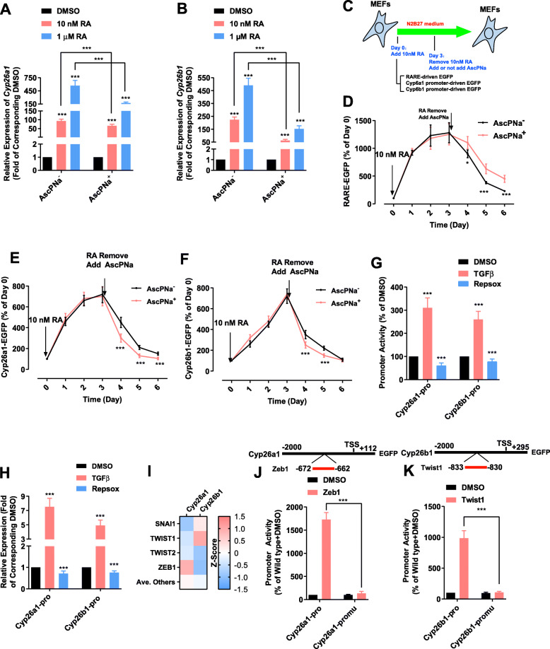
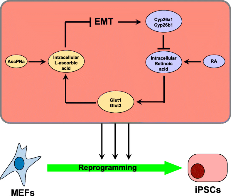
Retinoic acid (RA) and 2-phospho-L-ascorbic acid trisodium salt (AscPNa) promote the reprogramming of mouse embryonic fibroblasts to induced pluripotent stem cells. In the current studies, the lower abilities of RA and AscPNa to promote reprogramming in the presence of each other suggested that they may share downstream pathways at least partially. The hypothesis was further supported by the RNA-seq analysis which demonstrated a high-level overlap between RA-activated and AscPNa activated genes during reprogramming. In addition, RA upregulated Glut1/3, facilitated the membrane transportation of dehydroascorbic acid, the oxidized form of L-ascorbic acid, and subsequently maintained intracellular L-ascorbic acid at higher level and for longer time. On the other hand, AscPNa facilitated the mesenchymal-epithelial transition during reprogramming, downregulated key mesenchymal transcriptional factors like Zeb1 and Twist1, subsequently suppressed the expression of Cyp26a1/b1 which mediates the metabolism of RA, and sustained the intracellular level of RA. Furthermore, the different abilities of RA and AscPNa to induce mesenchymal-epithelial transition, pluripotency, and neuronal differentiation explain their complex contribution to reprogramming when used individually or in combination. Therefore, the current studies identified a positive feedback between RA and AscPNa, or possibility between vitamin A and C, and further explored their contributions to reprogramming.

Keywords: L-ascorbic acid; Mesenchymal-epithelial transition; Positive feedback; Reprogramming; Retinoic acid.


Conflict of interest statement

The authors declare that they have no competing interests.

Figures

Fig. 1
Fig. 1
RA and AscPNa promoted reprogramming less in the presence of each other. a-d 10 nM RA or 1 μM RA were used during reprogramming of MEFs with or without 0.16 mM AscPNa (a). Three different media were used for reprogramming, conventional mES medium (b), N2B27 medium (c), and E8 medium (d). The Oct4-GFP+ colonies were counted on day 12 in the presence of AscPNa and on day 23–25 in the absence of AscPNa. By controlling the time points at which the Oct4-GFP+ colonies were counted, the numbers of Oct4-GFP+ colonies in “DMSO” groups without AscPNa were similar to those in “DMSO” groups with AscPNa. The comparisons were preformed between RA-treated groups and corresponding control (DMSO) groups or between indicated groups with two-way ANOVA (n ≥ 5, standard deviations were provided). (E-H) 10 nM or 1 μM RA were used during reprogramming of MEFs with N2B27 medium with or without 0.16 mM AscPNa. RNA-seq was performed on day 6. MEFs and ESCs were used as controls. Top 500 upregulated and downregulated genes were selected and their expression on day 6 during reprogramming was summarized in (e). Target genes of RA or AscPNa were selected (Log2 change over 1) and compared (f). The genes upregulated (g) and downregulated (h) by both RA and AscPNa were subjected to GO analysis
Fig. 2
Fig. 2
RA sustained the intracellular level of Asc. a The protein levels of Glut1 and Glut3 were determined by immunoblotting on day 6 during reprogramming with AscPNa. b-c The bind sites of RARA::RARG were identified on the promoters of Glut1 and Glut3 by Pscan software. Promoter assay confirmed the abilities of RA to affect the functions of the promoters of Glut1 and Glut3 in MEFs. d The intracellular level of Asc was determined on day 6 during reprogramming with N2B27 medium (24 h after replacing medium with fresh medium containing AscPNa). e-j MEFs were cultured with N2B27 medium containing 0.16 mM AscPNa or equivalent concentration of DHAA (E&H). Different concentrations of RA were also used. The intracellular levels of Asc was determined at different time points during the treatment with AscPNa (F&I) or DHAA (f), and the area under curve (intracellular levels of Asc multiple time length) were summarized in (g) and (j) respectively. k-n Different concentrations of RA were used to treat MEFs cultured in N2B27 medium in the presence or absence of 0.16 mM AscPNa (k). The expression of Glut1 (l), Glut3 (m), and Svct2 (n) was determined by qPCR. The comparisons were preformed between RA-treated groups and corresponding control (DMSO) groups or between indicated groups with one-way ANOVA except with two-way ANOVA in (c, f & j). Experiments were repeated for at least five times (n ≥ 5). Standard deviations were provided
Fig. 3
Fig. 3
AscPNa impaired the degradation of RA. a-b Different concentrations of RA were used during reprogramming of MEFs with N2B27 medium in the presence or absence of 0.16 mM AscPNa. The expression of Cyp26a1 (a) and Cyp26b1 (b) was determined by qPCR. c-f EGFP reporter genes driven by different promoters were delivered to MEFs via lentivirus system (c). N2B27 medium was used 24 h after the delivery. 10 nM RA was included in the medium for the next three days. After the removal of RA, 0.16 mM AscPNa and PBS were used in two different groups. EGFP fluorescence was determined at indicated time points. RARE-containing mini promoter was used to indicate RA concentration in (d). Promoters of Cyp26a1 (e) and Cyp26b1 (f) were also used. g-h MEFs were cultured in N2B27 medium. Different concentrations of RA were used for three days. The activities of the promoters of Cyp26a1 and Cyp26b1 (g) and the expression of Cyp26a1 and Cyp26b1 (h) were determined by FACS and qPCR, respectively. i-k The bind sites of ZEB1 and TWIST1 were identified on the promoters of Cyp26a1 and Cyp26b1 by Pscan software, respectively. Promoter assay confirmed the abilities of ZEB1 and TWIST1 to affect the functions of the promoters of Cyp26a1 and Cyp26b1. The comparisons were preformed between RA-treated groups and corresponding control (DMSO) groups or between indicated groups with one-way ANOVA except in (d-f). The comparisons in (d-f, j-k) were preformed between AscPNa+ and AscPNa groups or between pro or promu with two-way ANOVA. Experiments were repeated for at least five times (n ≥ 5). Standard deviations were provided
Fig. 4
Fig. 4
Schematic summary of the positive feedback between Va and Vc

Similar articles

References

    1. Austria R, Semenzato A, Bettero A. Stability of vitamin C derivatives in solution and topical formulations. J Pharm Biomed Anal. 1997;15:795–801. doi: 10.1016/S0731-7085(96)01904-8. - DOI - PubMed
    1. Bar-El Dadon S, Reifen R. Vitamin A and the epigenome. Crit Rev Food Sci Nutr. 2017;57:2404–2411. doi: 10.1080/10408398.2015.1060940. - DOI - PubMed
    1. Chen G, Gulbranson DR, Hou Z, Bolin JM, Ruotti V, Probasco MD, et al. Chemically defined conditions for human iPSC derivation and culture. Nat Methods. 2011;8:424–429. doi: 10.1038/nmeth.1593. - DOI - PMC - PubMed
    1. Chen J, Guo L, Zhang L, Wu H, Yang J, Liu H, et al. Vitamin C modulates TET1 function during somatic cell reprogramming. Nat Genet. 2013;45:1504–1509. doi: 10.1038/ng.2807. - DOI - PubMed
    1. Cui J, Gong M, He Y, Li Q, He T, Bi Y. All-trans retinoic acid inhibits proliferation, migration, invasion and induces differentiation of hepa1-6 cells through reversing EMT in vitro. Int J Oncol. 2016;48:349–357. doi: 10.3892/ijo.2015.3235. - DOI - PubMed