. 2020 Oct 1;11(1):107.
doi: 10.1186/s13244-020-00914-2.

Diagnostic accuracy of ultrasonography in the assessment of anterior knee pain

Affiliations

Diagnostic accuracy of ultrasonography in the assessment of anterior knee pain

Mohammad Abd Alkhalik Basha et al. Insights Imaging. .

Abstract

Background: Anterior knee pain (AKP) is a problematic complaint, considered to be the most frequent cause of orthopedic consultancy for knee problems. This study aimed to highlight diagnostic accuracy of ultrasonography as a fast imaging technique in assessment of patients with AKP.

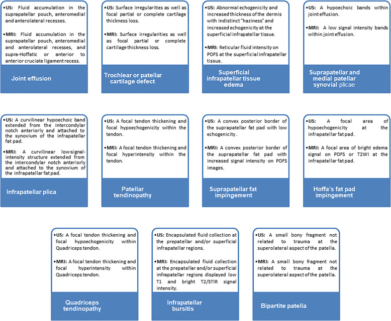
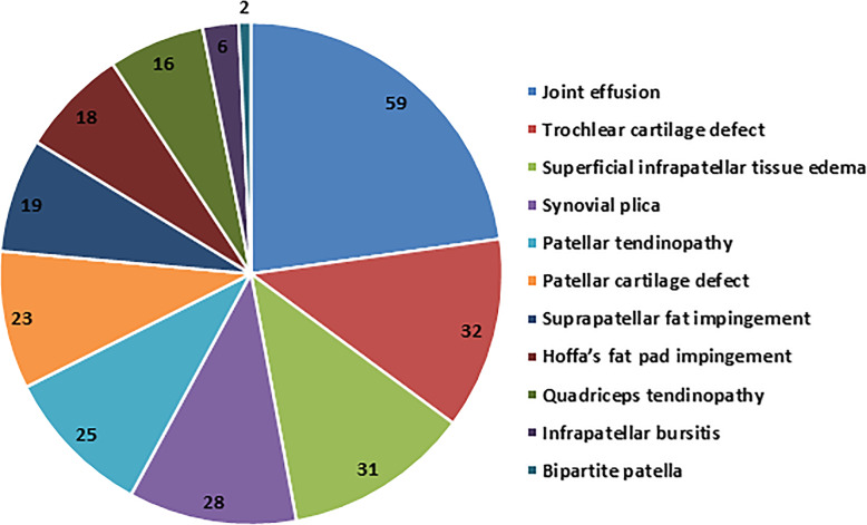
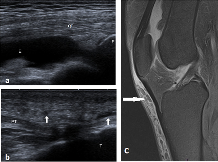
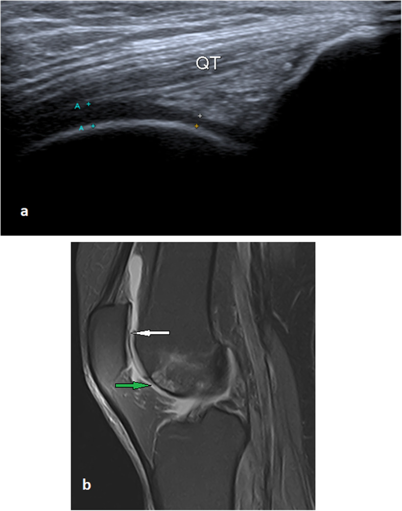
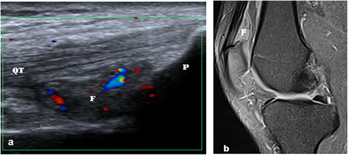
Methods and results: A prospective study was conducted on 143 patients with clinically confirmed AKP. All patients underwent ultrasonography and MRI examinations of the knee. The diagnostic accuracy of ultrasonography compared to MRI for evaluating different findings of possible causes of AKP were analyzed using receiver operating characteristic (ROC) curve and judged by area under curve (AUC). A total of 155 knees were included in the study; 26 knees showed no abnormalities, 19 knees showed positive MRI only, and 110 knees showed positive ultrasonography and MRI. Ultrasonography and MRI reported 11 different findings of possible causes of AKP or related to it. Joint effusion was the most common finding (38%) followed by trochlear cartilage defect (20.6%) and superficial infrapatellar subcutaneous edema (20%). The overall accuracy of ultrasonography was 85.3% sensitivity and 100% specificity. The ultrasonography provided the highest sensitivity (100%) in detecting bipartite patella, followed by 91.5% for joint effusion, and 87.5% for quadriceps tendinopathy. The ROC curve analysis of overall accuracy of ultrasonography showed an AUC of 0.93. The overall Kappa agreement between ultrasonography and MRI was good (k = 0.66).

Conclusion: Ultrasonography can be used to make a swift screening and assessment of painful anterior knee and as an alternative to MRI when it is unavailable or contraindicated.

Keywords: Anterior knee pain; Diagnostic accuracy; Knee joint; Magnetic resonance imaging; Ultrasonography.


Conflict of interest statement

The authors declare that they have no competing interests.

Figures

Fig. 1
Fig. 1
Flow chart of our study. AKP, anterior knee pain; n, number; MRI, magnetic resonance imaging
Fig. 2
Fig. 2
Ultrasonography and MRI findings of AKP. US, ultrasonography; MRI, magnetic resonance imaging; AKP, anterior knee pain; PDFS, proton density fat sat; STIR, short tau inversion recovery
Fig. 3
Fig. 3
Frequency of AKP findings as detected by MRI. AKP, anterior knee pain; MRI, magnetic resonance imaging
Fig. 4
Fig. 4
ROC curve analysis of ultrasonography for the identification of anterior knee findings compared to MRI. ROC, receiver operating characteristic curve; MRI, magnetic resonance imaging; AUC, area under the curve
Fig. 5
Fig. 5
A 44-year-old male with joint edema and anterior subcutaneous edema. a Long axis ultrasound image illustrates joint effusion in the suprapatellar pouch. b Long axis ultrasound scan illustrates hypoechoic subcutaneous reticulations anterior to the patellar tendon (arrows). c Sagittal PDFS MRI illustrates joint effusion, prepatellar, and superficial infrapatellar reticular fluid intensities of edema. QT, quadriceps tendon; PT, patellar tendon; E, effusion; P, patella; T, tibia
Fig. 6
Fig. 6
A 54-year-old male with focal patellar and trochlear partial thickness cartilage loss, mild joint effusion, and anterior infrapatellar subcutaneous edema of the left knee. a Long axis ultrasound image of the left knee illustrates a focal thinning of the anteroinferior aspect of the trochlear cartilage. b Sagittal PDFS MRI illustrates partial thickness cartilage loss of the patella (white arrow), partial-thickness cartilage loss of the anteroinferior surface of the trochlear cartilage with subchondral bone marrow changes (green arrow), joint effusion, and anterior subcutaneous edema. QT, quadriceps tendon
Fig. 7
Fig. 7
A 31-year-old male with a bipartite patella, joint effusion, and medial patellar plica. a Short axis ultrasound image illustrates the cleft between the patella and bipartite fragment at the superolateral pole of the patella. b Short axis ultrasound image illustrates joint effusion and medial patellar plica (arrow). c Axial T2WI MRI illustrates bipartite patella, joint effusion, and medial patellar plica. P, patella; E, effusion; B, bipartite patellar fragment
Fig. 8
Fig. 8
A 45-year-old female with right patellar tendinopathy and infrapatellar plica. a Long axis ultrasound image through the patellar tendon shows a focal thickening of the proximal part of the patellar tendon with a localized hypoechoic area sparing the anterior fibers. (White thick arrow). b and c Two subsequent sagittal PDFS MRI images through intercondylar notch show focal proximal tendon thickening and increased signal intensity (red arrow) sparing the anterior tendon fibers. Infrapatellar plica (white arrow) appears as a curvilinear high signal passing through Hoffa’s fat pad (not detected by ultrasound). P, patella; PT, patellar tendon
Fig. 9
Fig. 9
A 22-year-old male with left patellar tendinopathy. a Long axis ultrasound image demonstrates the thickened proximal part of the patellar tendon with cystic changes (*). Color Doppler mapping shows increased vascularity in and around the tendon. b Sagittal PDFS shows thickened proximal patellar tendon with fluid signal (*). P, patella
Fig. 10
Fig. 10
A 37-year-old male with left suprapatellar pad of fat impingement. a Long axis ultrasound image shows hypoechoic suprapatellar fat pad with convex posterior margin. Color Doppler ultrasound shows increased vascularity. b Sagittal PDFS MR image shows hyperintense suprapatellar fat pad with convex posterior margin. QT, quadriceps tendon; P, patella; F, suprapatellar fat pad

References

    1. Sanchis-alfonso V. Holistic approach to understanding anterior knee pain. Clinical implications. Knee Surg Sport Traumatol Arthrosc. 2014;22(10):2275–2285. - PubMed
    1. Whitlock K, Mosier B, Matzkin E (2018) Anterior Knee Pain: Diagnosis and Treatment. In: Katz J., Blauwet C., Schoenfeld A. (eds) Principles of Orthopedic Practice for Primary Care Providers. Springer, Cham. 19:313–327
    1. Samim M, Smitaman E, Lawrence D, Moukaddam H. MRI of anterior knee pain. Skeletal Radiol. 2014;43(7):875–893. - PubMed
    1. Sanchis-Alfonso V, Dye SF. How to deal with anterior knee pain in the active young patient. Sports Health. 2017;9(4):346–351. - PMC - PubMed
    1. Hosny S, McClatchie W, Sofat N, Hing BC (2012) Knee pain in adults & adolescents, diagnosis and treatment. Pain Perspect. October. IntechOpen

LinkOut - more resources