Review
. 2020 Oct 1;15(1):190.
doi: 10.1186/s11671-020-03418-6.

A Review on Enhancing the Antibacterial Activity of ZnO: Mechanisms and Microscopic Investigation

Affiliations
Review

A Review on Enhancing the Antibacterial Activity of ZnO: Mechanisms and Microscopic Investigation

Buzuayehu Abebe et al. Nanoscale Res Lett. .

Abstract

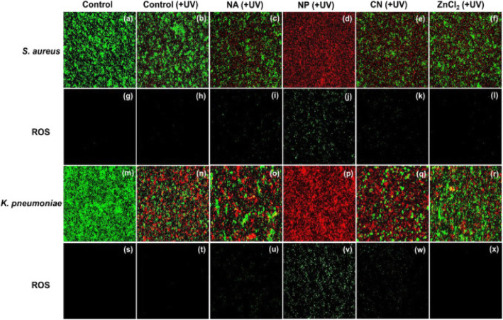
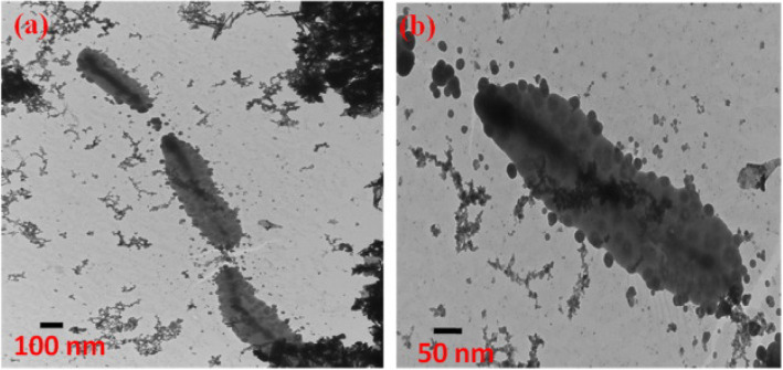
Metal oxide nanomaterials are one of the preferences as antibacterial active materials. Due to its distinctive electronic configuration and suitable properties, ZnO is one of the novel antibacterial active materials. Nowadays, researchers are making a serious effort to improve the antibacterial activities of ZnO by forming a composite with the same/different bandgap semiconductor materials and doping of ions. Applying capping agents such as polymers and plant extract that control the morphology and size of the nanomaterials and optimizing different conditions also enhance the antibacterial activity. Forming a nanocomposite and doping reduces the electron/hole recombination, increases the surface area to volume ratio, and also improves the stability towards dissolution and corrosion. The release of antimicrobial ions, electrostatic interaction, reactive oxygen species (ROS) generations are the crucial antibacterial activity mechanism. This review also presents a detailed discussion of the antibacterial activity improvement of ZnO by forming a composite, doping, and optimizing different conditions. The morphological analysis using scanning electron microscopy, field emission-scanning electron microscopy, field-emission transmission electron microscopy, fluorescence microscopy, and confocal microscopy can confirm the antibacterial activity and also supports for developing a satisfactory mechanism. Graphical abstract showing the metal oxides antibacterial mechanism and the fluorescence and scanning electron microscopic images.

Keywords: Antibacterial mechanism; Dopants; Metal oxide nanocomposites; Morphological investigation.


Conflict of interest statement

The authors declare that they have no competing interests.

Figures

Fig. 1
Fig. 1
Schematic illustration of the ZnO photocatalytic bacterial degradation mechanism
Fig. 2
Fig. 2
Different mechanisms of antimicrobial activity of ZnO NPs (represented by gray spheres). Reproduced from ref. [30] with permission from Springer Nature
Fig. 3
Fig. 3
Two-photon fluorescence microscopy and bright-field images of AZn2-labeled (aj) S. aureus and (kf) K. pneumoniae. Both species were treated with ZnO NPs (NA, NP, and CN) and ZnCl2 of 0.35 mM. Two-photon images were collected at 500–620 nm upon 780 nm excitation with femtosecond pulses. Reproduced from ref. [16] with permission from Elsevier
Fig. 4
Fig. 4
Dual immunofluorescence and ROS staining images for S. aureus (aj) and K. pneumoniae (kt) treated with nanoassemblies, NPs, conventional NPs, and ZnCl2 (0.35 mM) under dark conditions. Reproduced from ref. [16] with permission from Elsevier
Fig. 5
Fig. 5
Antibacterial activity mechanism of cerium oxide NPs. a Direct contact. b Indirect contact. Reproduced from ref. [18] with permission from Springer Nature
Fig. 6
Fig. 6
Fluorescence microscopy images of E. coli and S. aureus treated (a) without and (b) with ZnO–PVA NPs. Green fluorescence is characteristic of live cells, whereas red fluorescence is due to dead cells. Reproduced from ref. [14] with permission from The Royal Society of Chemistry
Fig. 7
Fig. 7
SEM images of untreated and treated bacterial strains using the prepared NRs; where ad are for P. aeruginosa, eh for S. aureus, il for C. Albicans, and mp for E. coli-GFP as a control (untreated) and treated with α-Mn2O3, γ–AlOOH, and γ–MnOOH NRs, respectively. Reproduced from ref. [9] with permission from The Royal Society of Chemistry
Fig. 8
Fig. 8
Typical Bio-TEM images of peanut-shaped ZnO with (a) E. coli, (b) K. pneumoniae, (c) S.Typhimurium, (d) S. aureus. Reproduced from ref. [59] with permission from Elsevier
Fig. 9
Fig. 9
a TEM images of E. coli mixed with Ag/Fe2O3/ZnO heterostructure. b Ag/Fe2O3/ZnO heterostructure anchored on the surface of E. coli. Reproduced from ref. [73] with permission from Elsevier
Fig. 10
Fig. 10
SEM images of bacterial cells. Samples of E. coli (a) untreated and (b) treated with 5NZO. Samples of A. baumannii (c) untreated and (d) treated with 5NZO. Red circles indicate areas of cell membrane disruption. Reproduced from ref. [21] with permission from The Royal Society of Chemistry
Fig. 11
Fig. 11
FESEM analysis of the bactericidal activity of nanobiocomposites and bare ESM. a, d Morphology of E. coli and S. aureus on bare ESM. b, e Efficiency of Ez towards E. coli and S. aureus; (c and f) efficiency of Eaz composites on the bacteria. Reproduced from ref. [23] with permission from the American Chemical Society

References

    1. Sukhdev A, Challa M, Narayani L, et al. Synthesis, phase transformation, and morphology of hausmannite Mn3O4 nanoparticles: photocatalytic and antibacterial investigations. Heliyon. 2020;6:e03245. doi: 10.1016/j.heliyon.2020.e03245. - DOI - PMC - PubMed
    1. Ohira T, Yamamoto O, Iida Y, Nakagawa ZE. Antibacterial activity of ZnO powder with crystallographic orientation. J Mater Sci Mater Med. 2008;19:1407–1412. doi: 10.1007/s10856-007-3246-8. - DOI - PubMed
    1. Kołodziejczak-Radzimska A, Jesionowski T. Zinc Oxide—From Synthesis to Application: A Review. Materials (Basel) 2014;7:2833–2881. doi: 10.3390/ma7042833. - DOI - PMC - PubMed
    1. Ong CB, Ng LY, Mohammad AW. A review of ZnO nanoparticles as solar photocatalysts: Synthesis, mechanisms and applications. Renew Sust Energ Rev. 2018;81:536–551. doi: 10.1016/j.rser.2017.08.020. - DOI
    1. Premanathan M, Karthikeyan K, Jeyasubramanian K, Manivannan G. Selective toxicity of ZnO nanoparticles toward Gram-positive bacteria and cancer cells by apoptosis through lipid peroxidation. Nanomed Nanotechnol Biol Med. 2011;7:184–192. doi: 10.1016/j.nano.2010.10.001. - DOI - PubMed

LinkOut - more resources