. 2021 Mar;73(3):520-529.
doi: 10.1002/art.41536. Epub 2021 Feb 8.

Dissecting the Cellular Mechanism of Prostacyclin Analog Iloprost in Reversing Vascular Dysfunction in Scleroderma

Affiliations

Dissecting the Cellular Mechanism of Prostacyclin Analog Iloprost in Reversing Vascular Dysfunction in Scleroderma

Pei-Suen Tsou et al. Arthritis Rheumatol. 2021 Mar.

Abstract

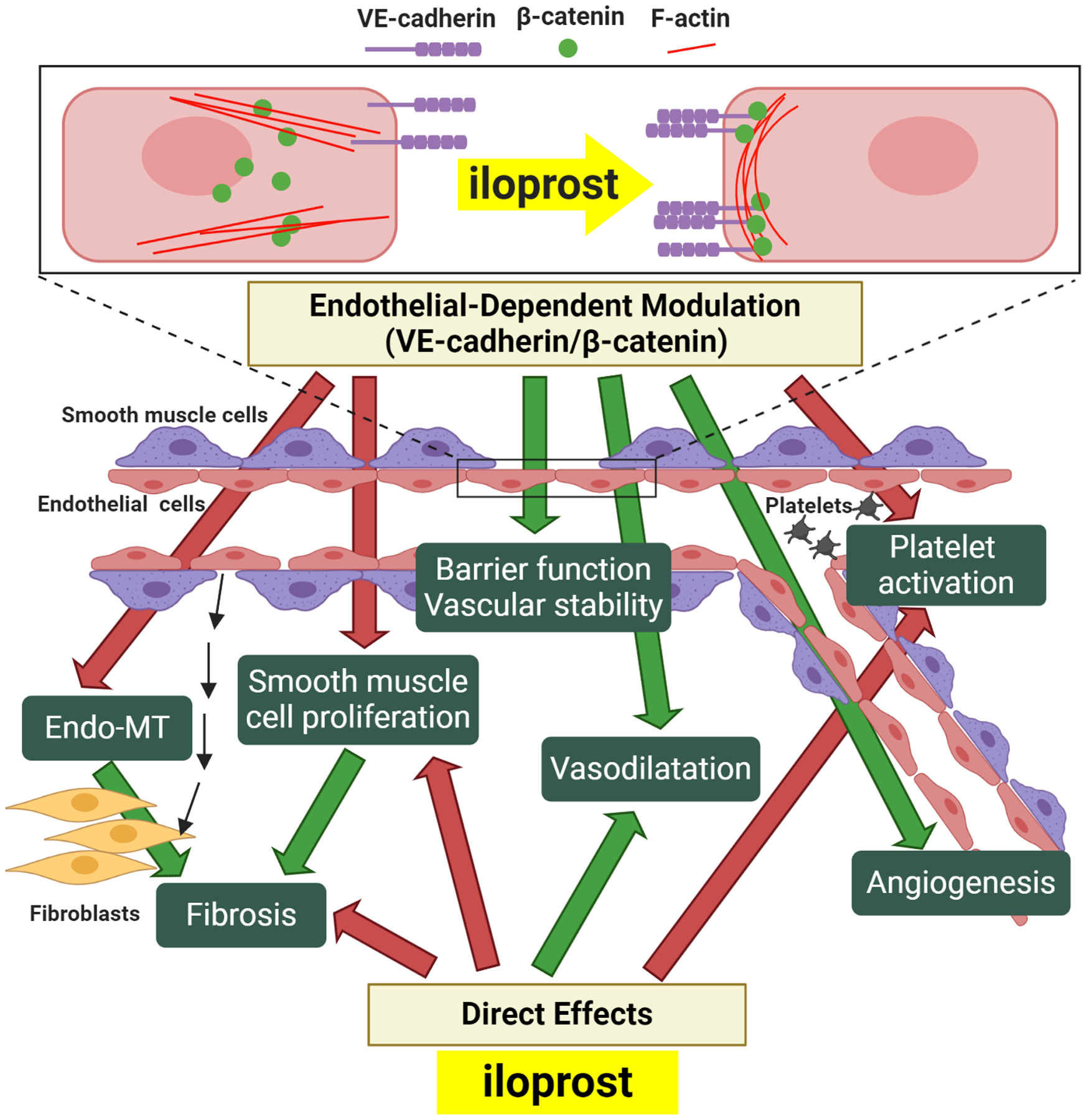
Objective: Intravenous iloprost improves Raynaud's phenomenon (RP) and promotes healing of digital ulcers in systemic sclerosis (SSc; scleroderma). Despite a short half-life, its clinical efficacy lasts weeks. Endothelial adherens junctions, which are formed by VE-cadherin clustering between endothelial cells (ECs), regulate endothelial properties including barrier function, endothelial-to-mesenchymal transition (EndoMT), and angiogenesis. We undertook this study to investigate the hypothesis that junctional disruption contributes to vascular dysfunction in SSc, and that the protective effect of iloprost is mediated by strengthening of those junctions.

Methods: Dermal ECs from SSc patients and healthy controls were isolated. The effect of iloprost on ECs was examined using immunofluorescence, permeability assays, Matrigel tube formation, and quantitative polymerase chain reaction.

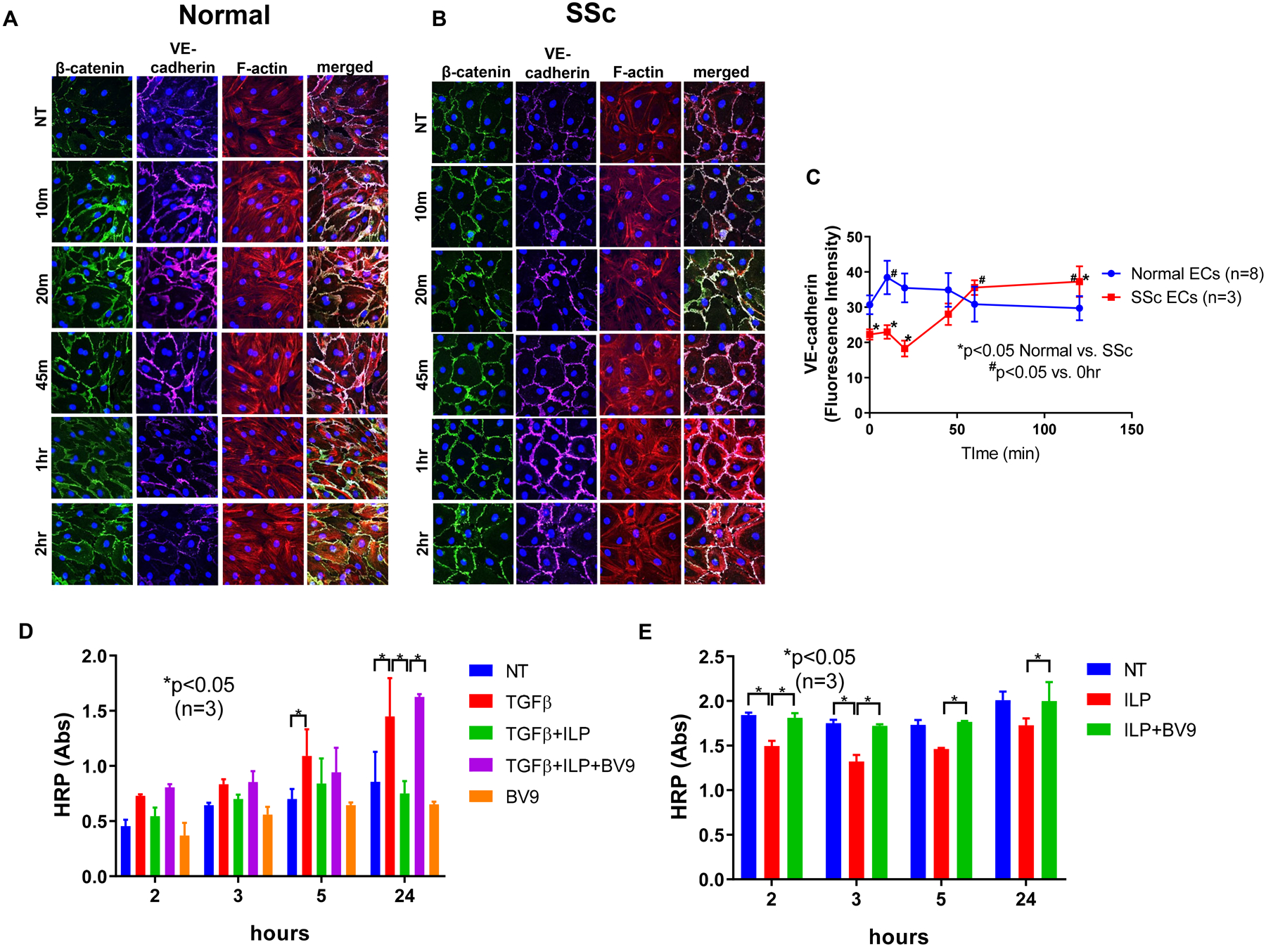
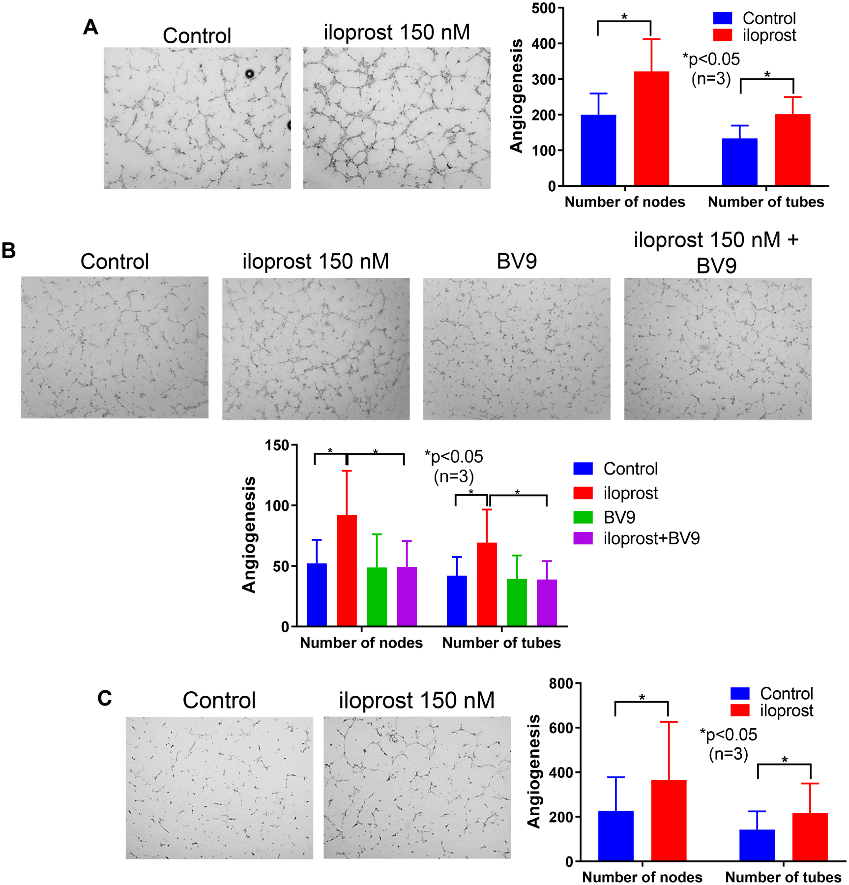
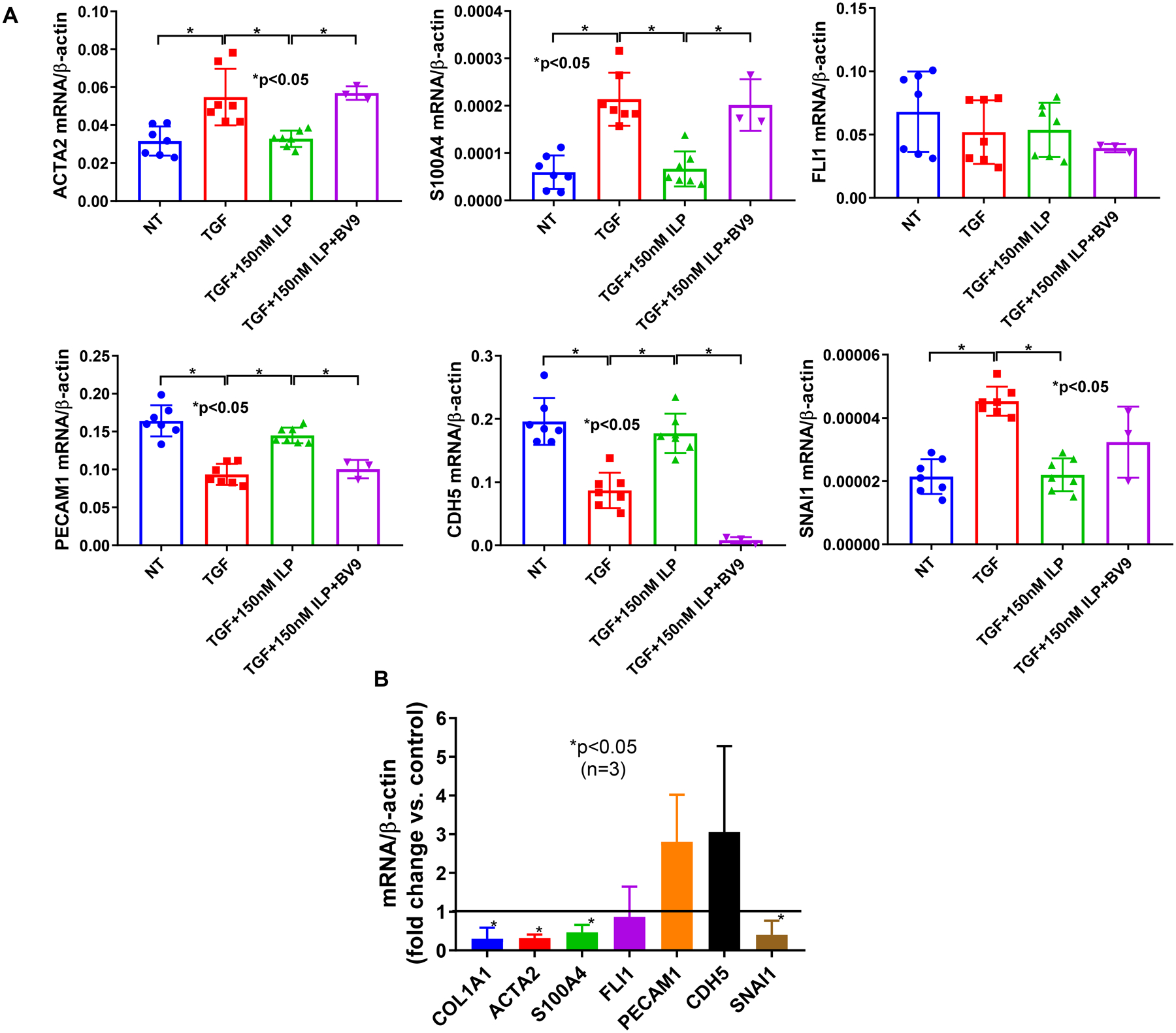
Results: Adherens junctions in SSc were disrupted compared to normal ECs, as indicated by reduced levels of VE-cadherin and increased permeability in SSc ECs (P < 0.05). Iloprost increased VE-cadherin clustering at junctions and restored junctional levels of VE-cadherin in SSc ECs (mean ± SD 37.3 ± 4.3 fluorescence units) compared to normal ECs (mean ± SD 29.7 ± 3.4 fluorescence units; P < 0.05), after 2 hours of iloprost incubation. In addition, iloprost reduced permeability of monolayers, increased tubulogenesis, and blocked EndoMT in both normal and SSc ECs (n ≥ 3; P < 0.05). The effects in normal ECs were inhibited by a function-blocking antibody that prevents junctional clustering of VE-cadherin.

Conclusion: Our data suggest that the long-lasting effects of iloprost reflect its ability to stabilize adherens junctions, resulting in increased tubulogenesis and barrier function and reduced EndoMT. These findings provide a mechanistic basis for the use of iloprost in treating SSc patients with RP and digital ulcers.


Conflict of interest statement

Competing interests: Dr. Khanna is the Chief Medical Officer and stock holder in Eicos Sciences, Inc, which is currently conducting Phase 3 clinical trial for intravenous iloprost in the treatment of digital ischemic episodes due to systemic sclerosis.

Figures

Figure 1.
Figure 1.. Effect of iloprost on VE-cadherin localization and cell permeability.
Immunofluorescence of VE-cadherin, β-catenin, and F-actin in ECs were visualized using a Nikon A1 confocal microscope and pictures were taken at 600x. Cell permeability was assessed by measuring HRP movement through EC monolayers using the Endothelial Transwell Permeability Assay Kit. Cells were treated with iloprost (150nM) and/or TGFβ (10ng/ml) at various time points. BV9 (25 μg/ml) was used to pre-treat the cells for 30 min before iloprost and TGFβ were added. (A) Iloprost increased junctional clustering of VE-cadherin and β-catenin as soon as 10 min of incubation in normal ECs; (B) Iloprost showed a delayed but more prolonged effect on VE-cadherin and β-catenin clustering in SSc ECs. Disorganized F-actin filaments in SSc ECs were also observed; (C) Quantification of fluorescent signal of VE-cadherin showed significant reduction of VE-cadherin in SSc ECs compared to normal ECs at baseline. After iloprost, VE-cadherin intensity was greater in SSc compared to normal ECs; (D) In normal ECs, TGFβ increased permeability as measured by HRP movement through EC monolayers. Iloprost inhibited permeability of EC monolayers while blockade of VE-cadherin by BV9 reversed it. (E) In SSc ECs, iloprost inhibited the increased permeability of these cells while the function blocking antibody to VE-cadherin (BV9) prevented the effects of iloprost. Experiments were done with at least 3 subject-derived lines. Results are expressed as mean +/− SD and p<0.05 was considered significant. NT: not treated; ILP: iloprost; Abs: absorbance
Figure 2.
Figure 2.. Effect of iloprost on EC angiogenesis.
Angiogenesis of ECs was measured using an in vitro Matrigel tube formation assay. For A and C, ECs were pre-treated with iloprost (150nM) overnight before they were plated on Matrigel. After 6 hours, cells were fixed and stained. For B, BV9 (25 μg/ml) was used to pre-treat the ECs for 30 min before iloprost was added. The cells were cultured for 8 hours before they were fixed and stained. (A) and (B) Iloprost increased tubulogenesis in normal ECs and this was reduced by a function blocking antibody to VE-cadherin (BV9); (C) Iloprost induced tubulogenesis in SSc ECs. Experiments were done with 3 subject-derived lines. Results are expressed as mean +/− SD and p<0.05 was considered significant.
Figure 3.
Figure 3.. Effect of iloprost on Endo-MT in ECs.
Normal ECs were treated with 10 ng/ml TGFβ and/or 150 nM of iloprost for 3 days. BV9 was added 30 min before the addition of various treatments. Cellular markers for Endo-MT were measured using qPCR. (A) Iloprost inhibited TGFβ-induced Endo-MT in normal ECs, and the effect was blocked by BV9, a VE-cadherin antibody; (B) Iloprost inhibited the Endo-MT phenotype in SSc ECs. Experiments were done with 3–7 subject-derived lines. Results are expressed as mean +/− SD and p<0.05 was considered significant.
Figure 4.
Figure 4.. Vascular protective effect of iloprost in SSc.
In SSc ECs, Iloprost increases VE-cadherin and β-catenin clustering at the adherens junction, accompanied with accumulation of peripheral F-actin. Increased interaction and signaling of VE-caderin/β-catenin will promote protective endothelial functions and vascular stability including an increase in barrier function, promotion of angiogenesis, inhibition of Endo-MT, and increased NO activity, which will contribute to vasodilation, inhibition of platelet activation, and blockade of smooth muscle cell proliferation. Endo-MT and smooth muscle proliferation can contribute to intravascular and extravascular fibrosis, and to microvascular rarefaction. In addition to these endothelial-dependent modulatory effects, iloprost can also act directly on platelets, fibroblasts, and smooth muscle cells to inhibit platelet activation and fibrosis, and promote vasodilation. Inhibitory effects are highlighted by red arrows, and positive effects are highlighted with green arrows.

References

    1. Nagaraja V, Cerinic MM, Furst DE, Kuwana M, Allanore Y, Denton CP, et al. Current and future outlook on disease modification and defining low disease activity in systemic sclerosis. Arthritis Rheumatol. 2020. - PMC - PubMed
    1. Olschewski H, Rose F, Schermuly R, Ghofrani HA, Enke B, Olschewski A, et al. Prostacyclin and its analogues in the treatment of pulmonary hypertension. Pharmacology & therapeutics. 2004;102(2):139–53. - PubMed
    1. Kowal-Bielecka O, Fransen J, Avouac J, Becker M, Kulak A, Allanore Y, et al. Update of EULAR recommendations for the treatment of systemic sclerosis. Ann Rheum Dis. 2017;76(8):1327–39. - PubMed
    1. Birukova AA, Zagranichnaya T, Fu P, Alekseeva E, Chen W, Jacobson JR, et al. Prostaglandins PGE(2) and PGI(2) promote endothelial barrier enhancement via PKA- and Epac1/Rap1-dependent Rac activation. Exp Cell Res. 2007;313(11):2504–20. - PMC - PubMed
    1. Baumer Y, Drenckhahn D, Waschke J. cAMP induced Rac 1-mediated cytoskeletal reorganization in microvascular endothelium. Histochem Cell Biol. 2008;129(6):765–78. - PubMed

Publication types

MeSH terms