Clinical Trial
. 2021 Jan 4;131(1):e141500.
doi: 10.1172/JCI141500.

Transcriptome-directed analysis for Mendelian disease diagnosis overcomes limitations of conventional genomic testing

Affiliations
Clinical Trial

Transcriptome-directed analysis for Mendelian disease diagnosis overcomes limitations of conventional genomic testing

David R Murdock et al. J Clin Invest. .

Abstract

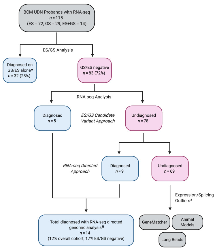
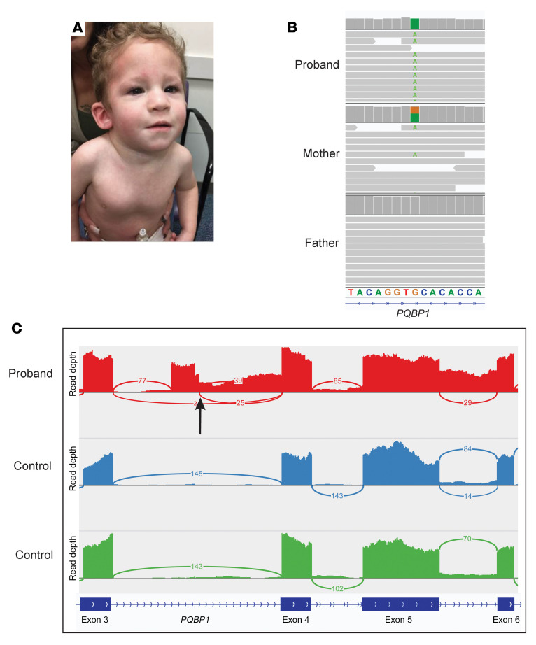
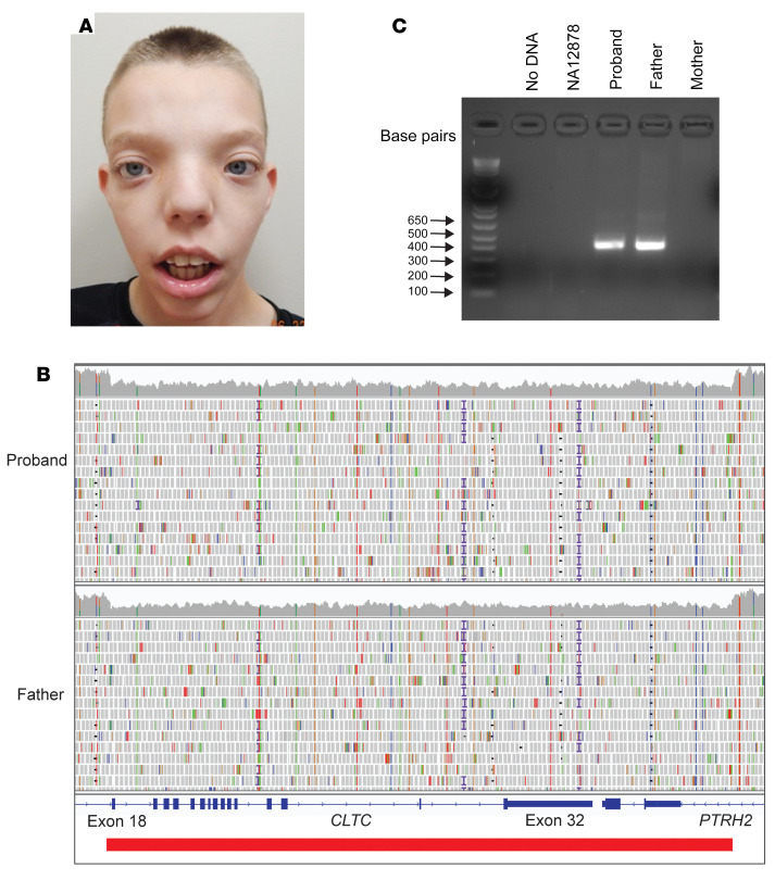
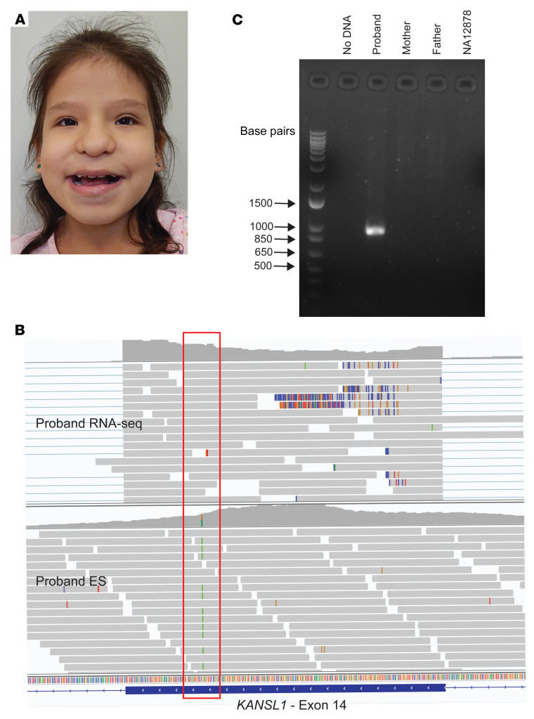
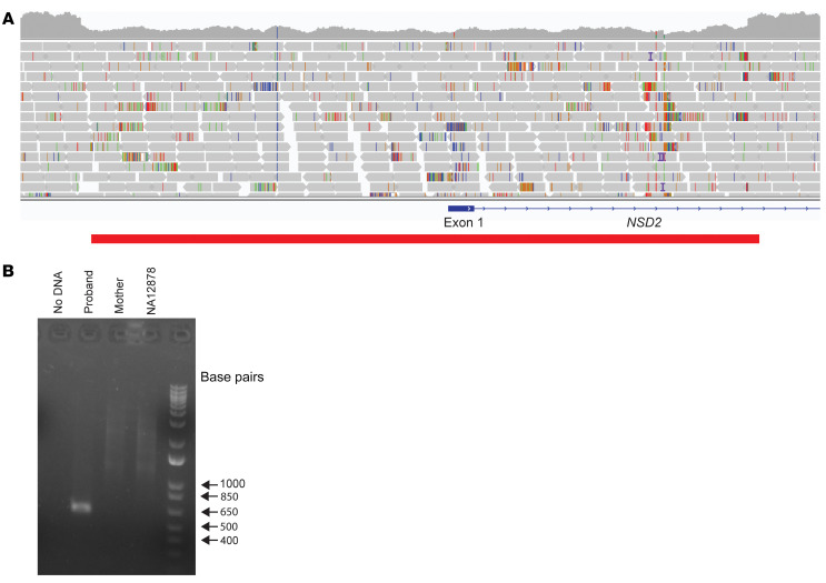
BACKGROUNDTranscriptome sequencing (RNA-seq) improves diagnostic rates in individuals with suspected Mendelian conditions to varying degrees, primarily by directing the prioritization of candidate DNA variants identified on exome or genome sequencing (ES/GS). Here we implemented an RNA-seq-guided method to diagnose individuals across a wide range of ages and clinical phenotypes.METHODSOne hundred fifteen undiagnosed adult and pediatric patients with diverse phenotypes and 67 family members (182 total individuals) underwent RNA-seq from whole blood and skin fibroblasts at the Baylor College of Medicine (BCM) Undiagnosed Diseases Network clinical site from 2014 to 2020. We implemented a workflow to detect outliers in gene expression and splicing for cases that remained undiagnosed despite standard genomic and transcriptomic analysis.RESULTSThe transcriptome-directed approach resulted in a diagnostic rate of 12% across the entire cohort, or 17% after excluding cases solved on ES/GS alone. Newly diagnosed conditions included Koolen-de Vries syndrome (KANSL1), Renpenning syndrome (PQBP1), TBCK-associated encephalopathy, NSD2- and CLTC-related intellectual disability, and others, all with negative conventional genomic testing, including ES and chromosomal microarray (CMA). Skin fibroblasts exhibited higher and more consistent expression of clinically relevant genes than whole blood. In solved cases with RNA-seq from both tissues, the causative defect was missed in blood in half the cases but none from fibroblasts.CONCLUSIONSFor our cohort of undiagnosed individuals with suspected Mendelian conditions, transcriptome-directed genomic analysis facilitated diagnoses, primarily through the identification of variants missed on ES and CMA.TRIAL REGISTRATIONNot applicable.FUNDINGNIH Common Fund, BCM Intellectual and Developmental Disabilities Research Center, Eunice Kennedy Shriver National Institute of Child Health & Human Development.

Keywords: Diagnostics; Genetic diseases; Genetics; Molecular genetics.


Conflict of interest statement

Conflict of interest: The authors have declared that no conflict of interest exists.

Figures

Figure 1
Figure 1. Flow diagram outlining BCM UDN RNA-seq diagnostic research process.
*Cases diagnosed on initial review of ES/GS without needing RNA-seq. #Undiagnosed but with expression/splicing outliers prompting follow-up studies for potentially novel disease gene discovery. §Five cases diagnosed with ES/GS candidate variant approach were validated using RNA-seq–directed approach. ES, exome sequencing; GS, genome sequencing.
Figure 2
Figure 2. Principal component analysis (PCA) plot of gene expression (TPM) in whole blood (blue) and skin fibroblasts (red).
Two distinct tissue clusters are visible; however, less variability is present in skin fibroblasts. This suggests that fibroblasts may be better for detecting clinically relevant differences in gene expression by RNA-seq. TPM, transcripts per million; FB, fibroblast; WB, whole blood.
Figure 3
Figure 3. Causative genomic variants identified through RNA-seq–directed genomic analysis.
Variant types included synonymous (n = 1), near intronic (3–50 bp from canonical exon boundary, n = 2), deep intronic (>50 bp away from canonical exon boundary, n = 4), promoter (n = 1), and canonical splice site SNVs (n = 1) as well as both coding (n = 3) and noncoding (n = 2) deletion CNVs. SNV, single-nucleotide variant; CNV, copy number variant.
Figure 4
Figure 4. Case 1 — Renpenning syndrome.
(A) Dysmorphic features, including microbrachycephaly, deep-set eyes, midface hypoplasia, broad nose, and low-set ears. (B) GS with hemizygous deep intronic PQBPQ1 variant (green) inherited from heterozygous mother. (C) RNA-seq sashimi plot from whole blood showing out-of-frame pseudoexon and distal intron retention in the proband (red) but absent from controls (blue/green). Black arrow indicates the location of PQBP1 intronic variant. GS, genome sequencing.
Figure 5
Figure 5. Case 2 — CLTC-associated ID syndrome.
(A) Dysmorphic features, including hypertelorism, broad forehead, and low posterior hairline. (B) CLTC deletion (red bar) on GS encompassing exons 18–32. (C) PCR confirmation of deletion in proband and father but absent from mother and control (NA12878). Expected size with deletion = 391 bp. GS, genome sequencing; ID, intellectual disability.
Figure 6
Figure 6. Case 3 — Koolen–de Vries syndrome.
(A) Dysmorphic features, including blepharophimosis, epicanthal folds, protruding ears, and a tubular nose with a broad tip. (B) Exon 14 SNP (red box) in ES but absent in RNA-seq consistent with loss of that allele. (C) PCR confirmation of de novo deletion in proband absent from parents and control (NA12878). Expected size with deletion = 926 bp. ES, exome sequencing; SNP, single-nucleotide polymorphism.
Figure 7
Figure 7. Case 4 — NSD2-associated ID syndrome.
(A) A 3.9 kb deletion (red bar) including the NSD2 transcription start site and upstream promoter/enhancer elements detected by GS. (B) PCR confirmation of deletion in proband absent from mother and control (NA12878). Expected size with deletion = 635 bp. GS, genome sequencing; ID, intellectual disability.

References

    1. Posey JE, et al. Molecular diagnostic experience of whole-exome sequencing in adult patients. Genet Med. 2016;18(7):678–685. doi: 10.1038/gim.2015.142. - DOI - PMC - PubMed
    1. Yang Y, et al. Molecular findings among patients referred for clinical whole-exome sequencing. JAMA. 2014;312(18):1870–1879. doi: 10.1001/jama.2014.14601. - DOI - PMC - PubMed
    1. Splinter K, et al. Effect of genetic diagnosis on patients with previously undiagnosed disease. N Engl J Med. 2018;379(22):2131–2139. doi: 10.1056/NEJMoa1714458. - DOI - PMC - PubMed
    1. Gonorazky HD, et al. Expanding the boundaries of RNA sequencing as a diagnostic tool for rare Mendelian disease. Am J Hum Genet. 2019;104(5):1007. doi: 10.1016/j.ajhg.2019.04.004. - DOI - PMC - PubMed
    1. Kremer LS, et al. Genetic diagnosis of Mendelian disorders via RNA sequencing. Nat Commun. 2017;8:15824. - PMC - PubMed

Publication types