

Comparison of virtual non-contrast dual-energy CT and a true non-contrast CT for contouring in radiotherapy of 3D printed lung tumour models in motion: a phantom study
- PMID: 33002387
- PMCID: PMC7715995
- DOI: 10.1259/bjr.20200152
Comparison of virtual non-contrast dual-energy CT and a true non-contrast CT for contouring in radiotherapy of 3D printed lung tumour models in motion: a phantom study
Abstract
Objectives: This work aims to investigate whether virtual non-contrast (VNC) dual-energy CT(DECT) of contrasted lung tumours can be used as an alternative for true non-contrast (TNC) images in radiotherapy. Two DECT techniques and a TNC CT were compared and influences on gross tumour volume (GTV) volume and CT number from motion artefacts in three-dimensional printed lung tumour models (LTM) in amotion phantom were examined.
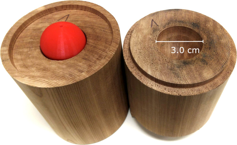
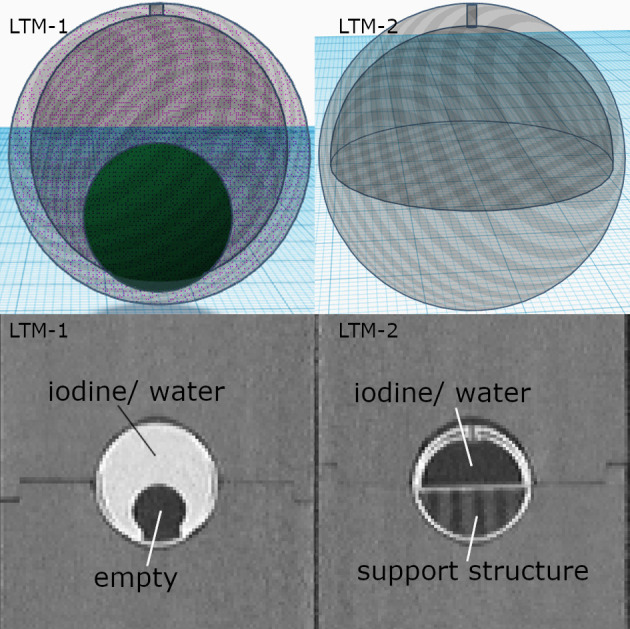
Methods: Two spherical LTMs (diameter 3.0 cm) with different inner shapes were created in a three-dimensional printer. The inner shapes contained water or iodine (concentration 5 mg ml-1) and were scanned with a dual-source DECT (ds-DECT), single-source sequential DECT (ss-DECT) and TNC CT in a respiratory motion phantom (15 breaths/min, amplitude 1.5 cm). CT number and volume of LTMs were measured. Therefore, two GTVs were contoured.
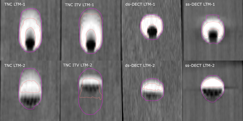
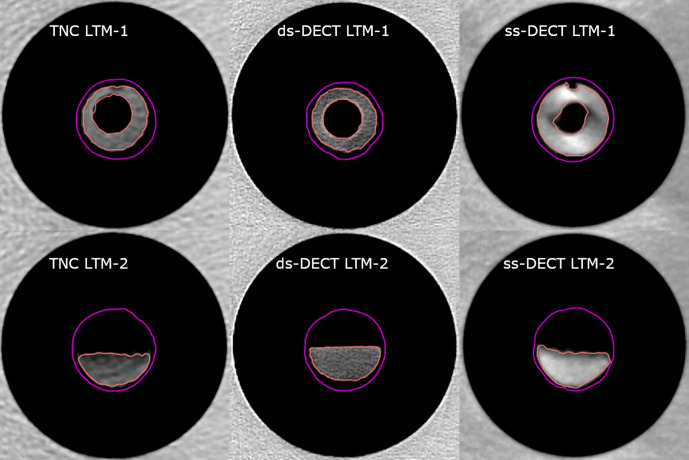
Results: Deviations in GTV volume (outer shape) of LTMs in motion for contrast-enhanced ss-DECT and ds-DECT VNC images compared to TNC images are not significant (p > 0.05). Relative GTV volume and CT number deviations (inner shapes) of LTMs in motion were 6.6 ± 0.6% and 104.4 ± 71.2 HU between ss-DECT and TNC CT and -8.4 ± 10.6% and 25.5 ± 58.5 HU between ds-DECT and TNC, respectively.
Conclusion: ss-DECT VNC images could not sufficiently subtract iodine from water in LTMs inmotion, whereas ds-DECT VNC images might be a valid alternative to a TNC CT.
Advances in knowledge: ds-DECT provides a contrasted image for contouring and a non-contrasted image for radiotherapy treatment planning for LTM in motion.
Figures
Similar articles
-
Deep learning-based virtual noncontrast CT for volumetric modulated arc therapy planning: Comparison with a dual-energy CT-based approach.Med Phys. 2020 Feb;47(2):371-379. doi: 10.1002/mp.13925. Epub 2019 Dec 3. Med Phys. 2020. PMID: 31733105
-
Virtual non-contrast images from dual-energy CT angiography of the abdominal aorta and femoral arteries: comparison with true non-contrast CT images.Br J Radiol. 2022 Sep 1;95(1138):20220378. doi: 10.1259/bjr.20220378. Epub 2022 Sep 8. Br J Radiol. 2022. PMID: 36039820 Free PMC article.
-
Virtual versus true non-contrast dual-energy CT imaging for the diagnosis of aortic intramural hematoma.Eur Radiol. 2019 Dec;29(12):6762-6771. doi: 10.1007/s00330-019-06322-5. Epub 2019 Jul 1. Eur Radiol. 2019. PMID: 31264015
-
Dual energy imaging in cardiothoracic pathologies: A primer for radiologists and clinicians.Eur J Radiol Open. 2021 Jan 20;8:100324. doi: 10.1016/j.ejro.2021.100324. eCollection 2021. Eur J Radiol Open. 2021. PMID: 33532519 Free PMC article. Review.
-
Dual-energy CT for gastrointestinal bleeding.BJR Open. 2023 Mar 22;5(1):20220054. doi: 10.1259/bjro.20220054. eCollection 2023. BJR Open. 2023. PMID: 37035764 Free PMC article. Review.
Cited by
-
Dual-Energy CT in Oncologic Imaging.Tomography. 2024 Feb 23;10(3):299-319. doi: 10.3390/tomography10030024. Tomography. 2024. PMID: 38535766 Free PMC article. Review.
-
Generation of deep learning based virtual non-contrast CT using dual-layer dual-energy CT and its application to planning CT for radiotherapy.PLoS One. 2024 Dec 30;19(12):e0316099. doi: 10.1371/journal.pone.0316099. eCollection 2024. PLoS One. 2024. PMID: 39775325 Free PMC article.
-
Split-filter dual energy computed tomography radiotherapy: From calibration to image guidance.Phys Imaging Radiat Oncol. 2023 Sep 25;28:100495. doi: 10.1016/j.phro.2023.100495. eCollection 2023 Oct. Phys Imaging Radiat Oncol. 2023. PMID: 37876826 Free PMC article.
-
Quantitative parameters of enhanced dual-energy computed tomography for differentiating lung cancers from benign lesions in solid pulmonary nodules.Front Oncol. 2022 Oct 6;12:1027985. doi: 10.3389/fonc.2022.1027985. eCollection 2022. Front Oncol. 2022. PMID: 36276069 Free PMC article.
References
-
- Sheen H, Shin H-B, Cho S, Cho J, Han Y. Feasibility of dual-energy computed tomography in radiation therapy planning. J Korean Phys Soc 2017; 71: 1056–63. doi: 10.3938/jkps.71.1056 - DOI
Publication types
MeSH terms
Substances
LinkOut - more resources
Full Text Sources
Medical
Miscellaneous
