

The HDAC Inhibitor Domatinostat Promotes Cell-Cycle Arrest, Induces Apoptosis, and Increases Immunogenicity of Merkel Cell Carcinoma Cells
- PMID: 33002502
- PMCID: PMC7987731
- DOI: 10.1016/j.jid.2020.08.023
The HDAC Inhibitor Domatinostat Promotes Cell-Cycle Arrest, Induces Apoptosis, and Increases Immunogenicity of Merkel Cell Carcinoma Cells
Abstract
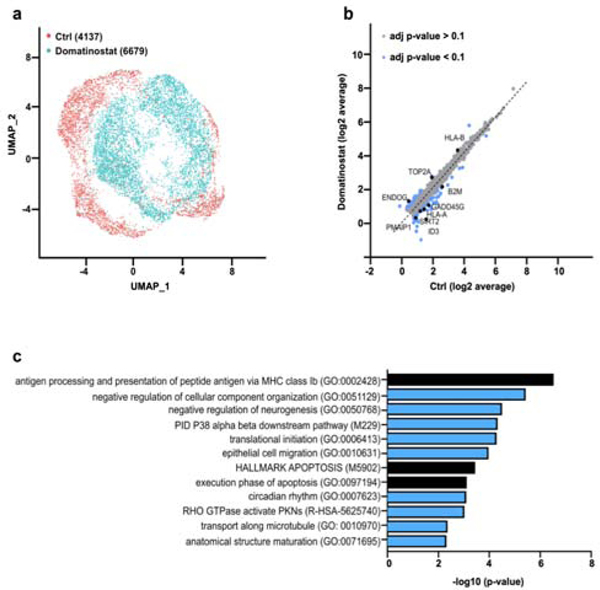
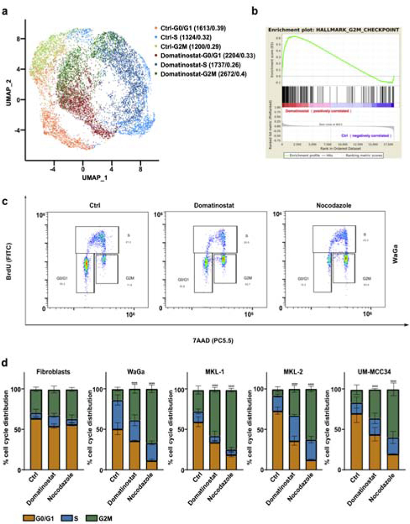
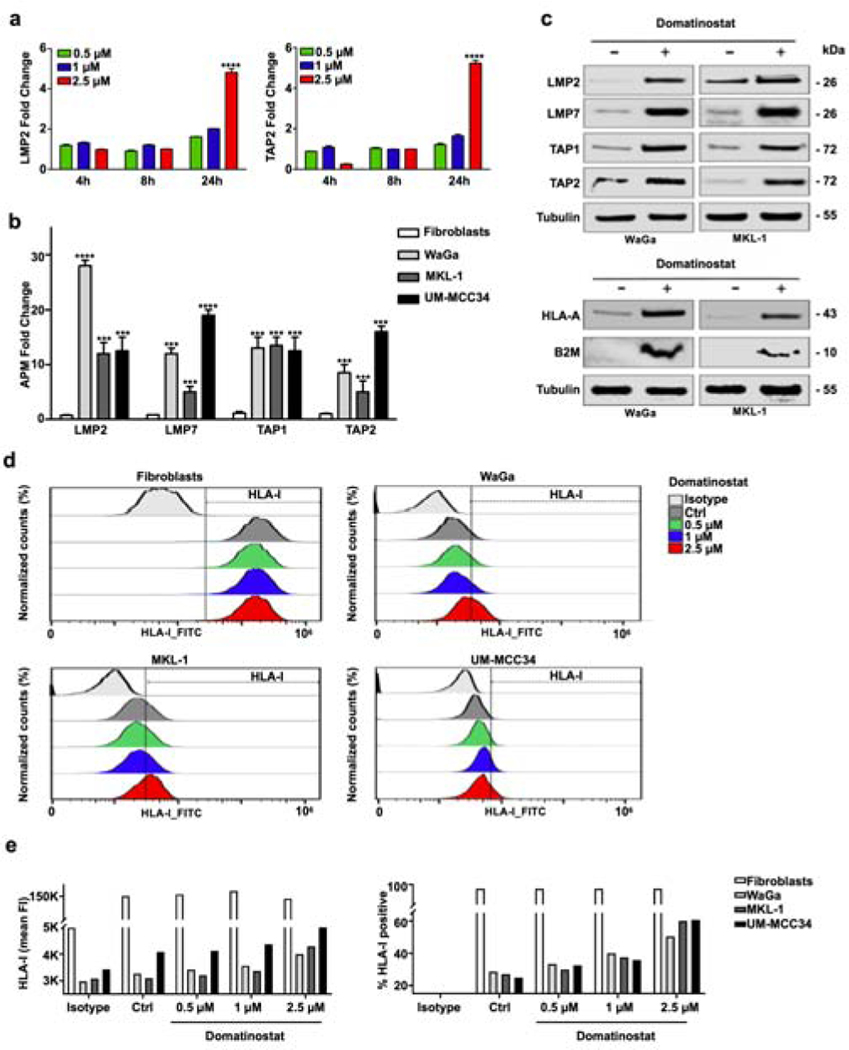
Merkel cell carcinoma (MCC) is a rare, highly aggressive skin cancer for which immune modulation by immune checkpoint inhibitors shows remarkable response rates. However, primary or secondary resistance to immunotherapy prevents benefits in a significant proportion of patients. For MCC, one immune escape mechanism is insufficient for recognition by T cells owing to the downregulation of major histocompatibility complex I surface expression. Histone deacetylase inhibitors have been demonstrated to epigenetically reverse the low major histocompatibility complex I expression caused by the downregulation of the antigen-processing machinery. Domatinostat, an orally available small-molecule inhibitor targeting histone deacetylase class I, is currently in clinical evaluation to overcome resistance to immunotherapy. In this study, we present preclinical data on domatinostat's efficacy and mode of action in MCC. Single-cell RNA sequencing revealed a distinct gene expression signature of antigen processing and presentation, cell-cycle arrest, and execution phase of apoptosis on treatment. Accordingly, functional assays showed that domatinostat induced G2M arrest and apoptosis. In the surviving cells, antigen-processing machinery component gene transcription and translation were upregulated, consequently resulting in increased major histocompatibility complex I surface expression. Altogether, domatinostat not only exerts direct antitumoral effects but also restores HLA class I surface expression on MCC cells, therefore, restoring surviving MCC cells' susceptibility to recognition and elimination by cognate cytotoxic T cells.
Copyright © 2021 The Authors. Published by Elsevier Inc. All rights reserved.
Conflict of interest statement
CONFLICT OF INTEREST
None of other authors indicated any potential conflicts of interest.
Figures

Similar articles
-
MHC class-I downregulation in PD-1/PD-L1 inhibitor refractory Merkel cell carcinoma and its potential reversal by histone deacetylase inhibition: a case series.Cancer Immunol Immunother. 2019 Jun;68(6):983-990. doi: 10.1007/s00262-019-02341-9. Epub 2019 Apr 16. Cancer Immunol Immunother. 2019. PMID: 30993371 Free PMC article.
-
Epigenetic priming restores the HLA class-I antigen processing machinery expression in Merkel cell carcinoma.Sci Rep. 2017 May 23;7(1):2290. doi: 10.1038/s41598-017-02608-0. Sci Rep. 2017. PMID: 28536458 Free PMC article.
-
Domatinostat favors the immunotherapy response by modulating the tumor immune microenvironment (TIME).J Immunother Cancer. 2019 Nov 8;7(1):294. doi: 10.1186/s40425-019-0745-3. J Immunother Cancer. 2019. PMID: 31703604 Free PMC article.
-
T-Cell Responses in Merkel Cell Carcinoma: Implications for Improved Immune Checkpoint Blockade and Other Therapeutic Options.Int J Mol Sci. 2021 Aug 12;22(16):8679. doi: 10.3390/ijms22168679. Int J Mol Sci. 2021. PMID: 34445385 Free PMC article. Review.
-
[Immune checkpoint inhibition in Merkel cell carcinoma].Hautarzt. 2019 Sep;70(9):684-690. doi: 10.1007/s00105-019-4465-x. Hautarzt. 2019. PMID: 31468071 Review. German.
Cited by
-
Viral Manipulation of the Host Epigenome as a Driver of Virus-Induced Oncogenesis.Microorganisms. 2021 May 30;9(6):1179. doi: 10.3390/microorganisms9061179. Microorganisms. 2021. PMID: 34070716 Free PMC article. Review.
-
Three-year survival, correlates and salvage therapies in patients receiving first-line pembrolizumab for advanced Merkel cell carcinoma.J Immunother Cancer. 2021 Apr;9(4):e002478. doi: 10.1136/jitc-2021-002478. J Immunother Cancer. 2021. PMID: 33879601 Free PMC article. Clinical Trial.
-
Genomic evidence suggests that cutaneous neuroendocrine carcinomas can arise from squamous dysplastic precursors.Mod Pathol. 2022 Apr;35(4):506-514. doi: 10.1038/s41379-021-00928-1. Epub 2021 Sep 30. Mod Pathol. 2022. PMID: 34593967 Free PMC article.
-
Selective Inhibition of Aurora Kinase A by AK-01/LY3295668 Attenuates MCC Tumor Growth by Inducing MCC Cell Cycle Arrest and Apoptosis.Cancers (Basel). 2021 Jul 23;13(15):3708. doi: 10.3390/cancers13153708. Cancers (Basel). 2021. PMID: 34359608 Free PMC article.
-
Perspectives in immunotherapy: meeting report from the immunotherapy bridge (December 2nd-3rd, 2020, Italy).J Transl Med. 2021 Jun 2;19(1):238. doi: 10.1186/s12967-021-02895-2. J Transl Med. 2021. PMID: 34078406 Free PMC article.
References
-
- Bolden JE, Peart MJ, Johnstone RW. Anticancer activities of histone deacetylase inhibitors. Nat Rev Drug Discov 2006;5(9):769–84. - PubMed
Publication types
MeSH terms
Substances
Grants and funding
LinkOut - more resources
Full Text Sources
Other Literature Sources
Medical
Research Materials
